STRESS REGULATES THE LYMPHOCYTE HOMING
RECEPTOR CD62L (L-SELECTIN)
Gisele Gus Manfro
1,2, Carlos Alexandre Netto¹, Mark Pollack²,
Kelin Maggioni Mezzomo³, Frederic Preffer², Richard Kradin²
ABSTRACT - Based on a previous study showing that panic disorder patients had increased expression of naïve phenotype lymphocytes (CD45RA+ and CD62L+), increased plasma cortisol, as well as decreased interleukin-2 (IL-interleukin-2) producion, we hypothesized that changes in the percentage of expression of these lymphocyte surface molecules could be related to the substances released by the hypothalamic-pituitary-adrenal (HPA) axis and possibly associated to panic disorder (cortisol, IL-2, serotonin and epinephrine). In order to study the altered expression, blood mononuclear cells of normal volunteers were stimulated with mitogen, in the presence of dexamethasone, IL-2, serotonin and epinephrin. CD62L is decreased by IL-2 in vitro. Serotonin and epinephrine did not promote changes in the expression of these surface molecules. The results of the ex vivo study are in agreement with a previous clinical study with panic patients. It could be suggested that stress is responsible for certain immunologic dysfunctions and new studies should be conducted.
KEY WORDS: corticosteroids, interleukin-2, lymphocyte migration, adhesion molecules, panic disorder.
Regulação do receptor de alojamento linfocitário CD62L (L Regulação do receptor de alojamento linfocitário CD62L (LRegulação do receptor de alojamento linfocitário CD62L (L
Regulação do receptor de alojamento linfocitário CD62L (LRegulação do receptor de alojamento linfocitário CD62L (L-selectina) pelo estresse-selectina) pelo estresse-selectina) pelo estresse-selectina) pelo estresse-selectina) pelo estresse
RESUMO - Baseado em estudo prévio que demonstrou que os pacientes com transtorno do pânico apresentavam aumento na porcentagem de expressão de linfócitos com fenótipo virgem (CD45RA+ e CD62L+), aumento no cortisol plasmático, assim como diminuição na produção de interleucinas, foi sugerido que as alterações na porcentagem de expressão dessas moléculas de superfície dos linfócitos poderia estar relacionada com a liberação de substâncias pelo eixo hipotálamo-hipófise-adrenal (HHA) e possivelmente associada ao transtorno do pânico (cortisol, IL-2, serotonina e epinefrina). Com o objetivo de estudar essas alterações, células mononucleares do sangue periférico de voluntários normais foram estimuladas com mitógeno, na presença de dexametasona, IL-2, serotonina e epinefrina. A expressão de CD62L “in vitro” é diminuida com IL-2. Serotonina e epinefrina não promovem alterações na expressão dessas moléculas de superfície. Os resultados desse estudo “in vitro” estão de acordo com o estudo clínico prévio com pacientes com transtorno do pânico. Pode-se sugerir que o estresPode-se é responsável por algumas disfunções imunológicas e novos estudos devem Pode-ser delineados para testar essa hipótese.
PALAVRAS-CHAVE: corticosteróides, interleucina-2, migração linfocitária, moléculas de adesão, transtorno do pânico.
1Departamento de Bioquímica, Universidade Federal do Rio Grande do Sul; Serviço de Psiquiatria do Hospital de Clínicas de Porto Alegre,
Porto Alegre RS, Brazil; 2Immunopathology Unit of the Departament of Pathology and the Psychopharmacology Unit of the Department
of Psychiatry, Massachusetts General Hospital, Harvard Medical School, Boston, MA, USA; 3Departamento de Psiquiatria e Medicina
Legal, Universidade Federal do Rio Grande do Sul, Brazil, Porto Alegre RS, Brasil. Received 6 April 2002, received in final form 8 August 2002. Accepted 29 August 2002.
Dra. Gisele Gus Manfra - Luiz Manoel Gonzaga, 630/11 - 90470-280 Porto Alegre RS, Brasil. FAX: 51 3328 9234. E-mail: [email protected]
The importance of the interactions between the nervous and the immune system has been the subject of a number of critical reviews1. These interactions
are expressed in many disorders and there is an ongoing literature suggesting abnormalities of the immune function in mentally ill patients, including panic disorder. Patients with panic disorder suffer from recurrent and frequently disabling panic attacks, as well as anticipatory anxiety accompanied by catastrophic cognitions and physiological changes that resemble a “fight or flight” response2. Panic
attacks are accompanied by activation of the autonomic nervous system and the hypothalamic-pituitary-adrenal (HPA) axis2,3. Changes in the
activation of the immune system can be observed as a consequence of the effects of neuroendocrine mediators generated during panic anxiety4. Women
with panic disorder showed increased serum levels of soluble interleukin-2 receptors (IL-2R) and increased natural killer (NK) activities5,6. However,
im-mune function in panic disorder patient7,8. Panic
disorder is also associated to changes in serotonergic and adrenergic system. Many clinical trials have shown the efficacy of serotonergic reuptake inhibitor in the treatment of panic disorder, suggesting that this neurotransmitter is associated to panic disorder9.
Lines of investigation also suggest that the central noradrenergic system might be “hiperactive” in panic disorders patients as demonstrated by sudies with the alpha-2 receptor10.
T-lymphocytes are motile cells that normally mi-grate between organized lymphoid tissues, the cir-culating blood and peripheral non-lymphoid organs, reflecting highly specific adhesive interactions of receptor-ligand pairs on lymphocytes and vessel walls11. CD62L is one of a family of adhesive
pro-teins designated as “selectins”. It is present on the surface membrane of all leukocytes, where it plays a critical role in the early weak adhesive “rolling” inte-ractions between white cells and vascular wall11.
L-selectin mediates homing of naïve T-lymphocytes (CD45RA+) to the high endothelial venules of peri-pheral lymph nodes and serve as the primary site of binding for circulating lymphocytes into organized peripheral lymphoid tissues12.
We have recently demonstrated that panic disor-der patients showed enrichment for naïve T lympho-cytes (CD45RA) and increased expression of CD62L compared to controls, as well as increased plasma cortisol13. The aim of this study is to evaluate if
peri-pheral blood lymphocytes of normal volunteers alte-red their surface membrane expression of molecules that characterize the homing and activation of the T-cells in the presence of substances related to stress in order to investigate if these substances could be involved in the phenotypic changes in L-selectins expression noticed in the previous clinical study.
METHOD
Peripheral blood was drawn by venipuncture into tubes with acid citrate dextrose from normal volun-teers who did not meet any diagnosis for major psychiatric disorder and were medically healthy and medication free. The study protocol was approved by the Human Studies Committee of the Massachu-setts General Hospital.
The blood mononuclear cells were purified on a Ficoll-Hypaque density gradient and stained with a panel of fluorescein isothiocyanate conjugated anti-bodies (FITC): anti-CD25 (anti-IL-2R)14, anti-CD45RO
(DAKO-UCHL-1), CD45RA (Leu 18), anti-CD62L (anti-Leu 8)15 and phycoerithrin conjugated
antibody (PE): anti-CD316 for 30 minutes at 4°C. After
the staining, cells were washed twice in phosphate buffered saline (PBS), pH 7.3 and fixed in 2% para-formaldehyde for 5 minutes. Subsequent analysis was performed in a FACS 440 cytofluorimeter (Becton Dickinson, Mountain View, Ca). The forward scatter (FCS) and side light scatter (SSC) characteristics were used to eletronically gate the lymphocyte population. Mononuclear cells (6 x 10⁶ cells) were divided into
4 groups (1.5 x 10⁶ cells in each one) and stimulated
with phytohemagglutinin (PHA, 5ug/ml) in RPMI 1640 supplemented with 10% fetal calf serum, 1M Hepes, 2mM/L glutamine (all from GIBCO) and 10mg/ ml gentamicine (SIGMA) in the presence of dexame-thasone (10-6 M, SIGMA), IL-2 (100 Cetus Unit),
epine-phrine 10-7 M and serotonin 10-4 M for up to 10 days
in a humidified chamber with 95% air and 5% carbon dioxide. The doses of dexamethasone, epinephrine and serotonin were based on the literature17 and
previous experiment done in our laboratory and represent the increase of cortisol generated during stress and the adrenergic stress respectively17,18. Cells
were subsequently harvested at 3 day intervals, wa-shed and stained with the same FITC and PE conju-gated monoclonal antibodies that react with the selected cell surface antigen.
Data were analysed by one way Analysis of Va-riance, ANOVA, followed by the Duncan multiple ran-ge test when indicated, using a statistical packaran-ge, SPSS version 6.0, for windows platform run in PC-compatible personal computer.
RESULTS
After 3 days of PHA stimulation, an increased expression of CD 25 was observed. As predicted, cells that were cultured in the presence of IL-2 showed incre-ased expression of the IL-2 receptor at days 3 (F=45.75; p<0.001), 6 (F=224.19; p<0.0001) and 9 (F=6,66; p<0.01). The addition of dexamethasone produced downregulation of CD25 expression (F=45.75; p<0.0001), but not in the presence or IL-2. (Fig 1).
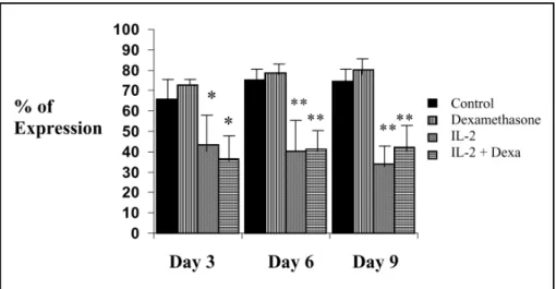
The results demonstrated that 3 days after treat-ment with IL-2 there was diminished expression of the percentage of the surface molecule CD62L (F=5.17; p<0.01). Such effect was sustained during the ten days of the experiment (F=13.58; p<0.001 at day 6 and F=21.44; p<0.0001 at day 9). Cells stimulated with PHA and cultured with dexametha-sone showed no significant changes in the expression of this molecule (Fig 2).
The addition of IL-2 to the cultured of cells stimu-lated with PHA produced no modification in the ex-pression of CD45RO, the memory T cells, at day 3 (F=1.24; p>0.05) and at day 6 (F=1.74; p>0.05). By day 9, on the other hand, cells stimulated with PHA and cultured with IL-2, showed increased ex-pression of CD45RO (F=5.19; p<0.01). The addition of dexamethasone to the culture of cells stimulated with PHA produced no changes in the expression of the isoforms CD45RA and CD45RO.
Interestingly, the addition of epinephrine and se-rotonin did not change the expression of the mole-cules studied (CD25, CD62L, CD45RA and CD45RO). Also, they did not interfere with the changes in the
expression of these molecules, promoted by IL-2, sug-gesting that these amines are probably not involved on the expression of CD25, CD45RA, CD45RO and CD62L observed in the experiment.
DISCUSSION
The aim of this study was to evaluate the effects of dexamethasone, IL-2, serotonin and epinephrine (substances that have been associated to panic disorder and to the HPA axis) on the expression of lymphocytes surface molecules expression (CD25, CD45RA, CD45RO, CD62L). Based upon the results of a previous clinical study demonstrating that panic disorder patients showed increased expression of
Fig 1. Surface membrane expression of CD 25 (IL-2R). Peripheral blood lymphocytes were activated with PHA (5mg/ml) and cultured in the presence of 100 units of human recombinant IL-2, dexamethasone (Dexa: 10-6 M). Cells were harvested at intervals up to day 10 and examined after immunostaining for
membrane expression of CD25. *p<0.0001; **p<0.01 as compared to the control group.
Fig 2. Surface membrane expression of CD 62L (L selectin). Peripheral blood lymphocytes were activated with PHA (5mg/ml) and cultured in the presence of 100 units of human recombinant IL-2, dexamethasone (Dexa: 10-6 M). Cells were harvested at intervals up to day 10 and examined after immunostaining for
CD62L+ and CD45RA+ lymphocytes as compared to controls13, we studied the expression of these
surface lymphocytes molecules “in vitro”.
Cells that were stimulated with PHA in the presence of IL-2 showed differences in the expression of CD25 and CD62L at day 3, but the changes in the expession of CD45RA and CD45RO could only be observed later, at days 6 and 9. This happens because the expression of CD25 (IL-2 receptor) increased soon after the lymphocytes were stimulates with PHA once small concentrations of IL-2 are enough for DNA syn-thesis and for stimulating cell cicle development19.
Steroids such as dexamethasone could inhibit IL-2 secretion20, but also lead to cell death by apoptosis21,
and this could explain why the diminished expression of CD25 after stimulation with dexamethasone occurred only at day 3 and no changes were observed at days 6 and 9.
Cells that were stimulated in the presence of IL-2 showed decreased expression of CD62L. Once the lymphocytes were activated, they lose the expression of L-selectin, the adhesion molecule that is present in naïve lymphocytes responsible for their migration to lymphonodes11. The lost of L-selectin is
accom-panied by the increased expression of other integrins such as LFA-1 and α1β2 that make the memory lym-phocytes migrate to the target-organs11. This change
in the L-selectin surface molecules is inversely related to the expression of CD25 (IL-2R). As it was described before, changes in the expession of CD45RA and CD45RO could be observed at days 6 and 9. It is known that activated T-cell lose the expression of CD45RA very slowly and those T-cells could present both isophorm (CD45RA and CD45RO) at the same time22. At day 6, cells stimulated with IL-2 showed
decreased of CD45RA and at day 9 they increased the expession of the isomorph CD45RO.
However, other substances implicated in the HPA axis, as the amines serotonin and epinephrine, could not change the expression of these studied surface molecule in cells stimulated with PHA. This result is consistent with previous studies showing that vasoconstrictor such as epinephrine has no influence on adhesion molecule expression23. On the other
hand, some studies suggested that adrenergic stress results in compartmental redistribution of T-lympho-cytes in vivo24. It is also known that epinephrine could
be responsible for immunosuppression and inhibition of lymphocyte activation once it can increase cortisol production25. Firing of neurons in the locus ceruleus
leads to the activation of nerve cells in the paraven-tricular nucleus of the hypothalamus that secrete
corticotropin-releasing hormone (CRH). This yields the release of ACTH by pituitary corticotrophs and subsequent corticosteroid production by the adrenal cortex24. Subsequently changes in the adhesion
molecules could be observed. Even though epineph-rine can be associated with decreased lymphocyte activation, in the present study, no changes in the expression of CD62L and CD45RA could be observed with the addition of this amine.
There is paucity of data addressing the relation-ship of serotonin and lymphocyte migration in the literature. The study of Yamaki et al. suggests that serotonin was less active than exogenous histamine in evoking venular polimorphonuclear leukocyte accumulation26. Our data suggest that the regulation
of CD45RA and CD62L is not related to the presence of serotonin or epinephrine, but depends on other mechanisms, such as the presence of IL-2 and the expression of IL-2R.
The present study has some limitations that should be aknowledged. Firstly, in vitro measures of immune function with the cultured cells were not performed, so there are no data related to prolife-ration lymphocyte. Also unstimulated cells were not analysed and the effects on the adhesion molecules could be related to the activation status of the cells. Our data suggest that IL-2 is involved in down-regulation of L-selectin. Even though this result is in agreement with our previous clinical study, we could not demonstrate that other components involved in panic disorder as epinephrine and serotonin are res-ponsible for the changes observed in lymphocyte phenotype expression. It is suggested that the in-creased expression of CD62L and CD45RA lympho-cytes in patients with panic disorder compared to controls are related to decrease cellular activation (demonstrated by decreased IL-2 production). As corticosteroids are potent inhibitors of IL-2 produc-tion, increased levels of cortisol in vivo could lead to diminished IL-2 production into memory lymphocytes following sensitization in vivo. The lymphocytes from panic patients could be less able to produce cytokines because these patients showed increased plasma cortisol (potent IL-2 production inhibitor)19 as it was
REFERENCES
1. Ader R, Cohen N. Psychoneuroimmunology. 2.Ed. San Diego: Academic Press, 1991.
2. Coryell W, Noyes R, Schelechte J. The significance of HPA axis disturbance in panic disorder. Biol Psychiatry1989;25:989-1002. 3. Avery D, Osgood T, Ishiki D, Wilson L, Kenny M, Dunner D. The DST
in outpatients with generalized anxiety disorder, panic disorder or primary affective disorder. Am J Psychiatry1985;142:844-848. 4. Blalock JE. The syntax of immune-neuroendocrine communication.
Immunol Today 1994;15:504-511.
5. Rapaport MH, Stein MB. Serum cytokine and soluble interleukin-2 re-ceptor in patients with panic disorder. Anxiety 1994;1:22-25. 6. McDaniel JS, Risby ED, Stipetic M, Jewart RD, Claude J. Natural killer cell
activity in patients with panic disorder. Anxiety 1994/1995;1:192-195. 7. Surman OS, Williams J, Sheehan DV, Strom TB, Jones KL, Coleman J.
Immunological response to stress in agoraphobia and panic attacks. Biol Psychiatry1986;21:768-774.
8. Weizman R, Laor N, Wiener Z, Wolmer L, Bessler H. Cytokine production in panic disorder patients. Clin Neuropharmacol1999; 22:107-109.
9. Pollack MH, Otto MW, Worthington JJ, Manfro GG, Wolkow W R. Sertraline in the treatment of panic disorder: a flexible dose multicenter srudy. Arch Gen Psychiatry1998;55:1010-1016.
10. Charney D, Woods SW, Goodman WK, Heninger GR. Neurobiological mechanisms of panic anxiety: biochemical and behavioral correlates of yohimbine-induced panic attacks. Am J Psychiatry1987;144:1030-1036. 11. Springer TA. Traffic signals for lymphocytes recirculation and leucocyte
emigration: the multistep paradigm. Cell1994;76:301-314.
12. Jung TM, Gallatin WM, Weissmen IL, Dailey MO. Down-regulation of homing receptor after T-cell activation. J Immunol1988;141:4110-4117. 13. Manfro GG, Pollack MH, Otto MW, et al. Cell-surface expression of L-selectin (CD62L) by blood lymphocytes: correlates with affective parameters and severity of panic disorder. Depress Anxiety2000;11:31-37.
14. Urdal DL, March CJ, Gillis S, Larsen A, Dower SK. Purification and chemical characterization of the receptor for interleukin-2 from activated human T-lymphocytes and from human T-cell lymphoma cell line. Proc Natl Acad Sci USA1984;81:6481-6485.
15. Camerini DS, James SP, Stamenkovic I, Seed B. Leu8/TQ1 is the human equivalent of the MEL-14 lymph node homing receptor. Nature1989; 342:78-82. 16. Kan EAR, Wang CY, Wang LC, Evans RL. Non-covalently bonded subunits of 22 and 28 Kd are rapidly internalized by T-cells reacted with anti-Leu4 antibody. J Immunol1983; 131:536-539.
17. Rupprecht R, Wodarrz P, Kornhuber J, et al. In vivo and in vitro effects of glucocorticoids on lymphocyte proliferation in depression. Eur Arch Psychiatry Clin Neurosci1991; 241:35-40.
18. Rodberg G, Kradin R. Epinephrine promotes T-lymphocyte traffic to the lung. Am J Respir Crit Care Med1994;49:A12.
19. Cantrell DA, Smith KA. The interleukin-2 T-cell system: a new cell growth model. Science1984;224:1312-1326.
20. Gillis S, Crabtree GR, Smith KA. Glucocorticoid-induced inhibition of T cell growth factor production: I. The effect on mitogen-induced lymphocyte proliferation. J Immunol1979;123:1624-1631.
21. Kroemer G. The pharmacology of T cell apoptosis. Am J Respir Crit Care Med1995;58:211-296.
22. Clement L, Yamashita N, Martin AM. The functionally distinct subpopulations of human CD4+ helper/inducer T lymphocytes defined by anti-CD45R antibodies derive sequentially from a differentiation pathway that is regulated by activation-dependent post-thymic differentiation. J Immunol1988;141:1464-1470.
23. Jilma B, Eichler HG, Stohlawtz P, et al. Effects of exercise on circulating vascular adhesion in healthy men. Immunobiology1997;197:505-512. 24. Ottaway CA, Husband AJ. The influence of neuroendocrine pathway
on lymphocyte migration. Immunol Today1994;15:511-517. 25. Chambers D, Cohen R, Perlman R. Neuroimmune modulation: signal
transduction and cathecolamine. Neurochem Int 1993;22:95-110. 26. Yamaki K, Thorlacius H, Xie X, Lindbom L, Hedqvist P, Raud J. Characteristics