Endodontics
Janaina Corazza Montero Graziela Garrido Mori
Endodontics Course, São Paulo Dental Association - APCD, Presidente Prudente, SP, Brazil.
Corresponding Author: Graziela Garrido Mori
E-mail: [email protected]
Received for publication on Mar 16, 2012 Accepted for publication on May 12, 2012
Assessment of ion diffusion from a
calcium hydroxide-propolis paste
through dentin
Abstract: This study evaluated the ability of ions from a non-alcoholic calcium hydroxide-propolis paste to diffuse through dentinal tubules. Thirty-six single-rooted bovine teeth were used. The tooth crowns were removed, and the root canals were instrumented and divided into 3 groups: Group 1 - calcium hydroxide-propylene glycol paste; Group 2 - calcium hydroxide-saline solution paste; Group 3 - calcium hydrox-ide-propolis paste. After the root canal dressings were applied, the teeth were sealed and placed in containers with deionized water. The pH of the water was measured after 3, 24, 72 and 168 hours to determine the dif-fusion of calcium hydroxide ions through the dentinal tubules. All of the pastes studied promoted the diffusion of calcium hydroxide ions through the dentinal tubules. Associating propolis to calcium hydroxide resulted in a pH increase, which occurred with greater intensity after 72 hours. The calcium hydroxide-propolis paste was able to diffuse in dentin.
Descriptors: Propolis; Calcium Hydroxide; Dentin.
Introduction
One of the most important factors for successful endodontic therapy is complete root canal cleaning.1 The dificulty involved in eliminating
microorganisms, as well as their residual presence, warrants the use of root canal dressings after biomechanical preparation.1
An adequate root canal dressing should have an antimicrobial poten-tial,2-4 the ability to diffuse through dentinal tubules,1,3,4
biocompatibil-ity,3,4 and, if possible, the ability to stimulate repair.3,4
Calcium hydroxide is one of the main root canal dressings used in Endodontics. It is a white, odorless powder, with low solubility in water, insolubility in alcohol, and a high pH. It also has extended clinical ac-tion. Moreover, it is biocompatible, has antimicrobial and anti-inlam-matory action, and activates the alkaline phosphatase enzyme, which induces mineralized tissue formation and acts in the repair process.4 It
is chemically classiied as a strong base, and its association with an ad-equate vehicle yields an alkaline paste.4
The success of calcium hydroxide paste as a root canal dressing is related to its dissociation into calcium and hydroxyl ions. The hydrox-yl ions alkalinize the environment.4 To be effective, the hydroxyl ions
should be able to diffuse in dentin and remain in pulp tissues in a suf-icient concentration to produce the pH level required to destruct
ria inside the root canal and dentinal tubules.4 The
antimicrobial action of calcium hydroxide and its ability to contain root resorption are related to this alkalizing action, which, in turn, is a consequence of its ionization into hydroxyl ions.4,5 The action of
these ions on tissues and bacteria explains the bio-logical and antimicrobial properties of calcium hy-droxide.4
Calcium hydroxide must be associated to a ve-hicle to be used as a paste.4 Such vehicles include
ol-ive oil, propylene glycol, saline, distilled water, and others.1,4,6-8
Lage-Marques et al.8 conducted a study to
evalu-ate the revalu-ate of ionic dissociation of calcium hydrox-ide associated to different vehicles—aqueous, vis-cous, and oily—and concluded that aqueous and viscous vehicles are better suited for paste use.
Propolis has been used in popular medicine for thousands of years because it is considered one of the most effective natural products discovered so far. In dentistry, propolis has been used to control the oral microbiota, e.g., in dentifrices.9
Propolis is dark in color. It is produced from material collected from plants by bees and used by them against pathogenic microorganisms.10 Its
anti-inlammatory properties have been described to act mainly against infection, rheumatism, tor-sions, muscular and articular diseases, as well as other types of inlammation.10,11 In addition to its
antibacterial and anti-inlammatory action, propolis promotes tissue reorganization.10 It is also
biocom-patible with pulp tissue11 and exerts antimicrobial
action against endodontic pathogens.11-13
The chemical composition of propolis varies richly: Over 200 substances have been identiied in the different propolis varieties from different geo-graphical regions, including phenolic acids, lavo-noids, esters, aromatic aldehydes, alcohols, amino acids, fatty acids, vitamins and minerals.14 Special
emphasis should be given to the lavonoids and phe-nolic acids, greatly responsible for the biological ac-tivity of propolis.14,15
Owing to the antimicrobial and anti-inlamma-tory properties of propolis, its association with cal-cium hydroxide has been suggested for use as a root
propolis pastes (with and without alcohol) associ-ated to calcium hydroxide. Even though both pastes were found to display antimicrobial action, the non-alcoholic paste produced greater inhibition halos than the paste with alcohol.
Considering the antimicrobial potential of a propolis-calcium hydroxide association, it becomes important to determine how well such a paste can promote the diffusion of ions through dentinal tu-bules, an attribute essential to the therapeutic effect of any calcium hydroxide paste.
The aim of this study was thus to evaluate the diffusion ability of ions from a non-alcoholic cal-cium hydroxide-propolis paste through dentinal tu-bules, compared to that of calcium hydroxide pastes prepared with saline or propylene glycol.
Methodology
This study was approved by the Institutional Review Board, Araçatuba Dental School, State University of São Paulo - UNESP (Process FOA 06021/2010).
Initially, 36 extracted single-rooted bovine teeth were kept in a 10% formaldehyde solution (Farma-Ticli Indústria Farmacêutica, São Paulo, Brazil). Next, the soft tissues and dental calculus that re-mained adhered to the root were removed with den-tal scalers (Trinity, São Paulo, Brazil), after which the teeth were stored in saline solution (Farmax, Divinópolis, Brazil).
Then, the crowns were transversally sectioned with a carborundum disc (Microdont, São Paulo, Brazil), at the cementoenamel junction level.
Root canal length was measured by inserting a #30 K-ile (Maillefer Instruments, Ballaigues, Switzerland) with a rubber stop. When the ile tip reached the apical foramen, the stop was leveled to the cervical edge of the root and the canal length was recorded.
The working length was established by subtract-ing one millimeter from the total root canal length. Apical preparation was performed up to this limit, up to ile #80, followed by a step-back instrumenta-tion up to ile #120.
wa-the aid of a Luer-Lock syringe (Dulex - S.S. White Artigos Dentários Ltda., Rio de Janeiro, Brazil) and a # 30 × 4 irrigation cannula (BD - Becton Dick-inson Indústrias Cirúrgicas Ltda., Rio de Janeiro, Brazil).
After instrumentation, a #30 ile was inserted to the total working length for apical cleaning, and the root canal was illed with an EDTA solution (Biodinâmica, Ibiporã, Brazil) for 3 minutes. After this period, the root canals were rinsed with sa-line solution and dried with absorbent paper points (Dentsply, Petrópolis, Brazil).
The teeth were randomly divided into 3 groups according to the following calcium hydroxide pastes used to ill the canals:
• Group I (n = 12): calcium hydroxide-propylene glycol paste, prepared by mixing 1 g of calcium hydroxide (Biodinâmica Química e Farmacêutica LTDA, Ibiporã, PR, Brazil) and 2 ml of propyl-ene glycol (Odontofarma, Londrina, PR, Brazil);
• Group II (n = 12): calcium hydroxide-saline paste, prepared by mixing 1 g of calcium hy-droxide and 1.5 ml of saline solution;
• Group III (n = 12): calcium hydroxide-propolis paste, prepared by mixing 1 g of calcium hy-droxide and 2 ml of propolis without alcohol (Apis Flora, Ribeirão Preto, SP, Brazil).
Complete illing of the root canals was checked by having the solution overlow through the api-cal foramen and low back through the root canal opening. After complete illing of the root canals,
their openings were sealed with temporary cement (Cimpat-Septodont, Louisville, USA). The apical fo-ramen and root canal openings (over the temporary cement) were sealed with epoxy resin (Araldite
-Maxepoxi Industrial e Comercial Ltda., São Paulo, Brazil). Next, the teeth were placed in containers with 50 ml of deionized water (pH = 6.17) and kept in an oven at 37°C, with 100% humidity.
After 3, 24, 72 and 168 hours, the pH values of the solutions in the lasks were measured with a pH meter (Hanna Instruments Brasil Copyright, São Paulo, Brazil), calibrated with standardized solu-tions at pH 4.0 and 7.0. Twelve measurements were performed for each group/period. For each measure-ment, the electrode of the pH meter was carefully rinsed with deionized water and dried with absor-bent paper to eliminate any residues that could in-terfere with the measurements.
The data was recorded in tables and the differ-ences between groups and periods were statistically analyzed by the Tukey test, at a signiicance level of 5%.
Results
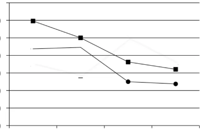
Figure 1 shows how the pH varied with time in the different study groups. The values indicate the measurement means obtained for each group.
Group 1 (propylene glycol) showed a mean pH of 7.992 three hours after preparing the paste and inserting it into the root canal. A signiicant pH re-duction with time (p < 0.05) was observed in this group (Table 1).
Figure 1 - pH variation over the experimental time for the study groups
6.800 7.000 7.200 7.400 7.600 7.800 8.000 8.200
3 hours 24 hours 72 hours 168 hours
Group 1
Group 2
Group 2 (saline) reached a mean pH of 7.675 and 7.692 respectively after 3 and 24 hours. There was no signiicant pH difference between these two timepoints. However, there was a signiicant pH re-duction (p < 0.05) after 72 and 168 hours, as ob-served in Table 2.
The pH in Group 3 (propolis) varied with time, with a greater pH increase (p < 0.05) observed be-tween 24 and 72 hours (Table 3).
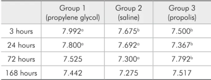
The pH in group 1 was higher than that observed in the other groups (p < 0.05) after 3 hours. Group 3 showed the lowest pH (p < 0.05) among the groups after 24 hours. Group 3 showed a pH higher than that observed in Group 2 after 72 hours (p < 0.05). Finally, there was no pH difference among the groups after 168 hours, as shown in Table 4, demon-strating that all pastes were able to diffuse through the dentinal tubules and that this ability did not change over time.
Discussion
The successful use of a calcium hydroxide paste as a root canal dressing is related to its dissociation into calcium and hydroxyl ions, the latter being re-sponsible for alkalizing the environment.1
The high pH of calcium hydroxide mitigates the inlammatory process,4 and its release of hydroxyl
ions makes it an excellent antibacterial agent.4
Calcium hydroxide must be associated to a
ve-mines its rate of dissociation into ions and diffusion through the dentinal tubules. Additionally, the vehi-cle inluences how well the paste can become soluble and be resorbed into the apical tissues.4 The vehicle
may also bear on the effectiveness of the calcium hy-droxide paste owing to its inluence on ionic disso-ciation and diffusion.4
Lage-Marques et al.8 concluded that calcium
hy-droxide pastes in aqueous and viscous vehicles are more effective than those in oily vehicles because the former reach higher pH levels more quickly and remain stable for a longer period of time. Other au-thors have also reported the greater effectiveness of aqueous and viscous vehicles.1,4,6-8 Similarly to
previous reports,1,4,6-8 this study evidenced that
sa-line- and propylene glycol-containing pastes diffuse well through dentin, as can be seen in Tables 1, 2 and 4. Similar levels of ion diffusion in dentin were observed for both groups, at different experimental times, conirming the indication of these substances as vehicles for calcium hydroxide pastes. The effec-tiveness of saline and propylene glycol as vehicles for calcium hydroxide was used as a comparative reference in this study.
According to the present results, the calcium hydroxide-propolis paste also diffused through the
Table 1 - Mean pH obtained for the calcium hydroxide-pro-pylene glycol paste (Group 1) after the experimental times.
3 hours 24 hours 72 hours 168 hours
Group 1 7.992 a 7.800 c 7.525b 7.442d
The values marked with letters “a” and “c” are significantly different from those marked with “b” and “d”.
Table 2 - Mean pH obtained for the calcium hydroxide-saline paste (Group 2) after the different experimental times.
3 hours 24 hours 72 hours 168 hours
Group 2 7.675a 7.692a 7.300b 7.275b
The values marked with letter “a” are significantly different from those marked with “b”.
Table 3 - Mean pH obtained for the calcium hydroxide-propolis paste (Group 3) after the different experimental times.
3 hours 24 hours 72 hours 168 hours
Group 3 7.500 7.367a 7.792b 7.517
The value marked with letter “a” is significantly different from that marked with “b”.
Table 4 - Mean pH obtained for the study groups after the different experimental times.
Group 1
(propylene glycol) Group 2(saline) (propolis)Group 3
3 hours 7.992a 7.675b 7.500b
24 hours 7.800a 7.692a 7.367b
72 hours 7.525 7.300a 7.792b
168 hours 7.442 7.275 7.517
face. This may be observed in Figure 1 and Table 3. The viscous consistency of propolis probably fa-vored this level of diffusion. Moreover, the compo-nents of the propolis solution did not impair or pre-vent the dissociation of the calcium hydroxide.
All of the pastes studied diffused through the dentinal tubules. After 168 hours, all of the pastes presented a similar ability to alkalize the external root surface (Table 4).
Using propolis as a vehicle for calcium hydroxide may thus be suggested. Even though the diffusion ability of the propolis-containing paste was similar to that of the saline- and propylene
glycol-contain-ing pastes, propolis may be a better indication be-cause it adds to the antimicrobial action of calcium hydroxide.11-14 Further studies are warranted to
in-vestigate the biocompatibility of this paste and con-irm the use of propolis without alcohol as a vehicle for calcium hydroxide.
Conclusion
The non-alcoholic calcium hydroxide-propolis paste tested in this study was able to diffuse through dentinal tubules. After 168 hours, all of the experi-mental pastes presented similar diffusion in dentin.
References
1. Mori GG, Ferreira FC, Batista FRS, Godoy AMS, Nunes DC. Evaluation of the diffusion capacity of calcium hydroxide pastes through the dentinal tubules. Braz Oral Res. 2009 Apr-Jun;23(2):113- 8.
2. Lima RK, Guerreiro-Tanomaru JM, Faria-Júnior NB, Tano-maru-Filho M. Effectiveness of calcium hydroxide-based intracanal medicaments against Enterococcus faecalis. Int Endod J. 2012 Apr;45(4):311-6.
3. Estrela C, Holland, R. Calcium hydroxide: study based on scientific evidences. J Appl Oral Sci. 2003 Dec;11(4):269- 82. 4. Mohammadi Z, Dummer PM. Properties and applications of
calcium hydroxide in endodontics and dental traumatology. Int Endod J. 2011 Aug;44(8):697-730.
5. Tronstad L, Andreasen JO, Hasselgren G, Kristersin L, Riis I. pH changes in dental tissues after root canal fillings with calcium hydroxide. J Endod. 1981 Jan;7(1):17-21.
6. Pacios MG, de la Casa ML, de los Angeles Bulacio M, Lopes ME. Calcium hydroxide’s association with different vehicles:
in vitro action on some dentinal components. Oral Surg Oral Med Oral Pathol Oral Radiol Endod. 2003 Jul;96(1):96-101. 7. Zmener O, Pameijer CH, Banegas G. An in vitro study of the
pH of three calcium dressing materials. Dent Traumatol. 2007 Feb;23(1):21-5.
8. Lage Marques JLS, Conti R, Antoniazzi JH, Guth I. Avaliação da velocidade de dissociação iônica do hidróxido de cálcio as-sociado a diferentes veículos/ In vitro assessment of the ionic dissociation velocity of clcium hydroxide to diferente vehicles. Rev odontol Univ São Paulo. 1994 Apr-Jun;8(2):81-7.
9. Ghisalbert EL. Propolis: a review. Bee Word. 1979;60(2): 59-83.
10. Marcucci MC. Propolis: chemical composition, biological properties and therapeutic activity. Apidologie. 1995;26(2):83-99.
11. Bretz WA, Chiego DJ, Marcucci MC, Cunha I, Custódio A, Schneider LGZ. Preliminary report on the effects of propolis on wound healing in the dental pulp. Z Naturforsch C. 1998 Nov-Dec;53(11-12):1045-8.
12. Kayaoglu G, Ömürlü H, Akca G, Gürel M, Gençay Ö, Sorkun K, et al. Antibacterial activity of Propolis versus conventional endodontic disinfectants against Enterococcus faecalis in in-fected dentinal tubules. J Endod. 2011 Mar;37(3):376-81. 13. Madhubala MM, Srinivasan N, Ahamed S. Comparative
evaluation of propolis and triantibiotic mixture as an intra-canal medicament against Enterococcus faecalis. J Endod. 2011 Sep;37(9):1287-9.
14. Gonsales GZ, Orsi RO, Fernandes Júnior A, Rodrigues P, Funari, S. Antibacterial activity of propolis collected in dif-ferent regions of Brazil. J Venom Anim Toxins Incl Trop Dis. 2006 Apr-Jun;12(2):276-84.
15. Mori GG, Nunes DC, Castilho LR, de Moraes IG, Poi WR. Propolis as storage media for avulsed teeth: microscopic and morphometric analysis in rats. Dent Traumatol. 2010 Feb;26(1):80-5.