Boniek Castillo Dutra Borges(a) Gabriela Voany Galdino Bezerra(a) Janaína de Almeida Mesquita(a) Márcia Rodrigues Pereira(b) Flávio Henrique Baggio Aguiar(c) Alex José Souza dos Santos(a) Isauremi Vieira de Assunção Pinheiro(a)
(a) Department of Dentistry, School of Dentistry, Federal University of Rio Grande do Norte (UFRN), Natal, RN, Brazil.
(b)Department of Chemistry, School of Chemistry, Federal University of Rio Grande do Norte (UFRN), Natal, RN, Brazil.
(c) Department of Restorative Dentistry, Piracicaba Dental School, State University of Campinas (UNICAMP), Piracicaba, SP, Brazil.
Corresponding Author Boniek Castillo Dutra Borges Rua Minas Novas, 390, cs. 18 Natal - Rio Grande do Norte - Brazil CEP: 59088-725
E-mail: boniek.castillo@gmail.com
Received for publication on Dec 17, 2010 Accepted for publication on Feb 08, 2011
Effect of irradiation times on the
polymerization depth of contemporary
fissure sealants with different opacities
Abstract: The aim of this study was to evaluate the depth of curing of 10 contemporary blue light-activated dental lowable materials at several opacities, inluenced by different irradiation times using FT-IR spectros-copy. Fifty-ive specimens (n = 5) with a 5-mm diameter and 1-mm thick-ness of translucent (Opallis Flow T), yellowed (Master Flow A2; Opallis Flow A2; Natural Flow A2; Fluroshield Yellowed), and opaque materials (Master Flow OA2; Natural Flow O; Opallis Flow OA3.5; Opallis Flow OP; Fluroshield White) were obtained at six curing times (10s, 20s, 30s, 40s, 50s, and 60s) using a high-intensity LED (Coltolux, Coltène/Whale-dent). The degree of conversion (DC) (%) was obtained using the Nexus 470 FTIR Spectrometer (Nicolet Instruments, USA). The FTIR-ATR spectra for uncured and cured samples were analyzed using a ZnSe crys-tal. The top and bottom surfaces of the cured specimens were analyzed to obtain the depth of curing. Two-way ANOVA was used to analyze the data. The highest curing depth was obtained by Natural Flow OA2, while the lowest was shown by Master Flow OA2. The shortest curing time generated similar depths of cure in comparison with the most ex-tensive for Opallis Flow A2 and Fluroshield Yellowed. Therefore, depth of curing, inluenced by the irradiation time, was dependent on the ma-terials. Using the Natural Flow OA2 opaque sealant and the 10-s curing time for Opallis Flow A2 and Fluroshield Yellowed may represent alter-native approaches to sealing tooth issures.
Descriptors: Pit and Fissure Sealants; Composite Resins; Pediatric Dentistry.
Introduction
Photoactivated dental composite resins are now the most widely used restorative material. The main advantage of this activation mode over the chemical one is the working time control by the operator.1,2
Low-viscosity light-cured materials such as traditional issure sealants or low-able composites are routinely used in minimal intervention procedures, preventing caries initiation and arresting caries progression by providing a physical barrier that inhibits microorganisms and food particles from collecting in pits and issures after polymerization.3
denominated degree of conversion (DC). The polym-erization of the monomer BisGMA occurs through the carbon–carbon double bond of the two meth-acrylate groups.5 Depth of curing is a crucial
fac-tor in obtaining the optimal physical properties and clinical performance of dental composite resins.6
Low DC values on the top and bottom surfaces of the material placed on tooth issures are associated with bacterial growth7 and sealant debonding,8
re-spectively, leading to clinical failure. Therefore, the optimal depth of curing for these materials is crucial in obtaining satisfactory clinical performance.
Factors such as exposure time, monomer compo-sition, and opacity of the material have signiicant ef-fects on the depth of curing of composite resins.2,3,9
The materials’ DC is proportional to the amount of light to which they are exposed.2 Concerning material
opacity, translucent, yellowed, or opaque issure seal-ants are commercially available. The more opaque it is, the lower the light absorption and transmittance to the bottom surface of the material will be,8 so short
curing times may provide insuficient depth of curing. Since materials of all opacities are usually utilized in children, the investigation of the shortest curing time that is suficient to provide the highest depth of cur-ing could facilitate their application without generat-ing undesirable weak physical properties after polym-erization. However, the literature is scarce in research studies evaluating the effect of different curing times on the depth of curing of several issure sealants with various opacities. Therefore, this study aimed to evaluate the depth of curing of 10 translucent, yel-lowed, and opaque contemporary issure sealants as inluenced by various polymerization times. The null hypothesis tested was that there would be no differ-ences among the materials and curing times.
Materials and Methods
Experimental design
The factors under study were: • materials at 10 levels:
-
Opallis Flow T (FGM, Joinville, Brazil),-
Opallis Flow A2 (FGM, Joinville, Brazil),-
Master Flow A2 (Biodinâmica, Ibiporã, Brazil),-
Natural Flow A2 (DFL, Rio de Janeiro, Brazil),-
Fluroshield Yellowed (Dentsply/Caulk, Rio deJaneiro, Brazil),
-
Master Flow OA2 (Biodinâmica, Ibiporã, Brazil),-
Natural Flow O (DFL, Rio de Janeiro, Brazil),-
Opallis Flow OA3.5 (FGM, Joinville, Brazil),-
Opallis Flow OP (FGM, Joinville, Brazil),-
Fluroshield White (Dentsply/Caulk, Rio deJa-neiro, Brazil);
• and curing time at six levels:
-
10s,-
20s,-
30s,-
40s,-
50s, and-
60s.Depth of curing by means of degree of conver-sion (DC) analysis for the top and bottom surfaces of the samples was performed.
The materials’ composition, opacities, and batch numbers are shown in Table 1. The physical charac-teristics of the light-curing unit used in the study are listed in Table 2.
Sample preparation
Cylinder Telon molds (5 mm diameter x 1 mm height) (Ferramentas ALFA, São Paulo, Brazil) were used to fabricate 330 specimens (n = 5 per group). The low-viscosity materials were injected into the center of the matrix using the disposable tip sup-plied by the manufacturer under controlled temper-ature and relative humidity conditions. The material surfaces were covered with a Mylar strip (K-Dent – Quimidrol, Joinville, Brazil) and then photoacti-vated using the light-emitting diode (LED) Coltolux (Coltène/Whaledent, Altstätten, Switzerland) at a 3-mm distance.3 After polymerization, the
speci-mens were removed from the matrices and stored dry in light-proof containers at 37ºC for 24 hours.8
DC analysis
4 cm−1 resolution, and from 300 to 4000 cm−1
wave-number. The top and bottom surfaces of the cured specimens were analyzed to determine the depth of curing. The percentage of unreacted carbon–carbon double bonds (% C=C) was determined from the ratio of the absorbance intensities of aliphatic C=C (peak at 1638 cm–1) against an internal standard
be-fore and after the curing of the specimen: aromatic C–C (peak at 1608 cm–1). The degree of conversion
was determined by subtracting the % C=C from 100%, according to the following equation:
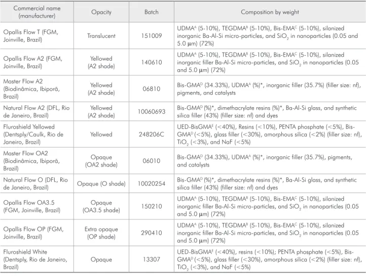
Table 1 - Composition and batch number of the dental flowable materials used in this study.
Commercial name
(manufacturer) Opacity Batch Composition by weight
Opallis Flow T (FGM,
Joinville, Brazil) Translucent 151009
UDMAA (5-10%), TEGDMAB (5-10%), Bis-EMAC (5-10%), silanized inorganic Ba-Al-Si micro-particles, and SiO2 in nanoparticles (0.05 and 5.0 µm) (72%)
Opallis Flow A2 (FGM, Joinville, Brazil)
Yellowed
(A2 shade) 140610
UDMAA (5-10%), TEGDMAB (5-10%), Bis-EMAC (5-10%), silanized inorganic filler Ba-Al-Si micro-particles, and SiO2 in nanoparticles (0.05 and 5.0 µm) (72%)
Master Flow A2 (Biodinâmica, Ibiporã, Brazil)
Yellowed
(A2 shade) 06810
Bis-GMAD (34.33%), UDMAA (%)*, inorganic filler (35.7%) (filler size: nf), pigments, and catalysts
Natural Flow A2 (DFL, Rio de Janeiro, Brazil)
Yellowed
(A2 shade) 10060693
Bis-GMAD (%)*, dimethacrylate resins (%)*, Ba-Al-Si glass, and synthetic silica filler (43%) (filler size: nf) and dyes
Fluroshield Yellowed (Dentsply/Caulk, Rio de Janeiro, Brazil)
Yellowed 248206C
UED-BisGMAE (<40%), Resins (<10%), PENTA phosphate (<5%), Bis-GMAD (<5%), glass filler (<30%), amorphous silica (<2%) (filler size: nf), TiO2 (<3%), and NaF (<5%)
Master Flow OA2 (Biodinâmica, Ibiporã, Brazil)
Opaque
(OA2 shade) 06010
Bis-GMAD (34.33%), UDMAA (%)*, inorganic filler (35.7%), pigments, and catalysts
Natural Flow O (DFL, Rio
de Janeiro, Brazil) Opaque (O shade) 10020254
Bis-GMAD (%)*, dimethacrylate resins (%)*, Ba-Al-Si glass, and synthetic silica filler (43%) (filler size: nf) and dyes
Opallis Flow OA3.5 (FGM, Joinville, Brazil)
Opaque
(OA3.5 shade) 150210
UDMAA (5-10%), TEGDMAB (5-10%), Bis-EMAC (5-10%), silanized inorganic filler Ba-Al-Si micro-particles, and SiO2 in nanoparticles (0.05 and 5.0 µm) (72%)
Opallis Flow OP (FGM, Joinville, Brazil)
Extra opaque
(OP shade) 290410
UDMAA (5-10%), TEGDMAB (5-10%), Bis-EMAC (5-10%), silanized inorganic filler Ba-Al-Si micro-particles, and SiO2 in nanoparticles (0.05 and 5.0 µm) (72%)
Fluroshield White (Dentsply, Rio de Janeiro, Brazil)
Opaque 13307
UED-BisGMAE (<40%), resins (<10%); PENTA phosphate (<5%), Bis-GMAD (<5%), glass filler (<30%), amorphous silica (<2%) (filler size: nf), TiO2 (<3%), and NaF (<5%)
AUrethane Dimethacrylate; BTriethylene Glycol Dimethacrylate; CEtoxilated Bisphenol A. Diglicidil Methacrylate; DBisphenol A-Glycidyl Methacrylate; EUrethane
modified Bis-GMA dimethacrylate. *The manufacturer did not provide the percentage of UDMA; nf: not furnished by the manufacturer.
Light-curing
unit Manufacturer PD
A PD3B Wavelength
range
Wavelength peak
Coltolux LED (serial 091215032)
Coltène/Whaledent, Altstätten, Switzerland
> 1000 mW/ cm²
900 mW/
cm² 450-470 nm 460 nm
APower density published by the manufacturer; BPower density at 3-mm distance. The radiance was measured
with a curing radiometer (model 100; Demetron Research Corporation, Danbury, CT, USA). A 3-mm distance was established using a digital caliper coupled to a metallic support.
Table 2 - Physical characteristics of the curing-light unit used in the study.
DC (%) = 1 − (1638 cm × 100
−1− 1608 cm−1) cured
Statistical analysis
The data were irst submitted to two-way analy-sis of variance (ANOVA). The top, bottom, and bot-tom-to-top conversion ratios were considered sepa-rately. Multiple comparisons among the groups were analyzed by means of Tukey’s test. All statistical tests were executed using the ASSISTAT Software (Federal University of Campina Grande, Campina Grande, Brazil) at the level of 5%.
Results
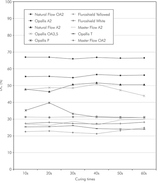
Top surface DC (%)
Figure 1 shows DC means of the materials’ top surfaces at each curing time. In all of them, the Nat-ural Flow O composite presented the highest DC, while the lowest DC values were shown by the
Mas-ter Flow OA2 composite. The shortest curing time evaluated in this study (10 s) provided similar DC means to those of Natural Flow O, Fluroshield Yel-lowed, and Fluroshield White. Only Master Flow OA2 had the highest DC means at 60 s. Opallis Flow T, Opallis Flow A2, Natural Flow A2, Master Flow A2, Opallis Flow OA3.5, and Opallis Flow OP, re-spectively, presented the highest DC at the following photoactivation times: 30s, 40s, 40s, and 20s.
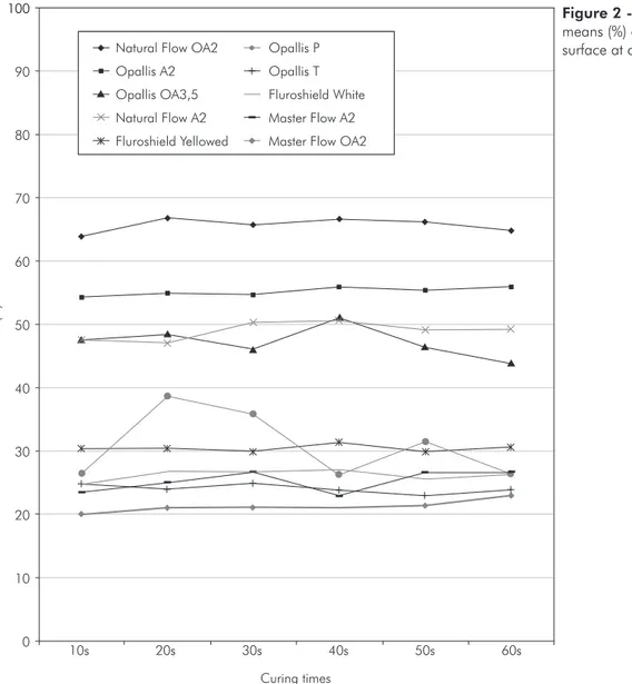
Bottom surface DC (%)
Figure 2 shows DC means of the materials’ bot-tom surface at each curing time. In all of them, the Natural Flow O composite presented the highest DC, while the lowest DC values were shown by the Master Flow OA2 composite, as also occurred on
Figure 1 - Degree of conversion means (%) of the materials’ top surface at different curing times.
D
C
(%)
Curing times Natural Flow OA2
Natural Flow A2
Master Flow OA2 Master Flow A2 Fluroshield Yellowed
Fluroshield White Opallis A2
Opallis T
Opallis P Opallis OA3,5 100
90
80
70
60
50
40
30
20
10
10s 20s 30s 40s 50s 60s
the top surface. The 10-s curing time provided simi-lar DC values to the 60-s results for Opallis Flow A2 and Fluroshield Yellowed. Also, only Master Flow OA2 had the highest DC means at 60s. Opallis Flow T, Natural Flow A2, Master Flow A2, Natural Flow O, Opallis Flow OA3.5, Opallis Flow OP, and Fluroshield White, respectively, presented the high-est DC at the following photoactivation times: 10s, 30s, 30s, 20s, 40s, 20s, and 20s.
Bottom-to-top DC ratio
Most ratio values were higher than 0.8, except for that of Opallis Flow OP at 10s (0.76) (Figure 3). For Opallis Flow A2, Natural Flow A2, Fluroshield Yellowed, and Natural Flow O, there were no
differ-ences among curing times, so the 10-s curing time provided similar bottom-to-top DC ratio values to the others. The highest value was found for Opal-lis Flow OP when polymerized for 30s. However, the lowest bottom-to-top DC ratios were shown by Opallis Flow OP at 10-s, 40-s, and 60-s curing times.
Discussion
The clinical success of sealants is well-document-ed in the literature and directly relatwell-document-ed to its capacity to remain bonded to occlusal pits and issures.10 The
hardened material forms a strong micromechanical bond to etched tooth enamel, thus physically obtu-rating susceptible areas of the tooth surface and pre-venting dental caries. Therefore, ensuring suficient
Figure 2 - Degree of conversion means (%) of the materials’ bottom surface at different curing times.
D
C
(%)
Curing times Natural Flow OA2
Natural Flow A2
Master Flow OA2 Master Flow A2
Fluroshield Yellowed
Fluroshield White
Opallis A2 Opallis T
Opallis P
Opallis OA3,5
100
90
80
70
60
50
40
30
20
10
10s 20s 30s 40s 50s 60s
curing is an integral requirement for the success and longevity of a tooth issure sealant.8
A direct relationship exists between the light intensity and DC of different composite resins.11
In this sense, it was found that an extended irra-diation time (60s) to polymerize a dental lowable material (Permalo, Ultradent, USA) increased the DC on its top surface,3 which could improve the
clinical performance of tooth issure sealer materi-als. Nevertheless, in the above-mentioned study, the bottom surfaces of the samples were not analyzed to assess the depth of curing. Moreover, consider-ing that these materials are usually used in pediatric dentistry, there is a need to investigate a shorter cur-ing time that is suficient to provide similar physical
properties to the 60-s curing time for other lowable materials with several opacities. This would facili-tate the application and decrease the drawbacks of a polymer with low DC.
The results obtained in this study showed that the major opaque sealants presented poorer con-version on either the top or bottom surface than the yellowed sealants. This inding corroborates other indings showing higher curing susceptibility for clearer/yellowed sealants in comparison with opaque sealants.8 The difference in curing
charac-teristics between clear and opaque sealants most likely is related to the opacifying agents present in the opaque sealant, which probably cause substan-tial relection, scattering, and absorption of light, Figure 3 - Bottom-to-top surface
conversion ratio means (standard deviations) of the materials at different curing times.
D
C
(%)
Curing times
1.10
1.05
1.00
0.95
0.90
0.85
0.80
0.75
0.70
10s 20s 30s 40s 50s 60s
Natural Flow OA2
Natural Flow A2
Master Flow OA2
Master Flow A2
Fluroshield Yellowed
Fluroshield White
Opallis A2 Opallis T
preventing more curing through the surface of the sealant.8 As a result, the polymerization reaction is
attenuated and the DC of these materials is lower than for non-opaque materials.
However, the better curing depth of Natural Flow O in comparison to its yellowed version (Nat-ural Flow A2) was found in this work. Probably, the chemical components of this material caused a contrary effect regarding relection, scattering, and absorption of light. Besides the irradiation time pro-vided by visible light sources and the opacity charac-teristics of the dental materials, the refractive index difference that exists between the base resin and in-organic components (especially the silica) inluences the transmission of visible light through the mate-rial. The higher the refractive index is, regardless of whether it increases during the polymerization phase of the monomers, the higher the light scattering and the lower the light transmission will be.12 Moreover,
light transmission can decrease with time as the re-fractive index of the curing resin rises above that of the iller.13 Therefore, possibly, although Natural
Flow O contains opacifying agents, the resultant refraction index before and after curing may favor higher monomer conversion, unlike Natural Flow A2 and other opaque materials. Although further studies are needed to conirm this supposition, the advantage of using an opaque material is the pos-sibility of masking dark coloration on the bottom of the tooth issures, increasing patient satisfaction.
In a clinical environment, the materials used to seal tooth issures usually present a 1-mm thick-ness.3 Depending on the cusp size and the
morphol-ogy of pits and issures, the light guide of the cur-ing unit may be placed at different distances from the sealant surface during occlusal sealing, such as 3 mm, increasing the dispersion and decreasing the irradiance of the light that reaches the material.3
Therefore, both distance and sample thickness had to be considered during the sample preparation to approximate clinical conditions. Since a high-inten-sity LED was used, the radiance decrease after posi-tioning the light tip at 3 mm from the material prob-ably did not affect the DC. The fact that, for some brands (Opallis Flow A2 and Fluroshield Yellowed), even the 10-s irradiation time could provide a DC
similar to that provided by the 60-s curing time on the bottom surface might be attributed to the afore-mentioned points. Also, a probable refraction index of the resin matrix that was close to the inorganic components, which may have not changed during polymerization, would explain this inding for both these materials.
Light-cured dental materials contain a light-sensitive initiator agent, usually an alpha-diketone. Among the diketones available, the most frequently used is camphorquinone (CQ).14 This
photosensitiz-er absorbs light at wavenumbphotosensitiz-ers between 460 and 480 nm, reaching a triplete excited state. CQ is the photosensitizing agent used in most of the brands available on the market. Camphorquinone is an in-tensely yellow-colored powder. Additionally, it has poor photobleaching, which means that the yellow color remains the same after light irradiation. Thus, CQ addition turns the material yellowish, making it dificult to incorporate when lighter shades are desired,15 so lower amounts of CQ are usually
pres-ent in translucpres-ent composites. In this case, alterna-tive photoinitiators such as monoacylphosphine oxide (MAPO), bisacylphosphine oxide (BAPO), or phenylpropanedione (PPD) may also be present in composites of light shades to decrease the yellowing generated by CQ.16 However, these chemical agents
exhibit the maximum light absorption centered in the ultra-violet A (UV-A) region. This may explain the inding that Opallis Flow T (translucent shade) presented lower conversion than other formulations made by the same manufacturer (FGM, Joinville, SC, Brazil). It is likely that the peak wavelength at 460 emitted by the light source used in this study did not excite all the photoinitiator molecules of this material, decreasing the DC.
Conclusions
The null hypothesis tested in this investigation was rejected. Opaque materials such as Natural Flow OA2 might properly be used to seal tooth is-sures. The use of a high-intensity LED to photoacti-vate dental lowable materials at curing times lower than 60 seconds may facilitate their placement in vivo without compromising their physical properties.
Acknowledgements
The authors thank Biodinâmica (Ibiporã, Brazil), DFL (Rio de Janeiro, Brazil), and FGM (Joinville, Brazil) for the courtesy of providing the lowable composites used in this study.
References
1. Voltarelli FR, dos Santos-Daroz CB, Alves MC, Peris AR, Marchi GM. Effect of different light-curing devices and aging procedures on composite knoop microhardness. Braz Oral Res. 2009 Oct-Dec;23(4):473-9.
2. Rastelli AN, Jacomassi DP, Bagnato VS. Effect of power den-sities and irradiation times on the degree of conversion and temperature increase of a microhybrid dental composite resin. Laser Phys. 2008 Sep;18(9):1074-9.
3. Borges BC, Souza-Júnior EJ, Catelan A, Lovadino JR, dos Santos PH, Paulillo LA, Aguiar FH. Influence of the extended light exposure time on the degree of conversion and plastici-zation of materials used as pit and fissure sealants. J Investig Clin Dent. 2010 Nov;1(2):151-5.
4. Costa SX, Martins LM, Franscisconi PA, Bagnato VS, Saad JR, Rastelli AN, et al. Effect of different light sources and photo-activation methods on degree of conversion and po-lymerization shrinkage of a nanocomposite resin. Laser Phys. 2009 Dec;19(12):2210-18.
5. Calheiros FC, Daronch M, Rueggeberg FA, Braga RR. Influ-ence of irradiant energy on degree of conversion, polymer-ization rate and shrinkage stress in an experimental resin composite system. Dent Mater. 2008 Sep;24(9):1164-8. 6. Imazato S, McCabe JF, Tarumi H, Ehara A, Ebisu S. Degree
of conversion of composites measured by DTA and FTIR. Dent Mater. 2001 Mar;17(2):178-83.
7. Takahashi Y, Imazato S, Russell RR, Noiri Y, Ebisu S. Influ-ence of resin monomers on growth of oral streptococci. J Dent Res. 2004 Apr;83(4):302-6.
8. Yue C, Tantbirojn D, Grothe RL, Versluis A, Hodges JS, Fei-gal RJ. The depth of cure of clear versus opaque sealants
as influenced by curing regimens. J Am Dent Assoc. 2009 Mar;140(3):331-8.
9. Aguiar FH, Lazzari CR, Lima DA, Ambrosano GM, Lovadino JR. Effect of light curing tip distance and resin shade on mi-crohardness of a hybrid resin composite. Braz Oral Res. 2005 Oct-Dec;19(4):302-6.
10. Rode KM, Kawano Y, Turbino ML. Evaluation of curing light distance on resin composite microhardness and polymeriza-tion. Oper Dent 2007 Dec;32(6):571-8.
11. Rueggeberg FA, Caughman WF, Curtis JW Jr, Davis HC. Factors affecting cure at depths within light-activated resin composites. Am J Dent. 1993 Apr;6(2):91-5.
12. Fujita K, Nishiyama N, Nemoto K, Okada T, Ikemi T. Ef-fect of base monomer’s refractive index on curing depth and polymerization conversion of photo-cured resin composites. Dent Mater J. 2005 Sep;24(3):403-8.
13. Shortall AC, Palin WM, Burtscher P. Refractive index mis-match and monomer reactivity influence composite curing depth. J Dent Res. 2008 Jan;87(1):84-8.
14. Brandt WC, Schneider LF, Frollini E, Correr-Sobrinho L, Sinhoreti MA. Effect of different photo-initiators and light curing units on degree of conversion of composites. Braz Oral Res. 2010 Jul-Sep;24(3):263-70.
15. Alvim HH, Alecio AC, Vasconcellos WA, Furlan M, de Oliveira JE, Saad JR. Analysis of camphorquinone in composite resins as a function of shade. Dent Mater. 2007 Oct;23(10):1245-9. 16. Neumann MG, Miranda Jr WG, Schmitt CC, Rueggeberg