ISSN 0102-695X DOI: 10.1590/S0102-695X2013005000032 Received 6 Jan 2013 Accepted 17 Mar 2013 Available online 10 May 2013
Rejane B. Oliveira,
1Daniela A. Chagas-Paula,
1Adriana Secatto,
1Thaís H. Gasparoto,
2Lúcia H. Faccioli,
1Ana P. Campanelli,
2Fernando B. Da Costa
*,11Faculdade de Ciências Farmacêuticas de Ribeirão Preto, Universidade de São
Paulo, Ribeirão Preto-SP, Brazil,
2Faculdade de Odontologia de Bauru, Universidade de São Paulo, Bauru-SP,
Brazil.
Abstract:Smallanthus sonchifolius (Poepp.) H. Rob., Asteraceae, known as yacon, is an herb that is traditionally used for the treatment of diabetes in folk medicine. However, recent studies have demonstrated that this plant has other interesting
properties such as anti-microbial and anti-inl ammatory actions. Thus, the purpose of this study was to evaluate the topical anti-inl ammatory property of different extracts
prepared from yacon leaves and analyze the role of different chemical classes in this
activity. Three yacon leaf extracts were obtained: aqueous extract, where chlorogenic acid derivatives and sesquiterpene lactones were detected; leaf rinse extract, rich in sesquiterpene lactones; and polar extract, rich in chlorogenic acid derivatives. All the extracts exhibited anti-edematogenic activity in vivo (aqueous extract: 25.9% edema inhibition at 0.50 mg/ear; polar extract: 42.7% inhibition at 0.25 mg/ear; and leaf rinse extract: 44.1% inhibition at 0.25 mg/ear). The leaf rinse extract furnished the best results regarding neutrophil migration inhibition, and NO, TNF-α and PGE2
inhibition. These data indicate that both sesquiterpene lactones and chlorogenic acid derivatives contribute to the anti-inl ammatory action, although sesquiterpene lactones seem to have more pronounced effects. In conclusion, yacon leaf extracts, particularly the sesquiterpene lactone-rich extract, has potential use as topical anti-inl ammatory
agent.
Keywords: Asteraceae chlorogenic acids inflammation
sesquiterpene lactones
Smallanthus sonchifolius
yacon
Introduction
Smallanthus sonchifolius (Poepp.) H. Rob., popularly known as yacon, is a medicinal herb belonging to the Asteraceae family. It is native to the Andean region, where its tuberous roots, rich in inulin, are consumed as
food (Goto et al., 1995; Pedreschi et al., 2003; Genta et al., 2005). Yacon cultivation has been expanded to several
countries such as Japan, New Zealand, Czech Republic,
and Brazil (Vilhena et al., 2000; Genta et al., 2010), and
its leaves are used in folk medicine for the treatment of
hyperglycemia as well as kidney and skin disorders (Goto et al., 1995; Pedreschi et al., 2003; Genta et al., 2005).
The hypoglycemic activity of different yacon leaf extracts has been investigated in normal and diabetic animals (Aybar et al., 2001; Miura et al., 2004; Miura, 2007; Baroni et al., 2008; Genta et al., 2010). In these studies, administration of polar extracts resulted in signii cant
hypoglycemic effect. Further investigations showed that
organic fractions of the foliar extracts have in vitro anti-oxidant activity and diminished glucose production in rat
hepatocytes (Valentová et al., 2005). This anti-oxidant
action has been attributed to the presence of higher concentrations of phenolic compounds such as chlorogenic acid and other caffeic acid derivatives in yacon leaves (Yan
et al., 1999; Simonowska, et al., 2003; Takenaka et al., 2003; Valentová & Ulrichová, 2003; Valentová et al., 2004; 2005). The existence of several chlorogenic acid derivatives (CGA) such as 3,4-dicaffeoylquinic, 3,5-dicaffeoylquinic, 4,5-dicaffeoylquinic, 2,3,5-tricaffeoylaltraric, and
2,4,5-tricaffeoylaltraric acids has been described in yacon leaves
and roots (Takenaka et al., 2003; Terada et al., 2009). The
two altraric acids isolated from yacon leaves elicit potent
inhibition of the enzyme α-glucosidase, and a patent for
these compounds as oral hypoglycemic agents has been
i led (Takenaka et al., 2003).
Additional studies have demonstrated that yacon display other interesting properties such as anti-fungal,
lactones (STL) (Lin et al., 2003; Pak et al., 2006), which
are present in higher concentrations in the leaves (Inoue et
al., 1995; Schorr & Da Costa, 2003). Enhydrin, uvedalin, sonchifolin, and polimatin B are the main STL identiied in yacon leaves (Schorr et al., 2007). Some of these STL, such as enhydrin and uvedalin, have been shown to exhibit anti-inlammatory activity through inhibition of the NF-κB transcription factor (Schorr et al., 2007; Siedle et al.,
2004).
Although several biological activities have been
reported for yacon leaf extracts, it has been recently demonstrated that the chronic oral consumption of aqueous yacon leaf extract is toxic to rats, culminating in kidney damage (Oliveira et al., 2011). The phytochemical analysis of this aqueous extract revealed that it contains both STL and CGA. In this same study, treatment with an STL-rich extract was compared with treatment using a CGA-rich extract. It was found that toxicity is due to the presence of STL, while the extract rich in CGA is safer.
Both STL and CGA have been described as anti-inlammatory agents in the literature (Shin et al., 2004; Siedle et al., 2004; Santos et al., 2006; Schorr et al., 2007), and yacon leaf extracts may be a promising therapeutic agent in topical applications. Thus, the aim of this work was to compare the effect of three different yacon leaf extracts using a topical model of anti-inlammatory activity as well
as in vitro assays, in order to gain insight into the classes
of compounds present in these extracts and the mode of
action involved in their activities.
Materials and Methods
Plant material
Smallanthus sonchifolius (Poepp.) H. Rob., Asteraceae, leaves were collected in a cultivated area of the university campus of Ribeirão Preto, University of Sao
Paulo, SP, Brazil. Intact leaves were air-dried at 40 °C, and a voucher specimen was deposited at the SPFR herbarium of the same university under the code R.B. Oliveira 495.
The identiication was conirmed by R.B. Oliveira.
Chemicals and drugs
Analytical grade MeOH and acetone (Dinamica)
were employed for preparation of the extracts. Glacial
acetic acid, acetonitrile (Merck), and ultra-pure water
(Millipore) were HPLC grade. Chlorogenic acid (95% of purity) and quercetin (95% of purity) (Sigma-Aldrich), as well as enhydrin (96% of purity by HPLC analyses) available in our laboratory (Schorr & Da Costa, 2003; Schorr et al., 2007) were utilized as HPLC standards. The following drugs were used for the anti-inlammatory tests:
acetone (Dinamica), croton oil (Sigma), glycerol (Synth),
indomethacin (99% of purity) (Sigma), dexamethasone
(0.001 g in 1.0 g of vehicle, Medley), Cremophor® EL
(Basf), hexadecyltrimethyl ammonium bromide (HTAB) (Sigma), TMB substrate reagent (BD-OptEIATM), DEMEM (Gibco), gentamicin (Gibco), bovine serum (Gibco), sulfanilamide (Sigma), N-(1-naphthyl)
ethylenediamine (Sigma), and PGE2 (Invitrogen) and TNF-α (Invitrogen) ELISA Kits.
Extract preparation
Three extracts from air-dried yacon leaves were
prepared as described previously (Oliveira et al., 2011).
Briely, the aqueous extract (AE) was obtained from 500 g of dried intact leaves. These leaves were divided in 24
portions of 20 g. In each portion of 20 g was added 1,000
mL of boiling water. The resulting extracts were cooled
at room temperature (ca. 26 °C), iltered, lyophilized,
and frozen at -20 °C until the experiment was conducted (total yield 45 g residue). The leaf-rinse extract (LRE) was
produced by rinsing 1 kg of dried yacon leaves with acetone
for 10 s, which furnished an extract rich in STL (STL are present in glandular trichomes on the leaf surface). This extract was then iltered, and the solvent was evaporated under vacuum. The solid material was re-suspended with
MeOH-H2O (7:3, v/v) and submitted to liquid-liquid
partition with n-hexane. The hydroalcoholic fraction was
evaporated under vacuum, lyophilized, and frozen at -20 °C (total yield 26.9 g) until the assay was performed.
The polar extract (PE) was prepared with powdered air-dried yacon leaves previously rinsed in acetone (LRE preparation). Extraction was carried out by three 24-h maceration procedures with 70% MeOH. The extract was iltered, the solvent was evaporated under vacuum, and the sample was submitted to liquid-liquid partition with
n-hexane. The hydroalcoholic fraction was dried under
vacuum, lyophilized, and frozen at -20 °C (total yield 95
g) until the experiment was accomplished.
Quantiication of major compounds
Phytochemical analysis of the three extracts was carried out by reversed-phase HPLC-UV-DAD proiling
as described previously (Oliveira et al., 2011). For the
quantiication procedure, the HPLC system consisted of a Shimadzu SCL 10Avp liquid chromatograph equipped
with a Shimadzu SPD-M10Avp photodiode array detector-DAD and a C-18 column (Shimadzu, ODS Shim-pack 5
µm, 4.6 x 250 mm). Elution was conducted in the following
way: initial gradient elution with a binary mobile phase consisting of H2O (0.5% AcOH) and MeCN (0.5% AcOH)
190 and 600 nm, and chromatograms were simultaneously
recorded at 254 and 325 nm. The chromatographic data were processed using Class VP software (version 5.02;
Shimadzu).
Quantiications were performed by using standard curves of enhydrin to STL, chlorogenic acid to CGA, and quercetin to lavonoids. The standard solutions were injected by means of an automatic injector, in triplicate. The concentrations were obtained by serial dilutions of 35.25-0.24 µg/mL for chlorogenic acid and quercetin, and 2,000-15.63 µg/mL for enhydrin, in a total of eight points for each curve. The peak areas were used for calculation of the concentration of the compounds in each extract using the obtained equations and by comparison of their UV
spectra at 254 nm.
Animals
Adult male Balb/c mice (20-25 g) were provided by the animal housing facility of Faculdade de Ciências
Farmacêuticas de Ribeirão Preto, USP, and were
maintained under standard laboratory conditions (25±2 °C
at 40-60% relative humidity and 12-h light-dark cycle). The animals were allowed free access to food and water.
All the animals were euthanized in a CO2 chamber. The study was approved on August 1, 2007 by the Institutional
Ethical Animal Committee of the University of Sao Paulo
(protocol number: 07.1.636.53.5), which followed the rules
of the Brazilian Committee on Animal Care (COBEA).
Croton oil-induced mouse ear edema
Topical anti-inlammatory activity was evaluated
as the inhibition of the croton oil-induced ear edema in
mice, using a methodology modiied from Tubaro et al. (1985). Briely, cutaneous inlammation was induced by application of 20 μL acetone solution containing 5%
irritant croton oil on the inner surface of the left ear of
the mouse (n = 6 per group). The right ears remained untreated. The target leaf extracts (0.125, 0.25, and 0.5
mg/ear) dissolved in glycerol were topically applied in the inner surface of the left ears 30 min after croton oil administration. Indomethacin (0.5 mg/ear) was used as reference compound, and control animals received irritant
and vehicle only. The vehicle was acetone-glycerol (1:8, v/v) for LRE, indomethacin, and control; and water-glycerol (1:8, v/v) for AE, PE, and control. The mice were euthanized six h later, and a six-mm diameter plug was
removed from both the treated and untreated ears with
the aid of a dermatologic punch. The edematous response
was measured as the weight difference between the two
plugs. The anti-inlammatory activity was expressed as the
percentage of edema reduction in treated mice as compared to control mice.
Myeloperoxidase assay
The myeloperoxidase (MPO)
kinetic-colorimetric assay was used for evaluation of leukocyte
migration to the subcutaneous tissue of mouse ears. Left
ear plugs obtained from the croton oil ear edema assay
(see above) were kept in 200 μL NaEDTA/NaCl buffer (pH 4.7) at -20 ºC until the experiment was initiated. The plugs were homogenized with Polytron (PT03100) and centrifuged at 956 x g for 15 min, at 4 ºC. The pellet was
re-suspended in 200 μL hexadecyltrimethyl ammonium bromide (HTAB) 0.5% buffer (pH 5.4) and homogenized again. The samples were re-centrifuged, and 20 μL supernatant was employed for MPO quantiication. MPO was quantiied by using 20 μL supernatant mixed with 30 μL NaPO4 0.08 M. The enzymatic reaction was
assessed with 50 μL TMB substrate reagent. After 10
min, the reactions were stopped with H2SO4 (2.5 M). The absorbance was measured at 450 nm (Bio-Rad model 680 - Microplate Reader, Brazil), and the MPO activity in the samples was compared to a standard curve of neutrophils.
The results are presented as the MPO activity (O.D./mg
of tissue).
In vitro anti-inlammatory activity
Murine macrophages of the RAW 264.7 cell
lineage were cultivated at 37 °C and 5% CO2 in DEMEM
(Dulbecco’s Modiied Eagle Medium) supplemented with 10% bovine serum and gentamicin. The cells were gently
detached from the bottle with cell scrapes, transferred to
50-mL tubes, and centrifuged for 10 min at 264 x g and 18
°C. The supernatant was discarded, and the cells pellet was re-suspended in 10 mL DEMEM. The cell concentration was adjusted to 5 x 10⁵ cells/mL, and the cells were
placed into 96-well plates at 37 °C for 24 h, to allow for
cell adhesion. The medium was then substituted with 100 µL medium containing the yacon leaf extracts at different concentrations (0.25, 0.5, and 1.0 µg/mL), as previously determined by means of the MTT viability cell assay, or the positive controls indomethacin or dexamethasone (1.0 µg/mL). Three independent experiments were conducted. Each independent experiment was performed in triplicate. After two hours, the cells were stimulated with LPS (lipopolysaccharide from E. coli 0111.B4 cellular wall) at 2 µg/mL. The cells were incubated for 24 h at 37 °C and 5% CO2. The supernatants were employed for NO (nitric
oxide), TNF-α, and PGE2 quantiication.
NO, TNF-α, and PGE2
sulfanilamide and N-(l-naphthyl)ethylenediamine 1%) for
5 min. Absorbance was measured at 550 nm, and the total nitrite concentration was determined by comparison with a standard NaNO2 curve. The remaining supernatants from
the RAW 264.7 culture were used for TNF-α and PGE2
quantiication by means of ELISA commercial kits, using the manufacturer's instructions. The results were compared with standard curves and are expressed in pg/mL.
Statistical analysis
Data are represented as mean±SEM. Results were analyzed by one-way ANOVA followed by Tukey's
multiple comparison test.
Results
The HPLC-UV-DAD phytochemical proiles of the extracts AE, PE, and LRE have been published
previously (Oliveira et al., 2011). Polar compounds
related to chlorogenic acid were identiied as the major components in AE and PE, whereas several STL and lavonoids were detected in LRE (Table 1). However, STL were also present in AE, albeit in smaller amounts. Herein,
we have constructed analytical curves for enhydrin (r2 =
0.992), chlorogenic acid (r2 = 0.999), and quercetin (r2
= 0.996) during the quantiication experiments. Table 1 shows that LRE contains high STL concentrations and low concentrations of lavonoids [3-O-methylquercetin (5) and
3,4’-di-O-methylquercetin (6)], while no CGA (1–4) is
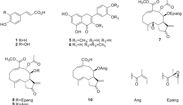
CO2H
HO
1R=H
2R=OH
R
O OH HO
OR2
OR3
OR1
5R1=CH3; R2=H; R3=H 6R1=H; R2=R3=Ch3
OEpang
O O
O
O O H3CO
7
OR
O O
O O H3CO
8R=Epang
9R=Ang
OAng CO2H
O
10
O O
O
Ang Epang
Table 1. Concentrations (µg/mL) of the compounds present in the three yacon leaf extracts investigated in this work as obtained by
HPLC-UV-DAD analytical curve.
Compound # Class Identiied as AE PE LRE
(1) CGA coumaric acid 0.3 3.5 0.0
(2) CGA caffeic acid 0.3 1.3 0.0
(3) CGA *CGA
1 2.0 9.9 0.0
(4) CGA *CGA2 1.3 9.6 0.0
(5) Flavonoid 3-O-methylquercetin 0.0 0.0 20.4
(6) Flavonoid 3,4’-di-O-methylquercetin 0.0 0.0 7.2
(7) STL enhydrin 99.7 0.0 1,997.7
(8) STL uvedalin 319.0 0.0 1,257.4
(9) STL polymatin B 129.3 0.0 616.2
(10) STL sonchifolin 102.7 0.0 519.1
The compounds were identiied by comparison with authentic standards available in our laboratory. *Chemical structures unidentiied. The UV proile suggests that these compounds correspond to CGA (UV max: 298 and 325 nm). CGA1 and CGA2 correspond to the most intense peaks in AE and PE
detected in this extract. The STL enhydrin (7), uvedalin
(8), polymatin B (9) and sonchifolin (10) appear at lower
concentrations in AE as compared to LRE, but their concentrations are still higher as compared to those of CGA in AE. PE, in turn, contains larger CGA concentrations as compared to AE, but this extract lacks STL.
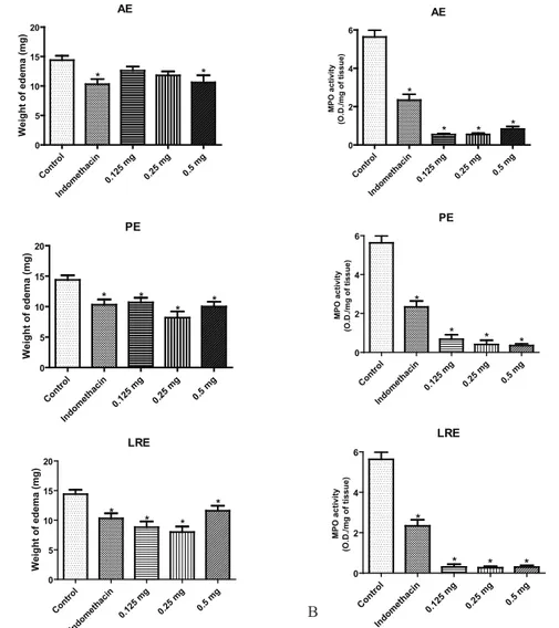
In the croton oil-induced ear edema assay, the
topical treatment of the animals with AE led to reduction of the ear edema, but a statically signiicant response (25.9% edema inhibition) was only veriied at the highest tested dose (0.5 mg/ear) (Figure 1A). This effect was statically similar to that elicited by indomethacin (28% edema inhibition). The topical effect of the extract PE was statically signiicant for all the assayed doses (25.2, 42.7, and 30.1 % ear edema inhibition for doses of 0.125, 0.25, and 0.5 mg/ear, respectively). Topical treatment with LRE showed statically signiicant outcome at all tested doses (0.125 mg/ear, 44.1% ear edema inhibition; 0.25 mg/ear, 38.5% ear edema inhibition; and 0.5 mg/ear, 18.9% ear
edema inhibition) (Figure 1A).
MPO activity was statically decreased as compared to the control group at all the investigated
extract doses as well as in the presence of indomethacin (Figure 1B). All the extracts had a more marked inluence
on MPO than indomethacin (0.5 mg/ear, 2.34 O.D./mg
of tissue). Meanwhile, PE (0.5 mg/ear, 0.35 O.D./mg of tissue) and LRE (0.5 mg/ear, 0.30 O.D./mg of tissue) had a more pronounced effect on MPO than AE (0.5 mg/ear,
0.83 O.D./mg of tissue).
Treatment of RAW 264.7 cells with AE and PE exercised no effect on NO production (Figure 2A). However, treatment of the cells with LRE resulted in a
statistically dose-dependent inhibition of NO production
(0.25 µg/mL: 23.2% inhibition; 0.5 µg/mL: 38.5% inhibition; 1.0 µg/mL: 72.96 % inhibition). The LRE activity at doses of 0.25 and 0.5 µg/mL was similar to the activity of the positive control dexamethasone (1.0 µg/mL: 33.6% inhibition), whilst LRE activity at a dose of 1.0 µg/ mL was statistically higher than that of dexamethasone.
Figure 1. Effect of the three yacon leaf extracts on the topical anti-inlammatory activity induced by croton oil. A. Weight of edema
measured as the weight difference between the two plugs of left ear (with edema induction) and right ear (without edema induction).
B. MPO activity in the ears with edema induction. Data are expressed as means±SEM. *p<0.05 in relation to the control (one-way
ANOVA following Tukey’s multiple comparison test. AE: aqueous extract, PE: polar extract, LRE: leaf-rinsed extract.
AE Con trol Indo met haci n 0.12
5 m
g 0.25 mg 0.5 mg 0 5 10 15 20 * * W e ig h t o f e d e m a (m g ) AE Con trol Indo met haci n 0.12 5 m
g 0.25 mg 0.5 mg 0 2 4 6 * * * * MPO a c ti v ity (O .D ./ m g o f ti s s u e ) PE Con trol Indo met haci n 0.12 5 m
g 0.25 mg 0.5 mg 0 5 10 15 20 * * * * W e ig h t o f e d e m a (m g ) PE Con trol Indo met haci n 0.12 5 m
g 0.25 mg 0.5 mg 0 2 4 6 * * * * MPO a c ti v ity (O .D ./ m g o f ti s s u e ) LRE Con trol Indo met haci n 0.12 5 m
g 0.25 mg 0.5 mg 0 5 10 15 20 * * * * W e ig h t o f e d e m a (m g ) LRE Con trol Indo met haci n 0.12 5 m
All the extracts were able to moderately inhibit TNF-α production (Figure 2B), and treatment with LRE evidenced a dose-dependent trend (0.25 µg/mL: 33.25% inhibition; 0.5 µg/mL: 42.13% inhibition; 1.0 µg/mL: 48.32% inhibition). LRE activity at a dose of 0.25 µg/mL
was statistically similar to that of indomethacin (1.0 µg/
mL: 27.41% inhibition), while LRE at doses of 0.5 and 1.0 µg/mL was more active than indomethacin (Figure 2B). AE (1.0 µg/mL: 38.42% inhibition) and PE (1.0 µg/mL: 36.37% inhibition) led to statistically similar effects at all
the tested doses (Figure 2B).
In the same way, all the extracts were able to
inhibit in vitro PGE2 production, being the actions of AE
(1.0 µg/mL: 73.45% inhibition) and LRE (1.0 µg/mL: 77.01% inhibition) more pronounced than that of PE (1.0 µg/mL: 41.57% inhibition). Treatment with LRE was the only one that presented a dose-dependent effect on PGE2 production (Figure 2 C). AE and LRE displayed activity
that was statistically similar to that of indomethacin (1.0
µg/mL: 79.11% inhibition).
Discussion
As previously reported (Oliveira et al., 2011),
HPLC analysis identiied PE as a polar extract rich in CGA and lacking STL, LRE as an extract rich in STL and lavonoids, and AE as an extract containing both CGA and STL. Here, we have quantiied the different classes of the main compounds in these three extracts. We veriied that AE displays lower STL concentrations as compared to LRE. However, STL concentrations in AE are larger if compared to CGA concentrations in this same extract. This suggests that STL play an important role in the biological activities observed for the aqueous extracts that are used orally by the population (Oliveira et al., 2011). PE contains high CGA concentrations, while STL are the major compounds in LRE. Thus, we have achieved our goal, which was to produce an extract rich in CGA and another one rich in STL, in order to gain insight into the role of these two classes of compounds in the anti-inlammatory activity of
yacon leaves.
Figure 2. In vitro anti-inlammatory assay in RAW 264.7 cells culture induced with LPS after treatment with the three different yacon leaf extracts. A. Nitric oxide determination. B. TNF-α determination. C. PGE2 determination. Data are expressed as mean±SEM.
*p<0.05 in relation to the control without LPS. AE: aqueous extract, PE: polar extract, LRE: leaf-rinsed extract.
AE Con trol Dex amet haso ne
0.25 0.5 1.0
0 50 100 150 200 * ______________________ LPS µg/mL N O ( µM) PE Con trol Dex amet haso
ne 0.25 0.5 1.0
0 50 100 150 200 * ______________________ LPS µg/mL N O ( µ M) RLE Con trol Dex amet haso
ne 0.25 0.5 1.0
0 50 100 150 200 * * * * ______________________ LPS µg/mL N O ( µ M) AE Con trol Indo met haci n
0.25 0.5 1.0
0 10000 20000 30000 * * * * _______________________ LPS µg/mL T N F -α (p g /m L ) PE Con trol Indo met haci n
0.25 0. 5 1.0 0 10000 20000 30000 * * * * _______________________ LPS µg/mL T N F
-α (p
g /m L ) RLE Con trol Indo met haci n
0.25 0. 5 1.0 0 10000 20000 30000 * * * * _______________________ LPS µg/mL T N F -α (p g /m L ) AE Con trol Indo met haci n
0.25 0.5 1.0 0 1000 2000 3000 4000 * * * * µg/mL _______________________ LPS PG E 2 (p g /m L ) PE Con trol Indo met haci n
0.25 0.5 1.0
0 1000 2000 3000 4000 * * * * µg/mL _______________________ LPS PG E2 (p g /m L ) RLE Con trol Indo met haci n
0.25 0.5 1.0 0 1000 2000 3000 4000 * * * * µg/mL _______________________ LPS PG E 2 (p g /m L )
that STL plays an important role in this activity. The effect of these extracts in decreased PGE2 levels can be related
with their effect on inhibition of leukocyte migration as demonstrated in the MPO assay, since it was demonstrated
that PGE2 production at the late phase in the TPA edema
model depends on the iniltrated leukocytes, while TNF-α levels are not related to cell iniltration in this model
(Murakawa et al., 2006).
Thus, our results have demonstrated that all the three tested yacon leaf extracts exhibit topical
anti-edematous activity in vivo. This activity may be a
consequence of an anti-inlammatory action, as evidenced by the fact that all the assayed extracts exert some effect on inlammatory mediators. This suggests that both STL and CGA contribute to the observed responses. However, the LRE extract, which is rich in STL, has a more pronounced effect on the anti-inlammatory mediators. In conclusion, our observations suggest that yacon leaf extracts, especially the one rich in STL, has potential application as a topical anti-inlammatory agent.
Acknowledgments
The authors thank FAPESP (proc. #2007/00844-3) and CAPES (proc. #314/09) for funds and grants, and
Prof. Ana M.S. Pereira for providing yacon rhizomes for
cultivation in the USP campus.
Authors’ Contributions
RBO (Ph.D. student) contributed in collecting
plant material, taxonomic identiication, confection of
herbarium vouchers, running the laboratory work, data
analysis and drafting the manuscript. DACP, THG and AS contributed to the biological studies. LHF, APC and
FBDC designed the study, supervised the laboratory work and contributed to critical reading of the manuscript. All
the authors have read the inal manuscript and approved
the submission.
References
Aybar MJ, Riera ANS, Grau A, Sánchez SS 2001. Hypoglycemic effect of the water extract of Smallanthus sonchifolius
(yacon) leaves in normal and diabetic rats. J Ethnopharmacol 74: 125-132.
Baroni S, Suzuki-Kemmelmeier F, Caparroz-Assef SM, Cuman RKN, Bersani-Amado CA 2008. Effect of crude extracts of leaves of Smallanthus sonchifolius (yacon) on glycemia in diabetics rats. Braz J Pharmaceutic Sci 44: 521-530.
Dirsch VM, Stuppner H, Ellmerer-Müller EP, Vollmar AM 2000. Structural requirements of sesquiterpene lactones to inhibit LPS-induced nitric oxide synthesis in RAW 264.7
macrophages. Bioorg Med Chem 8: 2747-2753.
We have found that all the prepared extracts have topical anti-edematous and anti-inlammatory activity, as conirmed by the fact that all the extracts decrease edema and neutrophil migration to the inlammatory site. The
edema was induced by topical application of the croton
oil, whose main inlammatory compound is the 12-O
-tetracanoylphorbol-3-acetate (TPA) (Hecker et al., 1968; Rao et al., 1993). Croton oil or puriied TPA application
in mouse skin has been used by several authors to access
the topical anti-inlammatory effects of non-steroidal and steroidal agents (Inoue et al., 1989). The TPA in
contact with the plasmatic membrane release arachidonic
acid that under action of the enzymes cyclooxygenase or lipoxygenase results in formation of cytokines or leukotrienes, respectively (Hecker et al., 1968; Rao et al.,
1993).
In the present study, AE was the least effective extract in decreased the edema induced by croton oil
application, since it is only able to reduce the edema
at a higher dose, while PE and RLE displays the
anti-edematous effect and decreases neutrophil migration at all
the tested doses. The in vitro experiments provided some
evidence about the action mechanisms involved in the
anti-inlammatory activity observed for the three extracts. AE and PE are not able to inhibit the NO production induced by LPS in macrophages, while LRE is a potent inhibitor. This suggests that STL has an important role in this effect. Indeed, several STL, e.g., parthenolide, isohelenin, and dehydrocostus lactone, were shown to
inhibit the expression of inducible nitric oxide synthase
(iNOS) in various cell systems (Dirsch et al., 2000). NO is over-produced endogenously by iNOS in response to
pro-inlammatory cytokines and LPS. There some evidence that STL inhibit iNOS expression via inhibition of the NF-κB. This is an interesting point, since it was demonstrated that enhydrin and uvedalin, two LST isolated from yacon leaves, were able to inhibit the NF-κB, a transcriptional
factor that has a central role in the transcription of the
genes related to the inlammatory process (Ghosh et al., 1998; Schorr et al., 2007).
Additionally, all the three yacon extracts are able
to inhibit the in vitro PGE2 production stimulated by LPS.
However, these extracts display low activity regarding TNF-α production, which is indication that neither STL nor CGA have signiicant activity on this inlammatory mediator. These indings can be useful to explain the in vivo anti-inlammatory effect of the extracts. Recently
it has been demonstrated that TNF-α and PGE2 levels
increased with edema formation after TPA application in mouse skin (Fürstenberger & Marks, 1980; Inoue et al., 1989, Murakawa et al., 2006). The increase of TNF-α peaked at ~5 h after TPA application, whereas the increase of PGE2 is biphasic with peaks at ~3 and 24 h after edema
Fürstenberger G, Marks F 1980. Early prostanglandin E synthesis
is an obligatory event in the induction of cell proliferation in mouse epidermis in vivo by the phorbol ester TPA. Biochem Biophys Res Commun 92: 749-756.
Genta SB, Cabrera WM, Grau A, Sánchez SS 2005. Subchronic 4-month oral toxicity study of dried Smallanthus sonchifolius (yacon) roots as a diet supplement in rats.
Food Chem Toxicol 43: 1657-1665.
Genta SB, Cabrera WM, Mercado MI, Grau A, Catalan CA,
Sánchez SS 2010. Hypoglycemic activity of leaf organic
extracts from Smallanthus sonchifolius: constituents of the most active fractions. Chem Biol Interact 185: 143-152.
Gonçalez E, Felicio JD, Pinto MM, Rossi MH, Medina C, Fernandes MJB, Simoni IC 2003. Inhibition of alatoxin
production by Polymnia sonchifolia and its in vitro cytotoxicity. Arch Inst Biol 70: 159-163.
Ghosh S, May MJ, Kopp EB 1998. NF-κB and Rel proteins:
evolutionary conserved mediators of immune responses.
Ann Review Immunol 16: 225-260.
Goto K, Fukai K, Hikida J, Nanjo F, Hara Y 1995. Isolation
and structural analysis of oligosaccharides from yacon (Polymnia sonchifolia). Biosci Biotech Biochem 59: 2346-2347.
Hecker E 1968. Cocarcinogenic principles from the seed oil of Croton tiglium and from other Euphorbiaceae. Cancer Res 28: 2338-234.
Inoue H, Mori T, Shibata S, Koshihara Y 1989. Modulation by glycyrrhetinic acid derivatives of TPA-induced mouse
ear oedema. Br J Pharmacol 96: 204 -210.
Inoue A, Tamogami S, Kato H 1995. Antifungal melampolides from leaf extracts of Smallanthus sonchifolius. Phytochemistry 30: 845-848.
Lin F, Hasegawa M, Kodama O 2003. Puriication and identiication of antimicrobial sesquiterpene lactones
from yacon (Smallanthus sonchifolius) leaves. Biosci Biotec Biochem 67: 2154-2159.
Miura T, Itho Y, Ishida T 2004. Hypoglycaemic and
hypolipidemic activity of the leaf of Samallhanthus sonchifolius in genetically type 2 diabetic mice. J Tradit Med 21: 275-277.
Miura T 2007. Antidiabetic activity of Fuscoporia oblique and
Smallanthus sonchifolius in genetically type 2 diabetic mice. J Trad Med 24: 47-50.
Murakawa M, Yamaoka K, Tanaka Y, Fukuda Y 2006. Involvement of tumor necrosis factor (TNF)-α in phorbol ester 12-O
-tetradecanoylphorbol-13-acetate (TPA)-induced skin
edema in mice. Biochem Pharmacol 71: 1331-1336.
Oliveira RB, Chagas-Paula DA, Rocha BA, Franco JJ, Gobbo-Neto L, Uyemura SA, Santos WF, Da Costa FB 2011. Renal toxicity caused by oral use of medicinal plants: the yacon example. J Ethnopharmacol 133: 434-441.
Pak A, Gonçalez E, Felício JD, Pinto MM, Rossi MH, Simoni IC, Lopes MN 2006. Inhibitory activity of compounds
isolated from Polymnia sonchifolia on alatoxin
production by Aspergillus lavus. Braz J Microbiol 37: 199-203.
Pedreschi R, Campos D, Noratto G, Chirinos R, Cisneros-Zevallos L 2003. Andean yacon roots (Smallanthus sonchifolius Poepp. Endl.) fructooligosaccharides as a
potential novel sources prebiotics. J Agricult Food Chem 51: 5278-5284.
Pinto MM, Gonçalez E, Rossi MH, Felicio JD, Medina CS, Fernandes MJB, Simoni IC 2001. Activity of the aqueous extract from Polymnia sonchifolia leaves on growth and
production of alatoxin B1 by Aspergillus lavus. Braz J Microbiol 32: 127-129.
Rao TS, Currie JL, Shaffer AF, Isakson PC 1993.
Comparative evaluation of arachidonic acid (AA)- and
tetradecanoylphorbol acetate (TPA)-induced dermal inlammation. Inlammation 17: 723-741.
Santos MD, Almeida MC, Lopes NP, Souza GEP 2006. Evaluation of the anti-inlammatory analgesic and antipyretic
activities of the natural polyphenol chlorogenic acid. Biol Pharm Bull 29: 2236-2240.
Schorr K, Da Costa F B. 2003. A proposal for chemical characterisation and quality evaluation of botanical
raw materials using glandular trichome microsampling of yacon (Polymia sonhifolia Asteraceae) an Andean medicinal plant. Rev Bras Farmacogn 13: 1-3.
Schorr K, Mefort I, Da Costa FB 2007. A novel dimeric
melampolide and further terpenoids from Smallanthus sonchifolius (Asteraceae) and the inhibition of the
transcription factor NF-κB. Nat Prod Commun 2: 367-374.
Shin K, Kim I, Park Y, Ha J, Choi J, Park H, Lee YS, Lee K 2004. Anti-inlamatory effect of caffeic acid methyl ester and
its mode of action through the inhibition of prostaglandin
E2 nitric oxide and tumor necrosis factor-α production.
Biochem Pharmacol 68: 2327-2336.
Siedle B, García-Piñeres AJ, Murillo R, Schulte-Mönting J, Castro V, Rüngeler P, Klaas CA, Da Costa FB, Kisiel W, Merfort
I 2004. Quantitative structure-activity relantionship of
sesquiterpene lactones as inhibitors of the transcription factor NF-κB. J Med Chem 47: 6040-6054.
Simonowska B, Vovk I, Andrenzek S, Valentová K, Ulrichová
J 2003. Investigation of phenolic acids in yacon (Smallanthus sonchifolius) leaves and tubers. J Chromatogr A 1016: 89-98.
Takenaka M, Yan X, Ono H, Yoshida M, Nagata T, Nakanishi T 2003. Caffeic acid derivatives in the roots of yacon
(Smallanthus sonchifolius). J Agricult Food Chem 51: 793-796 793.
Terada S, Itoh K, Noguchi N, Ishida T 2009. Alpha-glucosidase
inhibitor for blood glucose level elevation and functional food containing tricaffeoylaldaric acid and method for producing tricaffeoylaldaric acid. United States Patent Application Publication US 2009/0209649 A1.
Tubaro A, Dri P, Delbello G, Zilli C, Loggia RD 1985. The croton
Valentová K, Ulrichová J 2003. Smallanthus sonchifolius and
Lepidium meyenii - prospective Andean crops for the prevention of chronic diseases. Biomed Papers 147: 119-130.
Valentová K, Moncon A, Ulrichová J 2004. The effect of Smallanthus sonchifolius leaf extracts on rat hepatic
metabolism. Cell Biol Toxicol 20: 109-120.
Valentová K, Sersen F, Ulrichová J 2005. Radical scavenging and anti-lipoperoxidative activities of Smallanthus sonchifolius leaf extracts. J Agricult Food Chem 53: 5577-5582.
Vilhena SMC, Câmara FLA, Kakihara ST 2000. O cultivo de
yacon no Brasil. Hort Bras 18: 5-8.
Yan X, Suzuki M, Ohnishi-Kameyama M, Sada Y, Nakanishi
T, Nagata T 1999. Extraction and identiication of antioxidants in the roots of yacon (Smallanthus sonchifolius). J Agricult Food Chem 47: 4711-4713.
*Correspondence
Fernando Batista Da Costa
AsterBioChem Research Team
Faculdade de Ciências Farmacêuticas de Ribeirão Preto,
Universidade de São Paulo
Av. do Café s/n, 14040-903 Ribeirão Preto-SP, Brazil [email protected]