Review
Printed in Brazil - ©2017 Sociedade Brasileira de Química0103 - 5053 $6.00+0.00*e-mail: [email protected]; [email protected]
Silver and Silver Chloride Nanoparticles and their Anti-Tick Activity: a Mini Review
Nelson Durán,*,a,b,c Marcela Durán*,b,d and Celso E. de Souzae
aLaboratório de Química Biológica and bNanoBioss, Instituto de Química,
Universidade Estadual de Campinas, 13083-970 Campinas-SP, Brazil
cLaboratório Nacional de Nanotecnologia (LNNano-CNPEM), 13083-100 Campinas-SP, Brazil
dLaboratório de Carcinogenese Urogenital e Imunoterapia,
Departamento de Genética, Evolução e Bioagentes, Instituto de Biologia, Universidade Estadual de Campinas, 13083-970 Campinas-SP, Brazil
eLaboratório de Parasitoses por Flagelados, Superintendência de Controle de Endemias (SUCEN),
01027-000 São Paulo-SP, Brazil
This overview highlights the importance of characterization of biogenic nanoparticles of silver and silver chloride in order to understand the action on ticks or pathogens transmitted by them. These nanoparticles appear as important active principles in this area. They can act against ticks or against major pathogens transmitted by the bite of ticks such as bacteria, viruses or protozoa with equal or better efficacy of antibiotics, antiviral or antiparasitic agents. Anti-tick activities on Rhipicephalus (Boophilus) microplus, Hyalomma anatolicum, Hyalomma isaaci and Haemaphysalis bispinosa are discussed. Perspectives of these nanoparticles acting on bacteria, viruses and protozoa infections are also discussed.
Keywords: ticks, silver nanoparticles, silver chloride nanoparticles, anti-tick agents
1. Introduction
Ticks are vectors of many infections as they are able to carry bacteria, virus and also protozoan agents.1 Related to
the bacterial diseases transmitted by ticks are Lyme disease, rickettsioses, and tularemia.2-7
Tiboviruses (tick-borne viruses) cause many symptoms with severe effects that affect the central nervous system, such as meningitis, meningoencephalitis with high sequelae, and hemorrhagic diseases.8-10
Protozoan infections, for example, the babesiosis (Babesia bigemina), may also be transmitted by ticks.11-13 Toxin production can cause paralysis, from
Dermacentor andersoni (Rocky Mountain wood tick), Dermacentor variabilis (dog ticks), and Ixodes holocyclus (marsupial ticks).14-16 In this last and rare case, the mainstay
of treatment for tick’s paralysis, tick removal, and the time to full neurological recovery after tick removal are generally estimated to be around 1.5 days with initial improvement within hours.17,18
In Brazil, the main disease transmitted by ticks to humans is spotted fever (BSF) whose etiologic agent is Rickettsia rickettsii, which is an intracellular gram-negative bacterium. The main vector is Amblyomma sculptum and the disease is considered a major public health problem.19,20
The rise in the number of notified cases and the expansion of transmission area and elevated lethality rate have been observed in the country since 80’s. Lethality rate in the southeast region of Brazil ranges between 30 to 50%, and in São Paulo and Minas Gerais states one can found the most notifications of this disease.19,21
An excellent alternative for treatment of these diseases is the use of nanobiotechnology as a novel strategy in this area. One important nanostructure is the silver nanoparticles (Ag0) or also nanoparticles of the silver cloride (AgCl).
The silver nanoparticles biogenically synthesized are widely studied by many research groups in the world22-39
and Ag0 nanoparticles were quite efficient against
Aedes aegypti.40
It is important to be aware that the characterizations of these silver nanostructures are of paramount importance, since there are many reports in the literature in which erroneous characterization of silver structures were published. The X-ray diffraction (XRD) pattern for Ag0 as
well as for AgCl nanoparticles, which are the most common silver nanostructures that are biogenically synthesized, were discussed recently by Seabra et al.43 Based on
aforementioned discussion on XRD characterization of biogenic silver nanostructures, herein the anti-tick activities are to be discussed.
The XRD patterns for Ag0 nanoparticles (JCPDS
file No. 04-0783 or ICSD Code 64994) and for AgCl nanoparticles (JCPDS file No. 85-1355 or ICSD Code 64734) were used to construct Table 1.
2. Anti-Tick Activity
Marimuthu et al.42 biosynthesized silver nanoparticles
from leaf extract of Mimosa pudica Gaertn. (Mimosaceae) and tested those nanoparticles against the larvae of Rhipicephalus (Boophilus) microplus Canestrini, 1887 (Acari: Ixodidae) (R. (B.) microplus). In this case the silver nanostructure was classified as Ag0 nanoparticles.
Analyzing the XRD pattern data the nanostructure studied also exhibited values that correspond to AgCl nanoparticles (low presence) Ag0 and/or silver oxides as
spherical morphology. Reported nanoparticles exhibited an efficacy of LC50 of 8.98 µg mL-1 against the larvae of
R. (B.) microplus. At the concentration of 15 and 20 µg mL-1,
a 51 and 89% of mortality was found, respectively. The size of the nanoparticles by transmission electron microscopy (TEM) was around 25-60 nm and by scanning electron microscopy (SEM) were 25-50 nm.
The synthesis of mainly AgCl nanoparticles and with presence of low Ag0 nanoparticles silver nanoparticles
from leaf extract of Ocimum canum Sims (Labiatae) against the larvae of Hyalomma anatolicum (a.) anatolicum Koch, 1844 (actual name H . a n a t o l i c u m) and Hyalomma marginatum (m.) isaaci Sharif, 1928 (Acari: Ixodidae) (actual name H. isaaci) was achieved.41
SEM analyses were crucial to determine the size of the synthesized AgCl nanoparticles that was estimated to be 25-110 nm. The particles exhibited spherical and after drying formed rod and cubic morphology. These nanoparticles exhibited effect against H. anatolicum and H. isaaci with the LC50 values of 0.78 and 1.00 µg mL-1,
respectively, and caused 100% mortality at 2.5 µg mL-1.
Probably, in this case the cubic form of AgCl exerted a better activity than AgCl in its spherical morphology.47 It
is known that spherical and cubes as in the case of AgCl in this case, exhibits similar biological activities.
Stem aqueous extract of Cissus quadrangularis biosynthesis of AgCl nanoparticles and their effects
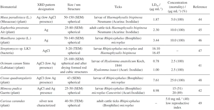
Table 1. Anti-tick activities of silver and silver chloride nanoparticles
Biomaterial XRD pattern designation
Size / nm
Structure Ticks
LD50 /
(µg mL-1)
Concentration (mortality) / (µg mL-1) (%)
Reference
Musa paradisiaca (L.) (Musaceae) (plant)
Ag (low AgCl presence)
50-150 (SEM) spherical
larvae of Haemaphysalis bispinosa
Neumann (Acarina: Ixodidae) 1.87 5.0 (100) 44
Euphorbia prostrata
Ait (plant) Ag
25-80 (SEM) spherical
adult cattle tick Haemaphysalis bispinosa
Neumann (Acarina: Ixodidae) 2.30 10.0 (100) 45
Manilkara zapota (L.)
(plant) Ag
70-140 (SEM) spherical
larvae Rhipicephalus (Boophilus)
microplus 3.44 10.0 (100) 46
Streptomyces sp. LK3
(bacteria) AgCl
5-20 (TEM) spherical
larvae Rhipicephalus microplus and
Haemaphysalis bispinosa
16.10
16.45 − 47
Ocimum canum Sims (Labiatae) (plant)
AgCl (low Ag presence)
25-100 (SEM) spherical and after drying formed rod and cubic structures
larvae of Hyalomma anatolicum Koch, 1844
Hyalomma isaaci (Acari: Ixodidae)
0.78
1.00
2.5 (100)
2.5 (100)
41
Cissus quadrangularis
(plant)
AgCl (low Ag presence)
43 (SEM) spherical
larvae of Rhipicephalus (Boophilus)
microplus 7.61 25.0 (100) 48
Mimosa pudica Gaertn (plant)
AgCl and Ag presence
25-50 (SEM) spherical
larvae Rhipicephalus (Boophilus)
microplus Canestrini (Acari:Ixodidae) 8.98
15 (51)
20 (89) 42
Carissa carandas
(plant)
silver non characterized
40-50 (TEM) spherical
adult cattle ticks Rhipicephalus
(Boophilus) microplus −
5.0 mg mL-1 (40)
low reproductive index
49
against the larvae of R. (B.) microplus were studied.48 The
size of the nanoparticles as determined by FESEM was 43 nm. The activity against the larvae of R. (B.) microplus exhibited LC50 values of 7.61 and at 25 µg mL-1 and 100%
of mortality for adult R. (B.) microplus was also observed. This nanostructure exhibited lower activity than any Ag0,
since this nanoparticle is spherical as in Marimuthu et al.42
Aqueous leaf extract from Musa paradisiaca (L.) (Musaceae) led to the synthesis of silver nanoparticles (mainly) and some AgCl nanoparticles were also present and their application against the larvae of Haemaphysalis bispinosa Neumann (Acarina: Ixodidae) (H. bispinosa) was reported.44 By SEM the size was
20-30 nm and exhibiting the LC50 of 1.87 µg mL-1 and at
concentration of 5 µg mL-1, those nanoparticles provoked
100% of mortality. The spherical morphology in Ag0
exerted an important biological activity.
Aqueous leaf extract from Euphorbia prostrata Ait. was used for the synthesis Ag0 nanoparticles (mainly with
traces of AgCl nanoparticles) and studied against the adult cattle tick H. bispinosa.45 SEM analysis showed a size of
25-80 nm. The LC50 value observed was 2.3 µg mL-1 and
100% of mortality at 10 µg mL-1 nanoparticles. In this
preparation the Ag0 presented a spherical morphology and
similar activities than in the work of Marimuthu et al.42
Rajakumar and Rahuman46 synthesized from aqueous
extract of Manilkara zapota (L.) mainly Ag0 nanoparticles
(spherical morphology) and traces of AgCl nanoparticles as seen by XRD pattern. A size of 70-140 nm by SEM was determined. LC50 values against R. (B.) microplus was
3.44 µg mL-1 and at a concentration of 10 µg mL-1 exhibited
a 100% mortality.
The synthesis of AgCl nanoparticles from Streptomyces sp. LK3 showed acaricidal activity against R. (B.) microplus and H. bispinosa with LC50 values of 16.10 and 16.45 µg mL-1,
respectively.47 In this case, the exhibited plasmon absorption
can be associated to a very low Ag0 nanoparticles
concentration present. The presence of AgCl in its spherical form exerts a low activity as compared with Ag0.
E va l u a t i o n o f a c a r i c i d a l e ffi c a cy ( a g a i n s t R. (B.) microplus) of plant mediated synthesis of silver nanoparticles using Carissa carandas leaf extract (40-50 nm by TEM) was reported. Adult immersion test was used to evaluate the efficacy of silver nanoparticles against cattle ticks. Results showed 40% mortality with silver nanoparticles (non characterized by XRD diffractions pattern and probably AgCl) using a high concentration (5 mg mL-1, 72 h). The effect was further observed on the
reproductive indices of ticks which exhibited significant decrease in this index with increasing concentrations of silver nanoparticles.49
Larvae and adult H. bispinosa were affected in the same extension (LD50 ca. 2 µg mL-1) and similar mortality
percentage from Ag0 nanoparticles from different sources
(Musa paeadisiaca)44 and Euphorbia prostate.45 However,
AgCl nanoparticles from Streptomyces47 showed to be
8-fold less effective than ones obtained from the plant sources.
Larvae R. (B.) microplus was affected by Ag0 nanoparticles
differently when different sources of the Ag0 nanoparticles
were used. The LD50 from Manilkara zapota (3.44 µg mL-1)46
and from Mimosa pudica (8.98 µg mL-1)42 were found. At
the same extension mortality were 3-fold more efficient in Manilkara than in Mimosa. In the presence of AgCl nanoparticles, the same value of LD50 (7.61 µg mL-1) was
obtained by the source of Cissus quadrangularis with a similar mortality48 against the ticks. AgCl nanoparticles
produced by Streptomyces exhibited a lower effect against the tested ticks (LD50 ca.16 µg mL-1) (Table 1).
Larvae of H. anatolicum and H. marginatum were affected significantly by AgCl nanoparticles synthesized by Ocimum canum Sims with LD50 of 0.78 and 1.0 µg mL-1,
respectively.41 Probably, in this case the spherical form that
after drying formed rod and cubic structures exerted a high biological activity in a similar or better profile than Ag0.
It is known that silver triangular nanoplates have basic {111} surface, nanospheres and nanocubes mainly have {100} planes, and nanorods have {100} side surfaces and {111} end facets. Then, the data said that {111} crystal plane of silver nanoparticles may play a prime role in antibacterial progress. Recently was reported that silver triangle nanoplates had the best antibacterial activity, while nanospheres, nanocubes and short nanorods showed similar efficacy.50
This was reinforced with MIC values of Ag0 nanoparticles
(60 nm) in which the cubic form and spherical one were 37.5 and 75 µg mL-1, respectively.51 The fact is, the particles
covered by {100} facets (cubic form) showed stronger antibacterial activity than those covered by {111} facets (spherical). This was demonstrated on Table 1.
The Table 1 shows a summary of all the anti-tick activities of biogenic silver nanoparticles. Fortunately, with the discrimination of the XRD pattern, it was possible to get some conclusions.
In conclusion, Ag0 and AgCl nanoparticles are efficient
anti-tick agents, however, their activities depend on the source of production of nanoparticles and from their morphologies. Due to this factor, AgCl in its spherical morphology exhibited less activity than spherical Ag0.
which probably due to the specificity of the protein corona interaction may strongly affect nanoparticles’ activities.26
3. Tick-Borne Diseases
3.1. Bacteria
Bacteria genera most common in the ticks’ infection are Borrelia (a genus of Gram-negative, aerobic, spirochete bacteria), Francisella (a genus Gram-negative, aerobic, coccobacillary or rod shaped, nonmotile bacteria), Coxiella (a genus of Gram-negative, aerobic, rod-shaped or spore-like particles, acidophilic bacteria), Rickettsia (a genus of non motile, Gram-negative, aerobic, obligate intracellular parasites, non spore-forming, highly pleomorphic bacteria that can present as cocci) and Ehrlichia (a genus of Gram-negative, aerobic, obligate intracellular parasites, rickettsia-shaped bacteria).
Burgdorfer,52 who is well known researcher in Lyme
diseases, tested in vitro proteinate silver (colloidal silver or Argyrol) up to 1500 ppm (1.5 mg mL-1) on bacteria that
cause Lyme disease (Borrelia burgdorferi spirochetes).53,54
These bacteria disappeared in less than five minutes and twenty-four hours later, none of the crops treated contained any living spirochetes. Similar results were observed with another bacterium, such as Borrelia hermsii (HS-1), the agent of relapsing fever.
As far as we know, no other bacteria in this area were studied under influence of silver nanoparticles. However, it is known from the literature,23,35-38,55 that Gram-negative
bacteria are effectively eliminated by Ag0 nanoparticles.
Then, the antibacterial studies regarding tick-borne infections are an open area for research.
3.2. Virus
The most known viruses from tick infections are the tick-borne viruses of the families Asfarviridae, Rhabdoviridae and Orthomyxoviridae, Reoviridae, Bunyaviridae and Flaviviridae, genus Flavivirus (around 200 viruses).8
Argovit (12 mg mL-1) that is spherical silver nanoparticles
of 35 ± 15 nm functionalized with poly(vinylpyrrolidone) (PVP, 10-30 kD) were tested on Rift Valley fever virus (RVFV) in a Vero cell cultures and on mice lacking the type I interferon receptor (IFNAR [−/−] mouse infection model). In cells infected with RVFV a viral reduction of 50% of the total virus was found. But, pre-incubation of RVFV with silver nanoparticles at concentrations of 0.2 mg mL-1, abolished almost completely viral
propagation, leading to 98% reduction of infectivity. Daily administration of silver nanoparticles by oral gavages
to lethally infected mice, previously incubated with 20 mg mL-1 of silver nanoparticles showed a
delayed-onset clinical disease and mortality, with a survival rate of 60%. The authors suggested that these results revealed the potential application of the microbiocide properties of silver nanoparticles to control the infectivity of this important zoonotic pathogen.56
In addition to the antimicrobial activity against bacteria of silver nanoparticles, there is strong evidence that they are also active against several viruses, including human immunodeficiency virus, hepatitis B virus, herpes simplex virus, respiratory syncytial virus and monkey pox virus. These metal nanostructures generate an important opportunity for the development of new antiviral therapies with a large number of targets, reducing viral resistance that can commonly happen with conventional antivirals.57
3.3. Protozoan
Protozoan infection babesiosis (Babesia bigemina) is transmitted by ticks13 and, in a similar way,
leptospirosis by Ixodes ricinus58 and leishmaniasis by
Rhipicephalus sanguineus.59
The effectiveness of silver nanoparticles as an alternative therapy for leishmaniasis, specifically by subcutaneous intralesional administration for cutaneous leishmaniasis was demonstrated.60 Biogenic silver nanoparticles (mainly
Ag0 nanoparticles) against Leishmania amazonensis
promastigotes showed to be 4-fold more effective than silver nanoparticles chemically synthesized. In vivo studies in infected mice demonstrated that the biogenic silver nanoparticles were equally effective as 300-fold higher doses of amphotericin B, and more effective than 4-fold higher doses of chemically synthesized one.61
The possibility of using silver nanoparticles on protozoan infections was summarized by Rai et al.62 These
facts demonstrated that we need more research in order to eliminate these types of contamination provoked by ticks.
4. Perspectives and Final Remarks
It is important to mention that it was clear from these analyzed data that reliable nanoparticles characterization and morphology are important in the biological activities. Then, the data said that {111} crystal plane of silver nanoparticles may play a prime role in antibacterial progress.
Another important conclusion is also that either Ag0 or
data indicated that the antibacterial and antiviral studies regarding tick-borne infections are an open area for research.
Then, we hope that this overview might incentive the researchers to study ticks-producer diseases more thoroughly in order to eradicate this problem globally.
Acknowledgments
Supports from FAPESP, CNPq, INOMAT (MCTI/ CNPq), NanoBioss (MCTI) and Brazilian Network on Nanotoxicology (MCTI/CNPq) are acknowledged.
Nelson Durán is a Professor of
Chemistry at the Universidade Estadual de Campinas (UNICAMP, Brazil). He received his PhD at University of Porto Rico (USA) working on photolysis and thermolysis of 1,2-dioxolanes (1972). Associated Professor at the Universidad Catolica de Valparaiso, Chile (1973-1975) and carried out Visiting Professorship at Universidade de São Paulo, Brazil (1975), investigating enzymatic generation of excited states intermediates. In 1978, he joined the Chemistry Institute of UNICAMP (Brazil) working in Biological Chemistry and Biotechnology. His present research interests are nanobiotechnology in cosmetics and in pharmaceuticals, besides metallic nanoparticles as antibiotics and anticancer carriers, and in carbon and silica nanocarriers. He is the Coordinator of the Brazilian Nanotoxicology Network; member of INOMAT (MCTI/ CNPQ) and Vice Coordinator of NanoBioss (MCTI) and member of Brazilian-NanoReg (European Community) in vivo nanotoxicology.
Marcela Durán is a pharmacist,
R e s e a r c h e r A s s o c i a t e d i n Carcinogenesis Laboratory Urogenital and Immunotherapy Anatomy, at Biology Institute of the Universidade Estadual de Campinas (UNICAMP). She is graduated at Universidade São Francisco (USF), São Paulo, Brazil. Currently she is developing research project related to Nanotechnology area, such as cell regeneration and stem cells. She has a Master in Food Technology (UNICAMP, 2004) and PhD in Medical Sciences (UNICAMP, 2014). She is member of Brazilian-NanoReg Toxicology group (European Community) working in in vivo study of carbon nanotubes by instillation on rats associated to NanoBioss (MCTI).
Celso Eduardo de Souza is a
Veterinarian of the Superintendence Control of Endemic Diseases of the Government of the State of São Paulo, Brazil. He graduated in Veterinary Medicine from the Universidade de Alfenas (1984-1988). Master in Animal Health area of concentration in Epidemiology by Universidade Federal Rural do Rio de Janeiro (2010). He holds a doctorate degree in Collective Health from the Faculty of Medical Sciences of the Universidade Estadual de Campinas (UNICAMP, 2015). He has experience in Preventive Veterinary Medicine, working mainly in research and diagnosis in the following subjects: Zoonosis and Public Health, epidemiology, parasitology and vector-borne diseases with emphasis on diseases transmitted by ticks. Technical advisor of the Program of Epidemiological Surveillance of Brazilian Spotted Fever and other Riquettoses of the Ministry of Health of Brazil.
References
1. Kon, K.; Rai, M. In Nanotechnology in Diagnosis, Treatment and Prophylaxis of Infections Diseases; Rai, M.; Kon, K., eds.; Elsevier: London, UK, 2015, ch. 3, p. 39.
2. Parola, P.; Raoult, D.; Clin. Infect. Dis. 2001, 32, 89. 3. Parola, P.; Paddock, C. D.; Raoult, D.; Clin. Microbiol. Rev.
2005, 18, 719.
4. Foley, J. E.; Nieto, N. C.; Vet. Microbiol. 2010, 140, 332. 5. Kung, F.; Anguita, J.; Pal, U.; Future Microbiol. 2013, 8, 41. 6. Portillo, A.; Santibañez, S.; García-Alvarez, L.; Palomar, A.
M.; Oteo, J. A.; Microbes Infect.2015, 17, 834.
7. Borchers, A. T.; Keen, C. L.; Huntley, A. C.; Gershwin, M. E.;
J. Autoimmun. 2015, 57, 82.
8. Labuda, M.; Nuttall, P. A.; Parasitology 2004, 129, S221. 9. Hubálek, Z.; Rudolf, I.; Parasitol. Res. 2012, 111, 9. 10. Pujalte, G. G. A.; Chua, J. V.; Prim. Care 2013, 40, 619. 11. Amsden, J. R.; Warmack, S.; Gubbins, P. O.; Pharmacotherapy
2005, 25, 191.
12. Schnittger, L.; Rodriguez, A. E.; Florin-Christensen, M.; Morrison, D. A.; Infect., Genet. Evol. 2012, 12, 1788. 13. Barto, D.; Brzozowski, J.; Nurs. Crit. Care2014, 9, 23. 14. Diaz, J. H.; J. Med. Toxicol. 2010, 6, 15.
15. Diaz, J. H.; Clin. Toxicol. 2015, 53, 874.
16. Teong, J. M. Y.; Adler, P. A.; Doggett, S. L.; Danehvar, D.; Shields, M. K.; Case Rep. Ophthalmol. 2015, 6, 120. 17. Pecina, C. A.; Semin. Neurol. 2012, 32, 531.
18. Taraschenko, O. D.; Powers, K. M.; Pediatr. Neurol. 2014, 50, 605. 19. Lima, V. L. C.; Souza, S. S. L.; Souza, C. E.; Vilela, M. F. G.;
20. Katz, G.; Camargo-Neves, V. L. F.; Angerami, R. N.; Nascimento, E. M. M.; Colombo, S.; Bol. Epidemiol. Paul. (BEPA)2009, 6, 4.
21. Nasser, J. T.; Lana, R. C.; Silva, C. M.; Lourenço, R. W.; da Cunha e Silva, D. C.; Donalicio, M R.; Rev. Bras. Epidemiol.
2015, 18, 299.
22. Durán, N.; Marcato, P. D.; Teixeira, Z.; Durán, M.; Costa, F. T. M.; Brocchi, M.; Curr. Nanosci. 2009, 5, 396.
23. Durán, N.; Marcato, P. D.; de Conti, R.; Alves, O. L.; Costa, F. T. M.; Brocchi, M.; J. Braz. Chem. Soc. 2010, 21, 949. 24. Durán, N.; Marcato, P. D.; Durán, M.; Yadav, A.; Gade, A.; Rai,
M.; Appl. Microbiol. Biotechnol. 2011, 90, 1609.
25. Durán, N.; Seabra, A. B.; de Lima, R. In Nanotoxicology: Materials, Methodologies, and Assessments; Durán, N.; Guterres, S. S.; Alves, O. L., eds.; Springer: New York, 2014, ch. 11, p. 245.
26. Durán, N.; Silveira, C. P.; Durán, M.; Martinez, D. S. T.;
J. Nanotechnol. 2015, 13, 55.
27. Durán, M.; Silveira, C. P.; Durán, N.; IET Nanobiotechnol.
2015, 9, 314.
28. Rai, M.; Yadav, A.; Gade, A.; Biotechnol. Adv. 2009,27, 76. 29. Marcato, P. D.; Durán, M.; Huber, S.; Rai, M.; Melo, P. S.;
Alves, O. L.; Durán, N.; J. Nano Res. 2012, 20, 99.
30. Ingal, A. G.; Chaudhari, A. N.; J. Nanomed. Nanotechol. 2013,
4, 2.
31. Abbasi, E.; Milani, M.; Aval, S. F.; Kouhi, M.; Akbarzadeh, A.; Nasrabadi, H. T.; Nikasa, P.; Joo, S. W.; Hanifehpour, Y.; Nejati-Koshki, K.; Samiei, M.; Crit. Rev. Microbiol. 2008, 42, 173. 32. Faramarzi, M. A.; Sadighi, A.; Adv. Colloid Interface Sci. 2013,
189-190, 1.
33. Gowramma, B.; Keerthi, U.; Rafi, M.; Rao, D. M.; 3 Biotech
2015, 5, 195.
34. Mittal, J.; Batra, A.; Singh, A.; Sharma, M. M.; Adv. Nat. Sci.: Nanosci. Nanotechnol. 2014, 5, 043002.
35. Mashwani, Z.; Khan, T.; Khan, M. A.; Nadhman, A.; Appl. Microbiol. Biotechnol. 2015, 99, 9923.
36. Natsuki, J.; Natsuki, T.; Hashimoto, Y.; Int. J. Mater. Sci. Appl.
2015, 4, 325.
37. Moghaddam, A. B.; Namvar, F.; Moniri, M.; Tahir, P.; Azizi, S.; Mohamad, R.; Molecules 2015, 20, 16540.
38. Keat, C. L.; Aziz, A.; Eid, A. M.; Elmarzugi, N. A.; Bioresour. Bioprocess. 2015,2, 47.
39. Picoli, S. U.; Durán, M.; Andrade, P. F.; Durán, N.; Front. Nanosci. Nanotechnol. 2016, 2, 107.
40. Durán, N.; Islan, G. A.; Durán, M.; Castro, G. R.; J. Braz. Chem. Soc. 2016,27, 1139.
41. Jayaseelan, C.; Rahuman, A. A.; Parasitol. Res. 2012,111, 1369.
42. Marimuthu, S.; Rahuman, A. A.; Rajakumar, G.; Santhoshkumar, T.; Kirthi, A. V.; Jayaseelan, C.; Bagavan, A.; Zahir, A. A.; Elango, G.; Kamaraj, C.; Parasitol. Res. 2011,108, 1541.
43. Seabra, A. B.; Nakazato, G.; Durán, N.; Appl. Microbiol. Biotechnol.2016, 100, 6555.
44. Jayaseelan, C.; Rahuman, A. A.; Rajakumar, G.; Santhoshkumar, T.; Kirthi, A. V.; Marimuthu, S.; Bagavan, A.; Kamaraj, C.; Zahir, A. A. S.; Elango, G.; Velayutham, K.; Rao, K. V. B.; Karthik, L.; Raveendran, S.; Parasitol. Res. 2012, 111, 921. 45. Zahir, A. A., Rahuman, A. A.; Vet. Parasitol. 2012, 187, 511. 46. Rajakumar, G.; Rahuman, A. A.; Res. Vet. Sci. 2012,93, 303. 47. Karthik, L.; Kumar, G.; Kirthi, A. V.; Rahuman, A. A.; Rao, K.
V. B.; Bioprocess Biosyst. Eng.2014, 37, 261.
48. Santhoshkumar, T.; Rahuman, A. A.; Bagavan, A.; Marimuthu, S.; Jayaseelan, C.; Kirthi, A. V.; Kamaraj, C.; Rajakumar, G.; Zahir, A. A.; Elango, G.; Velayutham, K.; Iyappan, M.; Siva, C.; Karthik, L.; Rao, K. V. B.; Exp. Parasitol. 2012, 132, 156. 49. Johari, P.; http://shodhganga.inflibnet.ac.in/handle/10603/2363,
accessed in November 2016.
50. Hu, G.; Jin, W.; Chen, Q.; Cai, Y.; Zhu, Q.; Zhang, W.; Appl. Phys. A 2016, 122, 874.
51. Hong, X.; Wen, J.; Xiong, X.; Hu, Y.; Environ. Sci. Pollut. Res.
2016, 23, 4489.
52. Burgdorfer, W.; Rev. Infect. Dis. 1986, 8, 932.
53. Burgdorfer, W.; From Penicillin to Mild Silver Protein - An Answer to Lyme Disease Without Antibiotics; Rocky Mountain Laboratories, National Institute of Health (Agency of the U.S. Department of Health), USA, 1995.
54. Schwan, T. G.; Burgdorfer, W.; Department of Health and Human Services, National Institutes of Health, Rocky Mountain Laboratories, January 13, 1995. Available at http://www.xpressnet. com/bhealthy/bhealthy.html, accessed in February 2017. 55. Lemire, J. A.; Harrison, J. J.; Turner, R. J.; Nature Rev.
Microbiol. 2013,11, 371.
56. Borrego, B.; Lorenzo, G.; Mota-Moralez, J. D.; Almanza-Reyez, H.; Mateos, F.; López-Gil, E.; de la Losa, N.; Burmistrov, V. A.; Pestryakov, A.; Brun, A.; Bogdanchikova, N.; Nanomedicine
2016, 12, 1185.
57. Galdiero, S.; Falanga, A.; Vitiello, M.; Cantisani, M.; Marra, V.; Galdiero, M.; Molecules 2011, 16, 8894.
58. Wójcik-Fatla, A.; Zajac, V.; Cisak, E.; Sroka, J.; Sawczyn, A.; Dutkiewicz, J.; Ann. Agric. Environ. Med. 2012,4, 656. 59. Campos, J. H. F.; Costa, F. A. L.; Rev. Inst. Med. Trop. São
Paulo 2014, 56, 297.
60. Islan, G. A.; Durán, M.; Cacicedo, M. L.; Nakazato, G.; Kobayashi, R. K. T.; Martinez, D. S. T.; Castro, G. R.; Durán, N.; Acta Trop.2017, 170, 16.
61. Rossi-Bergmann, B.; Pacienza-Lima, W.; Marcato, P. D.; de Conti, R.; Durán, N.; J. Nano Res. 2012, 20, 89.
62. Rai, M.; Kon, K. Ingle, A.; Durán, N.; Galdiero, S.; Galdiero, M.; Appl. Microbiol. Biotechnol. 2014,98, 1951.
Submitted: December 16, 2016