Hidden heterochromatin: Characterization in the Rodentia species
Cricetus
cricetus
,
Peromyscus eremicus
(Cricetidae) and
Praomys tullbergi
(Muridae)
Ana Paço, Filomena Adega, Henrique Guedes-Pinto and Raquel Chaves
Institute for Biotechnology and Bioengineering, Centre of Genetics and Biotechnology,
University of Trás-os-Montes and Alto Douro, Vila Real, Portugal.
Abstract
The use ofin situ restriction endonuclease (RE) (which cleaves DNA at specific sequences) digestion has proven to be a useful technique in improving the dissection of constitutive heterochromatin (CH), and in the understanding of the CH evolution in different genomes. In the present work we describe in detail the CH of the three Rodentia species,Cricetus cricetus, Peromyscus eremicus (family Cricetidae) and Praomys tullbergi (family Muridae) using a panel of seven REs followed by C-banding. Comparison of the amount, distribution and molecular nature of C-positive heterochromatin re-vealed molecular heterogeneity in the heterochromatin of the three species. The large number of subclasses of CH identified inPraomys tullbergi chromosomes indicated that the karyotype of this species is the more derived when com-pared with the other two genomes analyzed, probably originated by a great number of complex chromosomal rear-rangements. The high level of sequence heterogeneity identified in the CH of the three genomes suggests the coexistence of different satellite DNA families, or variants of these families in these genomes.
Key words:constitutive heterochromatin,in siturestriction endonuclease digestion, Rodentia.
Received: April 14, 2008; Accepted: July 14, 2008.
Introduction
Constitutive heterochromatin (CH) is a ubiquitous and abundant component of eukaryotic genomes that ac-counts for ~30% of the genome in humans and up to 50% in the kangaroo rat (Dipidomys ordii) (Singer, 1982; Dimitri et al., 2004, 2005; Rossiet al., 2007). The similarity in the genetic and molecular properties of CH among plants and animals, led to the traditional view of this genome fraction as a “genomic wasteland” or a repository of “junk” DNA (John, 1988). Nowadays this idea is becoming obsolete; in fact, in the past two decades molecular genetics studies have implicated CH in important cellular functions, in a re-markable structural and functional basis (Dimitri et al., 2004, 2005; Corradiniet al., 2007; Rossiet al., 2007).
Constitutive heterochromatin can occur as large blocks or discrete C-positive bands in any part of a chromo-some, but is most commonly found in large blocks near the centromere (Corradiniet al., 2007; Probst and Almouzni, 2008). Satellite DNA, the main constituent of this genomic fraction, usually occurs in the centromeric region of chro-mosomes (Chaveset al., 2000), but is also frequently found at telomeres (Shore, 2001). The occurrence of CH at inter-stitial positions is much less common, although large
blocks of interstitial CH have been found in the large chro-mosomes of some insects (John et al., 1985), plants (Bauchan and Hossain, 1999) and some mammals (Santos et al., 2004; Adegaet al., 2007; Meleset al., 2008).
Although present in almost all eukaryotes, the se-quence and chromosomal organization of CH is not well conserved among species. Indeed, there is strong evidence for the sharing of homologous satellite DNA sequences by closely related species (Waye and Willard, 1989; Jobseet al., 1995; Lee et al., 1999; Saitoet al., 2007), with spe-cies-specific sequences of satellite DNA occurring in al-most all taxonomic groups (Slamovits and Rossi, 2002).
It seems reasonable to accept that the presence of CH facilitates the occurrence of chromosome rearrangements, as it is in accordance with several authors that consider CH as hotspots for structural chromosome rearrangements (Yunis and Yasmineh 1971; Peacock et al., 1982; John, 1988; Chaveset al., 2004b). Wichmanet al.(1991) postu-lated that rapidly evolving families or variants of satellite DNA can promote chromosomal rearrangements via of their intragenomic movements among non-homologous chromosomes and between different chromosomal regions such as centromeres, arms and telomeres.
Sequences of CH can be easily detected by the prefer-ential “loss” of DNA from non-C-band regions of chromo-somes (Comings, 1973; Pathak and Arrighi, 1973), achieved by conventional C-banding, involving
depu-Send correspondence to Raquel Chaves. Institute for Biotechnol-ogy and Bioengineering, Centre of Genetics and BiotechnolBiotechnol-ogy, University of Trás-os-Montes and Alto Douro, Vila Real, Portugal. E-mail: rchaves@utad.pt.
rination and denaturation of chromosomal DNA (Arrighi and Hsu, 1971; Sumner, 1972) followed by its extraction during incubation in a saline solution (Holmquist and Dan-cis, 1979; Verma and Babu, 1995). Nevertheless other ana-lytical methodologies are indispensable when a detailed molecular characterization of CH is the central issue. The use of in situ restriction endonuclease (RE) digestion proved to be a very useful technique in improving the dis-section of CH, and in the understanding of the CH evolu-tion in different genomes (Gosálvezet al., 1997; Pieczarka et al., 1998). Besides the ability of REs followed by C-banding in demonstrating the C-heterochromatin heteroge-neity (Roccoet al., 2002; Schmidet al., 2002; Chaveset al., 2004b; Adegaet al., 2005).
In this work, we used seven restriction endonucleases followed by C-banding to study the heterochromatin of three Rodentia species, Cricetus cricetus, Peromyscus eremicus(family Cricetidae) andPraomys tullbergi (fam-ily Muridae). In rodents’ chromosomes,in situREs diges-tion was only applied without sequential C-banding and only inMicrotus savii(Galleniet al., 1992), species from the genusReithrodontomys(Van Den Busscheet al., 1993) (family Muridae) and from the genus Ctenomys (family Octodontidae) (Garcíaet al., 2000a, 2000b). The approach used here allowed a detailed CH characterization in terms of its location, detection of different CH subclasses, and revelation of its molecular composition.
Materials and Methods
Chromosome preparations
The material analyzed consisted of chromosomal preparations of Cricetus cricetus (CCR), Peromyscus eremicus (PER) and Praomys tullbergi (PTU), prepared from fibroblast cell lines obtained from the cell and tissue collection maintained at the Department of Systematics and Evolution, Muséum National d’Histoire Naturelle (MNHN), Paris. Standard cell culture was followed as de-scribed elsewhere (Chaveset al., 2004a) in order to prepare fixed chromosome spreads.
GTD-banding
Air dried slides were aged at 65 °C for 5 h or over-night and then subjected to standard G-banding procedures with trypsin (Seabright, 1971). The same slides were subse-quently fixed with formaldehyde and used for C-banding (Chaveset al., 2002). Slides were stained with DAPI for a better contrast. The inversion of the DAPI color in Adobe Photoshop (version 7.0) revealed the chromosomes G-banding pattern (GTD-banding, G-bands by trypsin with DAPI).
In situRE digestion
Air dried slides were aged at 65 °C for 6 h and then submitted toin siturestriction enzyme (RE) digestion. The
seven restriction enzymes used (AluI, ApaI, BamHI, DraI, HaeIII, PstI and RsaI) were diluted in buffers indicated by the manufacturer (Invitrogen Life Technologies) to give fi-nal concentrations of 30U per 100mL of solution. One
hun-dred microliters of the desired solution was placed on slides that were then covered with coverslips and incubated in a moist chamber for 16 h at 37 °C. Control slides were incu-bated only with buffer under the same conditions. Prior to C-banding, the slides were fixed with formaldehyde. Finally, the slides were stained with DAPI (the inversion of the DAPI color revealed the RE-banding). The residual bands obtained after the endonuclease digestion were suit-able for chromosome identification and karyotype organi-zation.
CBP-banding sequential to G-bands or RE-bands
The C-banding technique was performed sequentially to G-bands or to RE banding and was carried out after dis-taining the slides. CBP-banding (C-bands by barium hy-droxide using propidium iodide) was done using the standard procedure of Sumner (1972), but with propidium iodide as counterstain. The results presented below are rep-resentative of at least 35 metaphases from at least five inde-pendent experiments done for each endonuclease.
Chromosome observation
Chromosomes were observed with a Zeiss Axioplan 2 imaging microscope coupled to an Axiocam digital camera with AxioVision software (version Rel. 4.5). Digitized photos were prepared for printing in Adobe Photoshop (version 7.0); contrast and color optimization were the functions used and affected the whole of the image equally.
Results and Discussion
The karyotype ofCricetus cricetushas 22 chromo-somes, being the first description performed by Matthey (1952). This karyotype is composed by five meta/sub-metacentric chromosome pairs, four submeta/sub-metacentric and one acrocentric, being the X chromosome a large meta/sub-metacentric. The karyotype ofPeromyscus eremicushas 48 chromosomes, in agreement with the initial description by Hsu and Arrighi (1966). In this species, all of the chromo-somes are submetacentric, being the X chromosome a large submetacentric and the Y a small submetacentric. The karyotype ofPraomys tullbergi has 34 chromosomes in which all of the autosomes are acrocentric, the Y chromo-some is a small acrocentric and the X chromochromo-some is a large submetacentric (Matthey, 1958; Qumsiyeh et al., 1990; Capannaet al., 1996; Meleset al., 2008). The first description of this karyotype was reported by Matthey (1958).
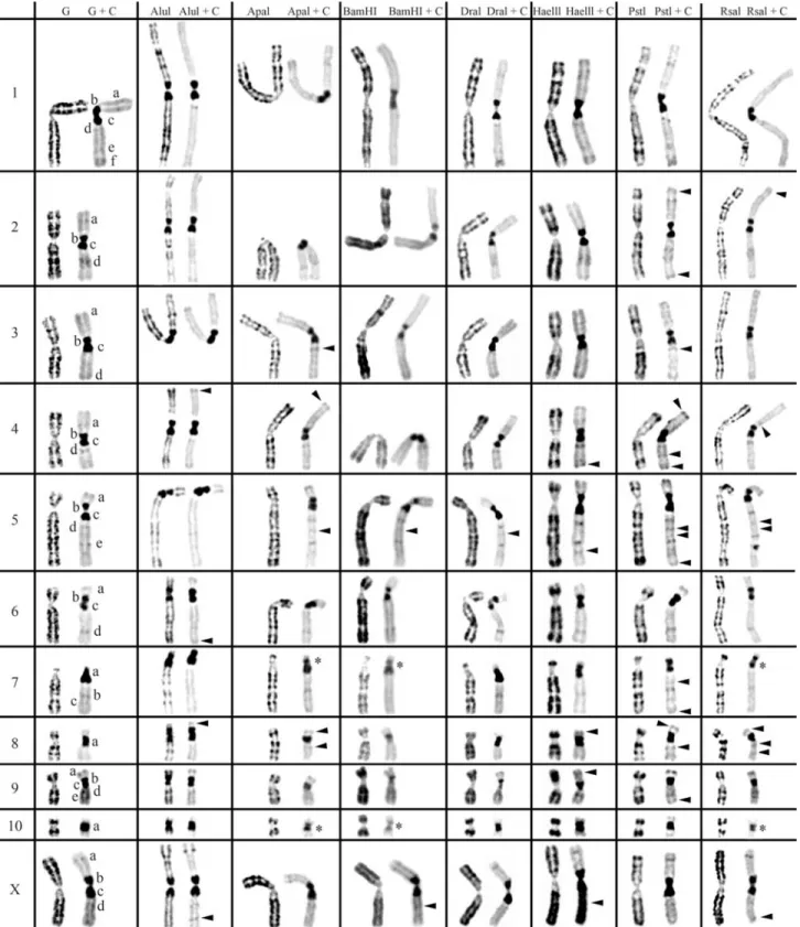
2 and 3, respectively. The residual bands seen after diges-tion with endonucleases AluI, ApaI, BamHI, DraI, HaeIII, PstI and RsaI (left column for each enzyme shown in Fig-ures 1-3) are mainly G-like and suitable for chromosome identification. Although each restriction endonuclease was expected to yield a specific banding pattern, in practice most of the banding patterns overlapped. Nevertheless some endonucleases (e.g.ApaI, PstI and RsaI in chromo-somes of Cricetus cricetus, BamHI, PstI and RsaI in Peromyscus eremicusand HaeIII, PstI and RsaI inPraomys tullbergi) produced a higher banding contrast. AluI was, perhaps, the used enzyme that produced the smallest num-ber of bands but the higher contrast banding pattern. It is important to refer that the banding patterns produced by each RE are reproducible and can be used in sequential ex-periment procedures without loss of chromosome morphol-ogy (Chaveset al., 2002; Adegaet al., 2005).
In a general overview, the C-positive hetero-chromatin (Figures 1-3, right chromosome in each column, showing control C-banding and RE+C-banding) is mainly found at the centromeres of most chromosomes, although some C-bands can also be seen at interstitial and telomeric locations. In the individuals analyzed, some hetero-chromatin polymorphism of minor significance were de-tected,i.e., variation in the banding patterns of homologous chromosomes of the same pair, as also reported for pig (Adegaet al., 2005) and some Tayassuidae species (Adega et al., 2007) chromosomes. The heterochromatin poly-morphisms detected in the chromosomes of the studied spe-cies were not considered for the analysis relatively to the characterization of CH here presented, because they might not be representative of the population.
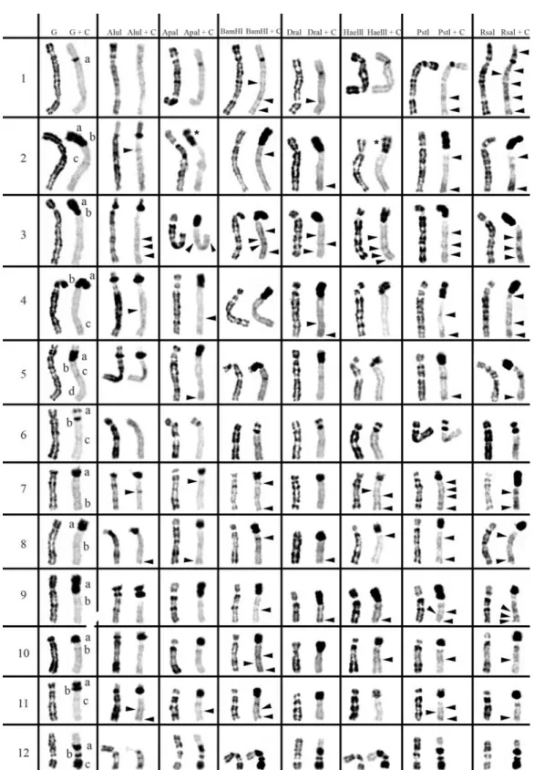
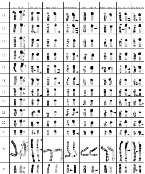
At least three major classes of CH were identified in the species studied in this work: (peri)centromeric, intersti-tial and telomeric (Figures 1-3). With RE+C-banding treat-ment, these major C-positive heterochromatin blocks could be discriminated in at least 26 C-positive heterochromatin subclasses in the autosomal complement of Cricetus cricetus[seven in (peri)centromeric regions, 13 in intersti-tial regions and six in telomeric regions] and three C-positive heterochromatin subclasses in the CCRX chromo-some [one (peri)centromeric and two in interstitial regions] (cf. Figure 1). InPeromyscus eremicuschromosomes (Fig-ure 2), the RE+C-banding treatment discriminated at least 26 C-positive heterochromatin subclasses in the autosomal complement [seven in (peri)centromeric regions, 13 in in-terstitial regions and six in telomeric regions], three C-positive heterochromatin subclasses in the PERX chromo-some (one in the centromeric region and two in interstitial regions) and two in the PERY chromosome (one centro-meric and one subtelocentro-meric). Finally, inPraomys tullbergi, the RE+C-banding treatment (Figure 3) discriminated the major C-positive heterochromatin blocks into at least 45 C-positive heterochromatin subclasses in the autosomal complement (two in centromeric regions, 35 in interstitial
regions and eight in telomeric regions), four C-positive heterochromatin subclasses in the PTUX chromosome (one in the centromeric region and three in interstitial regions) and three in the PTUY chromosome (one in the centromeric region and two in interstitial regions).
Constitutive Heterochromatin (C-positive heterochromatin) characterization inCricetus cricetus
Control experiment (G+C-banding) show that all the
chromosomes of Cricetus cricetus exhibit large
(peri)centromeric C-bands that in most cases consist of two blocks of CH (exception goes to CCR7, CCR8 and CCR10 chromosomes which show only one block of CH). Notice the very large centromeric CH block of the only acrocentric chromosome of the karyotype, CCR7. All the chromo-somes except CCR3, CCR8 and CCR10 exhibit interstitial C-positive heterochromatin. Telomeric C-bands can be seen on chromosomes CCR1, CCR3, CCR5, CCR6, and CCR9.
Incubation of this species chromosomes with restric-tion endonucleases followed by banding revealed C-bands heterogeneity (Figure 1), being verified that (peri)centromeric, interstitial or telomeric C-bands present a different molecular nature, exhibiting different restriction patterns when submitted to the same panel of REs. This is not surprising as similar results have been reported for other species (Babu, 1988; Fernández-Garcíaet al., 1998; Chaveset al., 2004b; Adegaet al., 2005, 2007).
The arrowheads in Figure 1 indicate C-bands re-vealed only after RE treatment (cryptic C-bands). Of the endonucleases used here, BamHI+C-banding was the one that produced the most evident effect in CH sequences of theCricetus cricetuschromosomes. See for instance chro-mosomes CCR7, CCR8, CCR9 and CCR10, being ob-served less intense bands in comparison with the control chromosomes. This enzyme, along with ApaI+C-banding and RsaI+C-banding, produced the partition of the (peri)centromeric CH band at chromosomes CCR7 and CCR10 into two distinct CH blocks, thus revealing the oc-currence of two instead of one (peri)centromeric CH block [bands identified with an asterisk in Figure 1]. Some en-zymes seem to have a drastic effect resulting in a more ac-centuated contrast pattern in the (peri)centromeric regions of some chromosomes. See, for example, chromosomes CCR1 and CCR6 with DraI+C-banding, CCR5 with BamHI+C-banding, CCR9 with BamHI+C-banding and DraI+C-banding.
Constitutive heterochromatin (C-positive
heterochromatin) characterization inPeromyscus eremicus
some of these chromosomes, the C-banding spreads from the centromeric region to the p arm telomere, apparently covering all the p arm, e.g., chromosomes PER9 and
PER17. In some chromosomes, this band seems to be split in two C-bands, one clearly centromeric and the other cov-ering the chromosome p arm (chromosomes PER2, PER3
and PER4). Chromosomes PER11 and PER16 display two well-defined bands of (peri)centromeric CH, although this
may have been an artifact caused by the small size of the p
arms. Chromosomes PER1 and PERY apparently display the lowest amount of heterochromatin in control
G+C-banding, showing PER1 only a small centromeric CH band.
The situation observed in the PERY is not usual for most of the mammals’ species, once this chromosome is usually the more heterochromatic of the whole complement. Some of the chromosomes exhibit C-bands at interstitial locations, presenting chromosome PERX the highest number of these bands (at least six). Telomeric C-bands can be observed in some chromosomes of this species, e.g., PER6, PER11, PER12, PER15 and PER16 (Figure 2).
When C-banding was applied afterin situREs diges-tion to the chromosomes of this species, it was possible to verify that its CH shows some degrees of heterogeneity
(Figure 2). The arrowheads in Figure 2 indicate C-bands re-vealed only after treatment with endonucleases (cryptic C-bands). From the REs used in this work, RsaI+C-banding, PstI+C-banding and BamHI+C-RsaI+C-banding, were the enzymes that revealed the greatest number of CH bands not previously detected by the control G+C-banding.
In a general analysis, AluI was the enzyme that pro-duced the most divergent effects on the CH ofPeromyscus eremicuschromosomes. In some cases, such as in chromo-somes PER1 and PER6, some C-bands seem to have under-gone a greater reduction or even have, apparently
peared when compared with control experiment, while in other cases, such as chromosomes PER7 and PER16, the CH was apparently unaffected by treatment with this en-zyme.
The p arms CH of PER2, PER3 and PER4 chromo-somes are particularly interesting in what respects to its molecular nature. In these heterochromatic arms the CH re-veals a high heterogeneity, what is verified by the different restriction patterns produced by the enzymes at these CH regions. For instance in the p arm of PER 2 there were rec-ognized two C-bands in the control G+C-banding; after AluI+C restriction a lesser intensity of one of these bands was observed and ApaI+C-banding and HaeIII+C-banding seem to reveal an extra C-band, by splitting one of the pre-vious in two [bands evidenced with an asterisk (*) in Fig-ure 2].
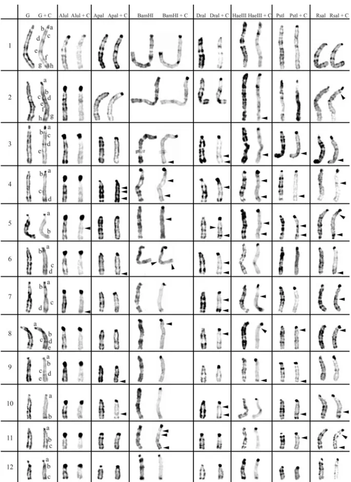
Constitutive heterochromatin (C-positive heterochromatin) characterization inPraomys tullbergi
From the studied species,Praomys tullbergi(Figure 3) is the one whose chromosomes exhibit the lower amount of centromeric CH in the control experiment (G+C-banding). In some chromosomes, centromeric CH is almost as abundant as interstitial CH, in contrast to the observed for the majority of the chromosomes from the other species here analyzed. However, the chromosomes PTU5 and PTU10 in the control experiment, present a small centro-meric CH band and apparently do not reveal interstitial bands. The majority of the chromosomes display several in-terstitial CH bands, presenting the chromosomes PTU1 and PTU2 the greatest number of these bands. Telomeric C-bands are clearly distinguishable in some chromosomes, e.g., chromosomes PTU10, PTU12 and PTU15. The PTUX chromosome presents three distinct classes of CH, centro-meric, interstitial and telomeric. PTUY chromosome ex-hibits a centromeric band and two interstitial C-bands.
When C-banding was applied afterin situREs diges-tion to the chromosomes of this species, it was possible to verify that its CH shows some degrees of heterogeneity, just as it was described for the other two rodent species studied in this work. AluI+C-banding produced the higher contrast between the centromeric versus interstitial/telo-meric CH classes; digestion with AluI greatly decreased the interstitial/telomeric CH while, simultaneously, evidenced the centromeric heterochromatin. See for instance, chromo-somes PTU15 or PTU16, whose centromeres showed in the control G+C-banding an almost absence of CH, and after the AluI+C-banding the centromeres showed large centro-meric CH blocks. Digestion with DraI seems to highlight the telomeric CH after C-banding, e.g., chromosomes PTU7 and PTU16. RsaI+C-banding seems to produce the most similar results with the control G+C-banding, how-ever also discriminating cryptic C-bands, such as the ones observed in chromosomes PTU4, PTU5, PTU10 or PTUX.
Other endonucleases also disclosed cryptic C-bands, espe-cially DraI+C-banding, BamHI+C-banding or HaeIII+C-banding.
These special bands are very interesting from the CH molecular nature point of view, since their disclosure is probably dependent on sequence modifications (not yet clearly understood) induced by the REs, leading for in-stance, to an increase of the stain capacity to bind a specific chromosome region (Gosálvezet al., 1997; Niedduet al., 1999; Chaveset al., 2004b). Whatever the mechanism be-hind these sequences modification, RE digestion triggers it, revealing “hidden” C-bands. Curiously, and from several different works in different species, these sequences not de-tected by classical C-banding have proven to correspond to clinical (Sus scrofa, Adega et al., 2005) or evolutionary breakpoints (Tayassuidae, Adegaet al., 2007).
Inter-species constitutive heterochromatin (C-positive heterochromatin)
A general comparison of the amount, distribution and molecular nature of C-positive heterochromatin in the three Rodentia species, suggests that the CH of these karyotypes is extremely different. Evidence comes from the detailed combined analysis of the different REs+C-banding patterns disclosed on the karyotypes of these species. The applica-tion of a seven REs panel to the chromosomes of three dif-ferent rodent species, Cricetus cricetus, Peromyscus eremicus(Cricetidae) andPraomys tullbergi(Muridae), al-lowed a characterization of its CH and the recognition of its molecular heterogeneity. These results are a clear reflex of the different C-positive heterochromatin composition of these karyotypes, possible to observe by the different REs actions on the respective chromosome’s bands.
Cricetus cricetus has an almost entirely meta/sub-metacentric karyotype (with only one acrocentric pair), with the CH primarily located in (peri)centromeric regions. Most of the chromosomes in this species exhibit two very large blocks at (peri)centromeric location, which suggested the occurrence of dicentric Robertsonian translocations or, alternatively, heterochromatin additions during the course of this karyotype evolution. The other Cricetidae species, Peromyscus eremicus, has a very distinct karyotype that comprises only submetacentric chromosomes. This karyo-type also displays great amounts of CH, especially located at the (peri)centromeric regions, being the p arms of some chromosomes composed entirely by this repetitive compo-nent of the genome. The heterochromatin of p arms revealed a great heterogeneity, what implies a different mo-lecular composition, which is certainly indicative of the co-existence of different satellite DNA families or variants at these chromosome regions.
terstitial heterochromatin is almost as abundant as centromeric heterochromatin. This uniform and scattered distribution, together with the higher number of CH sub-classes identified inPraomys tullbergi chromosomes (52 subclasses) suggests that this species has a more derivative karyotype than the other two genomes analyzed, probably originated by a great number of complex chromosomal re-arrangements. This is based on the assumption that hetero-chromatic rich regions act as hotspots for the occurrence of chromosome rearrangements (Yunis and Yasmineh, 1971; Peacocket al., 1982, John, 1988; Chaveset al., 2004b), ei-ther by promoting the chromosome structural rearrange-ments that reshape karyotypes or by being fragile regions prone to chromosome breakage, and consequently to chro-mosome rearrangement, representing remnants of these events. The suggestion that the karyotype of Praomys tullbergiwas originated by the occurrence of a high num-ber of complex chromosomal rearrangements is sup-ported by the work of Meleset al.(2008), where it was detected telomeric interstitial sequences in several chro-mosome arms of this species, probably the result of tan-dem fusions.
Finally, it is worth mentioning the value ofin situRE digestion with sequential C-banding as an alternative tool for the study of Rodentia chromosomes CH, especially when other techniques are not available, as fluorescentin situhybridization with different repetitive sequences.
Acknowledgments
This work was supported by a project
(POCI/BIA-BCM/58541/2004) and a PhD grant (SFRH/BD/
41574/2007) from the Science and Technology Foundation (FCT), Portugal. We are deeply grateful Dr. Vitaly Volo-bouev for providing the cell cultures of the Rodentia spe-cies.
References
Adega F, Chaves R and Guedes-Pinto H (2005) Chromosome re-striction enzyme digestion in domestic pig (Sus scrofa). Constitutive heterochromatin arrangement. Genes Genet Syst 80:49-56.
Adega F, Chaves R and Guedes-Pinto H (2007) Constitutive heterochromatin characterization of white-lipped and col-lared peccaries (Tayassuidae). J Genet 86:19-26.
Arrighi FE and Hsu TC (1971) Localization of heterochromatin in human chromosomes. Cytogenetics 10:81-86.
Babu A (1988) Heterogeneity of heterochromatin of human chro-mosomes as demonstrated by restriction endonuclease treat-ment. In: Verma RS (ed) Heterochromatin: Molecular and Structural Aspects. 1st edition. Cambridge University Press, New York, pp 250-275.
Bauchan GR and Hossain MA (1999) Constitutive heterochro-matin DNA polymorphisms in diploidMedicago sativassp.
falcata.Genome 42:930-935.
Capanna E, Codjia JTC, Chrysostome C and Civitelli MV (1996) Les chromosomes des rongeurs du Benin (Afrique de l’Ouest): 3 Murinae. Rend Fis Acc Lincei 8:25-37.
Chaves R, Guedes-Pinto H, Heslop-Harrison J and Schwarzacher T (2000) The species and chromosomal distribution of the centromeric alpha-satellite I sequence from sheep in the tribe Caprini and other Bovidae. Cytogenet Cell Genet 91:62-66.
Chaves R, Adega F, Santos S, Guedes-Pinto H and Heslop-Harrinson JS (2002)In situhybridization and chromosome banding in mammalian species. Cytogenet Genome Res 96:113-116.
Chaves R, Frönicke L, Guedes-Pinto H and Wienberg J (2004a) Multidirectional chromosome painting between the Hirola antelope (Damaliscus hunteri, Alcelaphini, Bovidae), sheep and human. Chromosome Res 12:495-503.
Chaves R, Santos S and Guedes-Pinto H (2004b) Comparative analysis (Hippotragini versus Caprini, Bovidae) of X-chromosome’s constitutive heterochromatin by in situ re-striction endonuclease digestion: X-chromosome constitu-tive heterochromatin evolution. Genetica 121:315-325. Comings DE (1973) Biochemical mechanisms of chromosome
banding and color banding with acridine orange. In: Casperson T and Zeck L (eds) Chromosome Identification -Techniques and Applications in Biology and Medicine. Ac-ademic Press, New York, pp 292-306.
Corradini N, Rossi F, Giordano E, Caizzi R, Vern F and Dimitri P (2007) Drosophila melanogasteras a model for studying protein-encoding genes that are resident in constitutive heterochromatin. Heredity 98:3-12.
Dimitri P, Corradini N, Rossi F and Verní F (2004) The paradox of functional heterochromatin. BioEssays 27:28-41. Dimitri P, Verní F, Mei E, Rossi F and Corradini N (2005)
Transposable elements as artisans of the heterochromatic genome. Cytogenet Genome Res 110:165-172.
Fernández-García JL, Martínez-Trancón M, Rabasco A and Pa-dilla JA (1998) Characterization of the heterochromatic chromosome regions in sheep. Genes Genet Syst 73:45-50. Galleni L, Stanyon R, Tellini A, Giordano G and Santini L (1992)
Karyology of the Savi pine vole,Microtus savii(De Sélys-Longchamps, 1838) (Rodentia, Arvicolidae): G-, C-, DA/DAPI, and AluI-bands. Cytogenet Cell Genet 59:290-292.
García L, Ponsá M, Egozcue J and García M (2000a) Comparative chromosomal analysis and phylogeny in fourCtenomys spe-cies (Rodentia, Octodontidae). Biol J Linn Soc 69:103-120. García L, Ponsá M, Egozcue J and García M (2000b) Cytogenetic
variation in Ctenomys perrensi(Rodentia, Octodontidae). Biol J Linn Soc 71:615-624.
Gosálvez J, López-Fernández C, Goyanes R and Mezzanotte V (1997) Chromosome differentiation using nucleases: An overview. In: Henriques-Gil N, Parker JS and Puertas MJ (eds) Chromosomes Today. Chapman & Hall, London, pp 23-49.
Holmquist GP and Dancis B (1979) Telomere replication, kine-tochore organizers, and satellite DNA evolution. Proc Natl Acad Sci USA 76:4566-4570.
Hsu TC and Arrighi FE (1966) Chromosomal evolution in the ge-nus Peromyscus (Cricetidae, Rodentia). Cytogenetics 5:355-359.
Jobse C, Buntjer JB, Haagsma N, Breukelman HJ, Beintema JJ and Lenstra JA (1995) Evolution and recombination of bo-vine DNA repeats. J Mol Evol 41:277-283.
John B (1988) The biology of heterochromatin. In: Verma RS (ed) Heterochromatin: Molecular and Structural Aspects. Cam-bridge University Press, CamCam-bridge, pp 1-128.
John B, King M, Schweizer D and Mendelak M (1985) Equi-locality of heterochromatin distribution and heterochro-matin heterogeneity in acridid grasshoppers. Chromosoma 91:185-200.
Lee C, Stanyon R, Lin CC and Ferguson-Smith MA (1999) Con-servation of human gamma-X centromeric satellite DNA among primates with an autosomal localization in certain Old World monkeys. Chromosome Res 7:43-47.
Matthey R (1952) Chromosomes des Muridae (Microtinae et Cricetinae). Chromosoma 5:113-138.
Matthey R (1958) Les chromosomes et la position systématique de quelques Murinae africains (Mammalia-Rodentia). Acta Trop 15:97-117.
Meles S, Adega F, Guedes-Pinto H and Chaves R (2008) The karyotype ofPraomys tullbergi(Muridae, Rodentia): A de-tailed characterization. Micron 39:559-568.
Nieddu M, Rossino R, Pichiri G, Rocchi M, Setzu MD and Mezzanotte R (1999) The efficiency ofin situhybridization on human chromosomes with alphoid DNAs is enhanced by previous digestion with AluI and TaqI. Chromosome Res 7:593-602.
Pathak S and Arrighi FE (1973) Loss of DNA following C-banding procedures. Cytogenet Cell Genet 12:414-422. Peacock WJ, Dennis ES and Gerlach WL (1982) DNA sequence
changes and speciation. Prog Clin Biol Res 96:123-142. Pieczarka JC, Nagamachi CY, Muniz JAPC, Barros RMS and
Mattevi MS (1998) Analysis of constitutive heterochro-matin ofAotus(Cebidae, Primates) by restriction enzyme and fluorochrome bands. Chromosome Res 6:77-83. Probst AV and Almouzni G (2008) Pericentric heterochromatin:
Dynamic organization during early development in mam-mals. Differentiation 76:15-23.
Qumsiyeh MB, King SW, Arroyo-Cabrales J, Aggundey IR, Schlitter DA, Baker RJ and Morrow KJ (1990) Chromo-somal and protein evolution in morphologically similar spe-cies of Praomyssensu lato (Rodentia, Muridae).J Hered
81:58-65.
Rocco L, Morescalchi MA, Costagliola D and Stingo V (2002) Karyotype and genome characterization in four cartilagi-nous fishes. Gene 295:289-298.
Rossi F, Moschetti R, Caizzi R, Corradini N and Dimitri P (2007) Cytogenetic and molecular characterization of hetero-chromatin gene models inDrosophila melanogaster. Genet-ics 175:595-607.
Saito Y, Edpalina RR and Abe S (2007) Isolation and character-ization of salmonid telomeric and centromeric satellite DNA sequences. Genetica 131:157-166.
Santos S, Chaves R and Guedes-Pinto H (2004) Chromosomal lo-calization of the major satellite DNA family (FA-SAT) in the domestic cat. Cytogenet Genome Res 107:119-122. Schmid M, Haaf T, Steinlein C, Nanda I and Mahony M (2002)
Chromosome banding in Amphibia: XXV. Karyotype evo-lution and heterochromatin characterization in Australian
Mixophyes (Anura, Myobatrachidae). Cytogenet Genome Res 97:239-253.
Shore D (2001) Telomeric chromatin: Replicating and wrapping up chromosome ends. Curr Opin Genet Dev 11:189-198. Singer MF (1982) Highly repeated sequences in mammalian
ge-nomes. Int Rev Cytol 76:67-112.
Slamovits CH and Rossi MS (2002) Satellite DNA: Agent of chromosomal evolution in mammals. A review. J Neotrop Mammal 9:297-308.
Sumner AT (1972) A simple technique for demonstrating centro-meric heterochromatin. Exp Cell Res 75:304-306.
Van Den Bussche RA, Honeycutt RL and Baker RJ (1993) Re-striction endonuclease digestion patterns of harvest mice (Reithrodontomys) chromosomes: A comparison to G-bands, C-G-bands, andin situhybridization. Genetica 87:141-149.
Verma RS and Babu A (1995) Human Chromosomes – Principles and Techniques. 2nd edition. McGraw-Hill, New York, 419 pp.
Waye JS and Willard HF (1989) Concerted evolution of alpha sat-ellite DNA: Evidence for species specificity and a general lack of sequence conservation among alphoid sequences of higher primates. Chromosoma 98:273-279.
Wichman HA, Payne CT, Ryder OA, Hamilton MJ, Maltbie M and Baker RJ (1991) Genomic distribution of heterochro-matic sequences in equids: Implications to rapid chromo-somal evolution. J Hered 82:369-377.
Yunis JJ and Yasmineh WG (1971) Heterochromatin, satellite DNA, and cell function. Structural DNA of eukaryotes may support and protect genes and aid in speciation. Science 174:1200-1209.
Associate Editor: Yatiyo Yonenaga-Yassuda