A Case of Megacolon in Rio Grande Valley as a Possible Case of
Chagas Disease
Karl Reinhard/
+, T Michael Fink*, Jack Skiles**
School of Natural Resource Sciences, 214 Bessey Hall, University of Nebraska - Lincoln, Lincoln, NE 68588-0340, USA *Arizona Department of Health Services, Phoenix, Arizona, USA ** Texas Departament of Health, TX 78871, USA
We have been searching for evidence of Chagas disease in mummified human remains. Specifically, we have looked for evidence of alteration of intestinal or fecal morphology consistent with megacolon, a condition associ-ated with Chagas disease. One prehistoric individual recovered from the Chihuahuan Desert near the Rio Grande exhibits such pathology. We present documentation of this case. We are certain that this individual presents a profoundly altered large intestinal tract and we suggest that further research should focus on confirmation of a diagnosis of Chagas disease. We propose that the prehistoric activity and dietary patterns in Chihuahua Desert hunter/gatherers promoted the pathoecology of Chagas disease.
Key words: Trypanosoma cruzi - archaeoparasitology - archaic - Texas
Archaeoparasitology is the integration of evidence of ancient parasites with archeological reconstructions of passed human activities. This includes direct evidence (eggs, larvae, DNA) and the pathology caused by para-sites (cysts, organ alterations, soft tissue lesions). For most of the history of the field, evidence of protozoa in-fection has come from characteristic lesions consistent with pathology caused by certain protozoa parasites. In the case of Trypanosoma cruzi, megacolon and/or megae-sophagus are the pathological indicators of the disease (Aufderheide & Rodríguez-Martín 1998). Now, DNA re-covery from ancient remains is augmenting diagnosis (Ferreira et al. 2000). However, because of variable taphonomic conditions, one can not anticipate that all pathological conditions evident in mummies or skeletal remains will respond to molecular analysis. Therefore, the search for gross pathology will continue to be a major goal in archaeoparasitology.
This paper summarizes the search for pathology diag-nostic of Chagas disease in the desert west of North America and on a specific case from the Chihuahuan Desert that appears to be a good candidate for this disease. Chagas disease is caused by T. cruzi. This flagellate pro-tozoan enters the blood stream, proliferates in the blood, and eventually invades nerve cells. Triatomine bugs of the family Reduviidae transmit the disease when feeding and defecating on humans.
BACKGROUND
The preservation of material in the desert west of North America, including states in Mexico and the USA, is ideal for bioarchaeological study (Reinhard 1992) and archaeoparasitology (Reinhard 1990). Rockshelters are abundant in the cliffs lining the canyons in the area and preserve dried mummies for study (El-Najjar et al. 1998). The general aridity of the region results in the preserva-tion of coprolites which provide important helminthological (Reinhard 1992), dietary (Minnis 1989), and medicinal (Reinhard et al. 1991) information.
Chagas disease is endemic to the region (Roberts & Janovy 1996). One of the syndromes caused by the dis-ease is megacolon, also called enlarged hollow viscera. This syndrome results when the infection causes the loss of nervous activity in the large intestine. Peristalsis is disrupted, eventually stops, and the colon fills with partly digested food. The intestine diameter becomes pro-foundly altered. As the colon fills, muscular strength or tonus is lost, and the colon diameter enlarges. Eventually death occurs, often due to toxic compounds and bacteria leaking from the enlarged colon.
In archaeological settings of South America, megaco-lon is evident as abnormal fecal accumulation in the comegaco-lon (Aufderheide & Rodríguez-Martín 1998). In well preserved mummies, the remains of the enlarged intestine surround-ing partially digested food have been found. Even if the epithelium is not preserved, the morphology of feces as-sociated with a mummy or skeleton is abnormal in appear-ance. Normal feces from the colon are small pellets of food that are defined in size by the haustra within which they form (Reinhard 1993). These pellets are not cylindri-cal except in the lower bowel. In general, the shape of normal pellets is discoidal or globular (Fig. 2). Dry weight of pellets ranges from 1-6 g. In size they are 2.5-3 cm in largest dimensions. In cases of megacolon, the feces in mummies will exceed the size and weight of normal intes-tinal fecal pellets. The feces can also become contiguous with each other and form larger, cylindrical forms. There-fore, examination of feces for abnormal morphology can be useful in identifying megacolon.
The views expressed herein do not necessarily reflect those of the Arizona Department of Health Service, nor the Texas De-partment of Health.
The Fulbright Commission supported final write-up of this paper.
+Corresponding author. Fax: +402-472.6858. E-mail: [email protected]
The analysis of skeletonized burials during the 1980s and 1990s showed that fecal residues and pellets could be found with burials even of no soft tissue was pre-served. The first such find was made in 1984 when fecal components were found with an Anasazi skeleton (Reinhard et al. 1992). In this case the fecal pellets were not preserved. The feces had fragmented and the fecal components were mixed in the sediments within the pel-vic girdle. In 1988, a Mimbres burial from New Mexico was found (Shafer et al. 1989). In the sediment of the pelvic girdle, distinct fecal pellets were found. These were of size and weight consistent with normal colonic feces. These studies demonstrated that feces could be found in burials and provided evidence of diet and medicine. How-ever, they did not indicate any pathology that affected the size and morphology of the fecal pellets.
In 1991, a skeleton of an Anasazi child was found in Utah (Dominguez et al. 1992). Large fecal pellets were found in the abdominal region. Although the pellets were of normal shape for the large intestine, they were of ab-normal amount. From this individual, 21 large fecal pellets were recovered. In the laboratory, the pellets were ori-ented in anatomical position. It appeared that most of the colon was filled with the pellets. The feces were com-posed almost entirely of grass seed of the type known as Indian Rice Grass (Oryzopsis). Palynological study re-vealed grass pollen (Poaceae) and Mormon tea pollen (Ephedra). Unfortunately, the feces were recovered from a disturbed burial and no soft tissue was preserved. Thus the nature of the feces was consistent with megacolon, but the anatomical association was not sufficiently pre-served in the field to warrant a diagnosis of this condi-tion. However, the laboratory evidence is highly sugges-tive of megacolon.
Radiographic analysis of two mummies from Chi-chuahua, Mexico show no distinctly identifiable fecal pellets (Luibel-Hulen 1985).
Profoundly abnormal fecal deposits were found in a prehistoric burial on the Coahuilan-Texan border. This individual is the focus of the remaining discussion.
SMM mummy
Bioarchaeology of Northern Coahuila and the Lower Pecos of Texas
The region of the Rio Grande (Rio Bravo) possesses a rich bioarchaeology, especially between the border towns of Ciudad Acuna, Coahuila and Del Rio, Texas and ex-tending to the tributaries of the Amistad International Recreation Area and on both sides of the international border (Fig. 1). In arid rockshelter environments, coproli-tes and human remains are excellently preserved (Turpin et al. 1986). Culturally and ecologically, the region has a heritage derived from Archaic hunter-gatherer cultures that extended over Northeastern Mexico (Shafer 1986, Tay-lor 1966). The hunter-gatherers who inhabited the area created art and artifacts that are some of the most impres-sive in the world. Dramatic rock art is common in the area (Shafer 1975, 1986, Turpin et al. 1982, Turpin 1986). Cer-tain other artifacts such as “painted pebbles” (Pearce & Jackson 1933, Shafer 1986) and clay figurines (Shafer 1975) are unique to the region. Although not unique to the
area, complex woven artifacts are commonly found in the area (Andrews & Adovasio 1980, Shafer 1986, Turpin et al. 1986).
Fig. 1: map of portions of Coahuila and Texas showing location where the mummy was discovered.
Previous parasitological study has been confined to the analysis of 116 coprolites. In one of these coprolites, pinworm eggs were found (Reinhard 1990). There is no evidence of any other intestinal parasites. There is no evidence of lice reported from the analysis of mummies (El-Najjar et al. 1998). Dietary analysis shows that the lower Pecos inhabitants occasionally ate millipedes. This habit would have exposed them to infection with Macrocanthorynchus ingens, an acanthocephalan. How-ever, eggs of this species have not been found in copro-lites. Thus, parasitological analysis to date has not re-vealed evidence of significant parasitic disease in the lower Pecos.
sis of the mummy with comparison to four others. A ra-diocarbon date indicates the individual died about 1,150 years ago. The mummy is referred to as SMM and was an adult male between 35 and 45 years of age. Dietary study of the intestinal contents of the mummy is presented by Turpin et al. (1986). Palynological analysis showed that grass dominated the pollen spectrum followed by Asteraceae pollen with low amounts of Pinus, Cheno Am, Celtis, and unknown types. Other taxa present include Onagraceae, Umbelliferae, Juniperus, Malvaceae (Mal-low), Dasylirion, Agave, Quercus, Prosopis, and fern spores. Grass pollen aggregates were present. Macro-scopically, Opuntia and grass seed (identified as Setaria) were found as well as fibers, probably from Agave. A surprising variety of animals were found in the intestinal remains including skeletal elements of several fish, a snake, a bat, Peromyscus (white footed mouse), Thomomys (pocket gopher) and 250 fragments of grasshoppers. It appears from the pollen and small animal evidence that death occurred in spring (Turpin et al. 1986). Hair and feather were also present.
Artifacts
The SMM mummy was found in an elaborate burial with many artifacts. Textiles buried with the mummy in-cluded five woven mats, two of which were also painted. The mats were woven of fibers from local succulent plants, probably Dasylirion. Stone beads were also buried with the mummy. The exact location of the beads relative to the corpse is unknown. The burial was wrapped in a fur robe and a rope reportedly made of human hair. Also wrapped around the body was a long strap made of deer hide painted red.
Of these artifacts, the rope and strap are most impor-tant. The rope consists of tightly woven hair ranging from 0.9 to 1.1 cm in diameter. In total, there are 44.03 m of rope. Four independent sections are tied into double or quadruple loops attached to a single rope strand. Al-though the rope has been identified previously as human (Turpin et al. 1986), microscopic analysis shows that the rope was made of animal hair of three different species. Hair rope, whether made of human or animal hair, is very rare. Only one other specimen has been found. Normally, rope and other cordage is made of vegetal fibers. The presence of the rope wrapping around the burial suggests that it had a special status in life.
The deer hide strap may have had a more utilitarian function. The rope is 1 cm wide and is painted red on both sides. It is now in many fragments. However, our examination of the fragments reveals only two cut ends, so it is probable that the rope was one long piece when it was used. Now it is in 17 segments ranging from 7 to 70 cm in length. The total length of the segments is 756 cm. When the mummy was found, the strap was found wrapped around the lower torso.
The mummy
The mummy is composed of a nearly complete skel-eton with hair, some connective tissue and a large portion of the digestive tract. The mummy was laid on its right side. There is a mass of material, possibly the contents of the small intestine, resting on the right side of the
tho-racic and abdominal area. Sections of this material are under study to determine their anatomical origin.
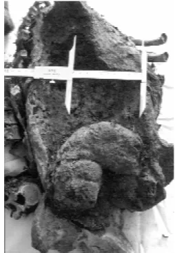
The large intestine is nearly complete (Figs 3-6). The appearance of this organ is consistent with megacolon. There are ten segments of the colon that are preserved today. Most segments exhibit a grossly enlarged diam-eter. The observations of diameter, length, weight, and volume for each segment are presented in Table I. The average diameter of the large intestine was 6 cm. There are 112.9 cm of the large intestine, compacted with par-tially digested food, which are “mummified”. The reason the intestine “mummified” is due to the fact that it was filled with compacted food remains that desiccated and retained the form of the colon (Fig. 3). Of the intestine segments that could be weighed, 1,170 g of feces were present. The total volume of the preserved large intestine was 3,710 cm3.
The SMM mummy contrasts with normal mummies. As noted above, normal fecal pellets are present ranging up to 3 cm in section. These are separated from each other in the haustra of the intestine. The diameter of a normal, mummified intestine is approximately 3 cm. Nor-mally, the amount of feces in the large intestine weighs less than 30 g. In contrast, the SMM mummy contains feces of 6 cm in diameter and at least 3,710 cm3 of feces are within the mummy (Figs 3-6). Never has a large intes-tine been found that is completely filled with partially di-gested food. Analysis of the contents of one intestinal segment revealed bones of fish, four rodents, a bat, 250 grasshoppers, plant fibers, seeds, and grass pollen. The entire colon is filled with these sorts of foods. Therefore, the findings of this study demonstrate that this mummy suffered from megacolon in life.
TABLE I
Observations of colon segments
Segment Weight (g) Diameter (cm) Length (cm) Volume (cm)
1 78.2 5.3 10.0 145
2 7.2 3.0 4.5 57
3 96.8 (minimum) 7.0 9.9 396
4 190.3 8.1 13.2 660
5 148.9 8.5 8.5 544
6 186.6 5.5 13.1 236
7 230.0 (minimum) 6.1 14.1 423
8 202.0 7.0 17.0 663
9 30.0 2.6 5.5 39
10 not measurable 6.5 17.1 547
Total 1,170 mean = 6.0 112.9 3,710
Fig. 4: parts of the colon reconstructed in the pelvic girdle. Feces literally filled this part of the body in life.
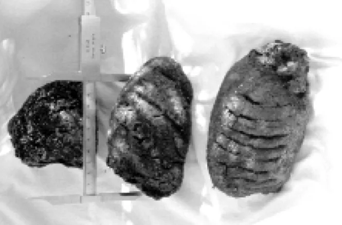
Fig. 5: three segments of the colon compared to a caliper opened to 10 cm. The grossly enlarged feces are evident when compared to Fig. 2.
Fig. 6: eight fragments composing most of the large colon com-pared to a caliper opened to 10 cm.
tions of the colon. The anterior iliac crests left their im-pressions on two other segments of colon. This indi-cates that the abdomen was distended. It is possible that the deer hide strap represents an attempt to support the distention of the abdomen. There are three grooves in the mass of feces from the right side of the body that suggest that the strap was wrapped around the abdomen. There would have been abnormal bladder function at the time of death because the pelvic girdle was entirely filled with food at the time of death.
Pathoecology of Chihuahuan Desert Chagas Disease
Vector Considerations Regarding Chagas Disease in the Rio Grande
Triatomine bugs - Triatomine bugs belong to the order Hemiptera, family Reduviidae, and of the subfamily Triatominae. They are obligate hemophagous ectopara-sites that feed on vertebrate blood. Triatomines are hemi-metabolous true bugs, and therefore go from the egg, nymphal, and adult stages without a pupal phase (i.e., incomplete metamorphosis). Adult and nymphal bugs generally feed on the same variety of vertebrate hosts as adults, and both are therefore capable of transmitting T. cruzi infections.
sec-The insects are also referred to in the entomological literature as triatomiid (or triatomid) and reduviid bugs. Other more common regional sobriquets include “conenose” and “kissing” bugs, “barbeiros,” and “vin-chuca” (Ryckman & Blankenship 1984:414-418, Schofield et al. 1987: 19). In the New World triatomine bugs are rec-ognized as the principle vector of Chagas disease and can also cause allergic reactions in individuals who are frequently bitten by the arthropods (Ryckman 1981). Al-though the bugs are found in Africa, Asia and Australia, neither T. cruzi or Chagas disease appears to be present in the Old World (Lent & Wygodzinsky 1979, Ryckman & Archbold 1981).
Adult and nymphal triatomiid bugs are generally noc-turnal and take their blood meals when vertebrate hosts are quiescent or sleeping. The rostrum of conenose bugs, i.e. the proboscis-like apparatus inserted into the skin to draw blood, is needle-like and causes little or no discom-fort to the host during the blood meal (Ryckman 1981). However, it is not able to penetrate blankets or clothing, and as a consequence bugs must feed on open areas of the skin such as arms, hands, legs, feet, and faces.
Triatomiid bites often produce localized reactions of intense itching and wheal formation. In some cases pa-tients may suffer more severe systemic allergic reactions or anaphylactic shock, especially when fed on frequently (Wood 1942b, Edwards & Lynch 1984). The reactions are attributed to foreign proteins in triatomine saliva injected into the wound to retard coagulation of the blood and to keep their feeding rostrums clear (Ryckman 1981).
However, T. cruzi infections are not transmitted through triatomine bites, but via the feces of infectious bugs. Triatomiids become infected with T. cruzi after feed-ing on vertebrate reservoirs, generally rodents and small mammals (Usinger 1944:18, Ryckman 1986). The parasites multiply in the gut and are passed in the feces of infected bugs. Competent triatomiid vectors defecate during or soon after feeding, directly contaminating the area of the bite wound. Contamination of skin abrasions, the mu-cosa of the mouth, or the conjunctiva of the eyes can also result in infection. Humans may contaminate the bite wound with feces while rubbing or scratching the site. Infections may also occur when infected triatomines are eaten. While this is probably the way most rodents and small mammals become infected (Burkholder et al. 1980), eating infected insects or even rodents is not considered epidemiologically important in modern human Chagas dis-ease (Usinger 1944:3, Lent & Wygodzinsky 1979:136).
While Chagas disease is recognized as hyperendemic in South America (even to the point that the disease is also referred to as South American trypanosomasis), in-fections are rare in the United States. It has been pointed out that this is probably due to North American trypano-somes being less virulent than South American strains (Schofield et al. 1987). However, naturally acquired hu-man cases of Chagas disease have occurred in North America, most notably in Texas.
Seven species of Triatoma are know to be present in Texas: T. gerstaekeri, T. indictiva, T. lecticularius (syn-onym T. heidemanni), T. neotomae, T. protracta, T. rubida, and T. sanguisuga (Elkins 1951b, Lent & Wygodzinsky
1979). Most have a wide range within the state. However, T. rubida is generally restricted to far west Texas near the New Mexico border while T. neotomae is found princi-pally in the Rio Grande Valley (Elkins 1951b, Eads et al. 1963). Regardless, all seven Texas species have be re-ported to be naturally infected with T. cruzi (Wood 1941a,b, Davis et al. 1943, DeShazo 1943, Sullivan et al. 1949, Elkins 1951a, Pippin et al. 1968) and all are consid-ered potential vectors of Chagas disease. These species have found naturally infected in other regions of North America as well (Wood 1941a,b, 1942a, Usinger 1944:6).
The primary vertebrate reservoir host for T. cruzi in Texas appears to be wood rats (Neotoma micropus, N. mexicana, Neotoma spp.) (Packchanian 1942, Sullivan et al. 1949, Buckholder et al. 1980, Ryckman 1986). However, several other rodents and small mammals have been im-plicated as probable reservoirs including two species of pocket mouse (Perognathus hispidus, Liomys irrorattus), the grasshopper mouse (Onychomys leucogaster), house mouse (Mus musculus), opossum (Didelphis virginiana), nine-banded armadillo (Dasypus noveminctus texanus), and Western Pipistrelle bat (Pipistrellus hesperus) (Wood 1941a, Packchanian 1942, Burkholder et al. 1980).
In the Rio Grande Valley, four separate studies con-ducted between 1941 and 1980 (Wood 1941, Sullivan et al. 1949, Eads et al. 1963, Burkholder et al. 1980) have shown T. cruzi to be enzootic in five species of conenose bugs. Between the four studies, a combined total of 861 triatomines were tested for the protozoa with 21.4% tive. The number of species tested and the number posi-tive in each study are presented in Table II. The known vertebrate reservoirs in the Rio Grande Valley include the Southern Plains wood rat (N. micropus), Hispid pocket mouse (Pg. hispidus), Mexican Spiny pocket mouse (L. irrorattus), and Northern grasshopper mouse (O. leucogaster) (Buckholder et al. 1980). Infections have also been detected in the Western Pipistrelle bat (P. hesperus) (Wood 1941a).
but infection was due to a foreign strain of T. cruzi. Re-gardless, the non-Texas evidence serves to further dem-onstrate the infectivity of North American trypanosome strains.
The probable case of Chagas disease represented by the SMM mummy was most likely due to inoculation with infectious feces from a conenose bug. While it is not pos-sible to determine the species involved, there is evidence to narrow the field of candidates. Infection rates for triatomiids from the Rio Grande Valley presented in Table IV suggests T. gerstaekeri and T. neotomae as the most likely vectors. However, T. neotomae maintains a strong preference for N. micropus for bloodmeals, whereas T. gerstaekeri is more opportunistic, feeding on a wide vari-ety of hosts, including humans (Packchanian 1939, Eads et al. 1963, Lent & Wygodzinsky 1979: 236, 275). Indeed, participants in one Texas serosurvey specifically stated
that they had been bitten by the latter species (Woody et al. 1961).
Fecal inoculation is not the only means by which this individual could have become infected. Indeed, the intes-tinal contents of the SMM mummy suggest that this indi-vidual may have inoculated himself through his diet. It has been reported that T. cruzi transmission can occur when animals eat either infected conenose bugs or in-fected rodents (Usinger 1944:3, Lent & Wygodzinsky 1979: 136). Among the intestinal contents of the SMM mummy were the remains of a white-footed mouse, pocket gopher, Western pipistrelle bat, and 250 grasshoppers. Analysis of coprolites from west Texas shows that people in this area ate a wide variety of rodents, the most common of which were Neotoma species (Table V). The food residue preserved in SMM mummy raises the possibility that he could have ingested an uncooked rodent or bat that was infected with T. cruzi. Similarly, the presence of grass-hoppers in his diet suggests he may have occasionally eaten other insects, including infected triatomiids. While ingesting infected vectors or reservoir hosts does not appear to be epidemiologically important in the spread of Chagas disease in extant human populations, the SMM mummy suggests it should be reconsidered with respect to societies exhibiting other than western dietary menus.
TABLE III
Serosurveys results for subclinical Trypanosoma cruzi infections, Texas, USA
Region Study No. of patients examined No. positive for T. cruzi %
Corpus Christy Woody et al. (1961) 500 9 1.8
San Antonio Woody et al. (1965) 117 3 2.6
Lower Rio Grande Valley Burkholder et al. (1980) 500 12 2.4
Total 1,117 24 2.1
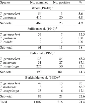
TABLE II
Trypanosoma cruzi infected triatoma species from counties in the Rio Grande Valley, Texas, USA
Species No. examined No. positive % Wood (1941b) a
T. gerstaeckeri 54 3 5.6
T. protracta 415 20 4.8
Sub-total 469 23 4.9
Sullivan et al. (1949) b
T. gerstaeckeri 57 7 12.3
T. protracta 2 2 100
T. rubida 2 2 100
Sub-total 61 11 18
Eads et al. (1963) c
T. gerstaeckeri 133 84 63.2
T. neotomae 31 27 87.1
T. sanguisuga 226 50 22.1
Sub-total 390 161 41.3
Burkholder et al. (1980) d
T. gerstaeckeri 49 13 26
T. neotomae 3 2 66.7
T. sanguisuga 35 6 17.1
Sub-total 87 21 22.6
Total 1,007 216 21.4
a: counties sampled: Brewster and Maverick; b: counties sampled: Brewster, Cameron, El Paso, Kinney, and Val Verde; c: counties sampled: Cameron; d: counties sampled: Cameron and Hidalgo
TABLE IV
Infection rate of Triatoma species from counties in the Rio Grande Valley, Texas, USA
Species No. examined No. positive %
T. gerstaeckeri 293 107 36.5
T. neotomae 34 29 85.3
T. protracta 417 22 5.3
T. rubida 2 2 100
T. sanguisuga 261 56 21.5
Total 1,007 216 21.4
Sourcer: Wood (1941b), Sullivan et al. (1949), Eads et al. (1963), Burkholder et al. (1980)
PREHISTORIC PATHOECOLOGY OF CHAGAS DISEASE
also believe that the lifestyle, especially cave dwelling, would have provided the behavioral conditions that pro-mote infection. Cave excavations in the region, for ex-ample, indicate that the fill is composed of a great quan-tity of vegetal fiber. Layers of vegetable refuse would have provided ideal habitat for heomphagous arthropods including triatomes. Also, grass “beds” have been found in the caves indicating that the local prehistoric popula-tion did sleep in the caves, at least on occasions (Shafer 1986). Thus, individuals sleeping in the caves infested with the nocturnally active triatomes could have been at risk of contracting Chagas disease.
Another aspect of the pathoecology of Chagas in the area is the human consumption of uncooked woodrats in prehistory. The fact that unburned bone and hair have been found in the coprolites from the area indicates that humans could have consumed infected rodents. The con-sumption of infected rodents increases the risk of infec-tion.
CONCLUSION
We present this as evidence of the gross pathology of Chagas disease. We believe that molecular and histology studies are needed to verify this as Chagas disease. We hope to include molecular studies in a battery of tests to document this mummy more fully.
Because autochthonous Chagas disease is rare in North America, documented human cases are unique and
worthy of note be they contemporary or ancient. Indeed, in representing a newly recognized North American case of Chagas disease, the SMM mummy is important to paleoepidemiologists and clinical epidemiologists alike.
ACKNOWLEDGEMENTS
To Beverlee Hall, Frank Ramberg, and Dave Engelthaler for their assistance in preparing this article. To Greg Pye, Texas Department of Health, Harlingen, for his help in obtaining up to date information on Chagas disease in Texas.
REFERENCES
Andrews RL, Adovasio JM 1980. Perishable Industries from Hinds Cave, Val Verde County, Texas, Ethnology Mono-graph 5, Department of Anthropology, University of Pitts-burgh.
Anonymous 1956. Found: two cases of Chagas disease. Tex Health Bull 9: 11-13.
Aronson PR 1962. Septicemia from concomitant infection with
Trypanosoma cruzi and Neisseria perflava: - first case of laboratory-acquired Chagas disease in the U.S. Ann Intern Med 57: 994-1000.
Auferderheide A, Rodrigues-Martin C 1998. The Cambridge Encyclopedia of Human Paleopathology, Cambridge Uni-versity Press, Cambridge, p. 228, 276.
Betz TG 1984. Chagas disease investigation. Tex Prevent Dis News 31: 1-4.
Burkholder JE, Allison TC, Kelly VP 1980. Trypanosoma cruzi
(Chagas) (Protozoa: Kinetoplastida) in invertebrate, reser-voir, and human hosts of the Lower Rio Grande Valley of Texas. J Parasitol 66: 305-311.
Davis DJ, McGregor T, DeShazo T 1943. Triatoma sanguisuga
(LeConte) and Triatoma ambigua Neiva as natural carries of Trypanosoma cruzi in Texas. Public Health Rep 58: 353-354.
DeShazo T 1943. A survey of Trypanosoma cruzi infection in
Triatoma spp. collected in Texas. J Bacteriol 46: 219-220. Diamond LS, Rubin R 1958. Experimental infection of certain farm mammals with a North American strain of Trypano-soma cruzi from the raccoon. Exp Parasitol 7: 383-390. Dominguez S, Reinhard KJ, Sandness KL, Edwards CA,
Danielson D 1992. The Dan Canyon Burial, 42A21339, a P III Burial in Glen Canyon National Recreation Area. Lin-coln: Midwest Archeological Center’s Occasional Studies Series 26.
El-Najjar MY, Mulinski TMJ, Reinhard KJ. 1998. Mummies and mummification practices in the southern and South-western United States. In E Cockburn & T Reyman (eds),
Mummies, Disease, and Ancient Cultures, Cambridge Uni-versity Press, Cambridge, p. 121-137.
Eads RB, Trevino HA, Campos EG 1963. Triatoma (Hemi-ptera: Reduviidae) infected with Trypanosoma cruzi in south Texas wood rat dens. Southwest Natural 8: 38-42. Edwards L, Lynch PJ 1984. Anaphylactic reaction to kissing
bug bite. Ariz Med 41: 159-161.
Elkins JC 1951a. Chagas disease and vectors in north central Texas. Field Lab 19: 95-99.
Elkins JC 1951b. The Reduviidae of Texas. Tex J Sci 3: 407-412.
Ferreira LF, Britto C, Cardoso A, Fernandes O, Reinhard K, Araújo A 2000. Paleoparasitology of Chagas disease re-vealed by infected tissues of Chilean mummies. Acta Trop 75: 79-84.
Grögl M, Kuhn RE, Davis DS, Green GE 1984. Antibodies to
Trypanosoma cruzi in coyotes in Texas. J Parasitol 70: 189-191.
TABLE V
The number of coprolites containing bone of the given taxa from Hinds Cave in the Chihuahuan Desert of Texas. The data
show that rodents, especially of the genus Neotoma, were commonly eaten. A total of 97 coprolites were studied from
Hinds Cave for animal remains. More than one species of animal were found in many coprolites.
Unidentifiable 14
Mammalia 8
Rodentia 32
Neotoma 19
Sigmodon 13
Peromyscus 2
Ondatra 1
Spermophilus 3
Sylvilagus 7
Lepus 7
Procyon 3
Urocyon 1
Odocoileus 1
Aves 9
Colinus 1
Zenaidura 2
Lizard 3
Sceloporus 1
Snake 3
Rana 1
Osteichthes 10
Aplodinatus 2
Kagan IG, Norman N, Allain D 1966. Studies on Trypanosoma cruzi isolated in the United States: a review. Rev Biol Trop 14: 55-73.
Kasa TJ, Lathrop GD, Dupuy HJ, Bonney CH, Toft JD 1977. An Endemic focus of Trypanosoma cruzi infection in a sub-human primate research colony. J Am Vet Med Assoc 171: 850-854.
Lent H, Wygodzinsky P 1979 Revision of the Triatominae (Hemiptera, Reduviidae), and their significance as vectors of Chagas Disease. Bull Am Mus Natur Hist 163: 125-520. Luibel-Hulen AM 1985. Use if computer assisted tomography in the study of a female mummy from Chihuahua, Mexico. In RA Tyson & DV Elerick (eds), Two Mummies from Chi-huahua, Mexico: Multidisciplinary Approach, San Diego Museum of Man, San Diego Museum Papers no. 19, USA. Meurs KM, Anthony MA, Slater M, Miller MW 1998. Chronic
Trypanosoma cruzi infection in dogs: 11 cases (1987-1996).
J Am Vet Med Assoc 213: 497-500.
Minnis PE 1989. Prehistoric diet in the northern southwest: macro-plant remains from four corner feces. Am Antiq 54: 543-563.
Packchanian A 1939. Natural infection of Triatoma gerstakeri
with Trypanosoma cruzi in Texas. Public Health Rep 54: 1547-1555.
Packchanian A 1940. Natural infection of Triatoma heidemanni
with Trypanosoma cruzi in Texas. Public Health Rep 55: 1300-1306.
Packchanian A 1942. Reservoir hosts of Chagas disease in the United States. Am J Trop Med 22: 623-631.
Packchanian A 1943. Infectivity of the Texas strain of Trypa-nosoma cruzi to man. Am J Trop Med 23: 309-314. Pearce JE, Jackson AT 1933. A Prehistoric Rock Shelter in Val
Verde County, Texas, University of Texas Bulletin 327, Austin.
Pippin WF, Law PF, Gaylor MJ 1968. Triatoma sanguisuga texana Usinger and Triatoma sanguisuga indictiva Neiva naturally infected with Trypanosoma cruzi Chagas in Texas.
J Med Entomol 5: 134
Reinhard KJ 1990. Archaeoparasitology in North America. Am J Phys Anthropol 82: 145-162.
Reinhard KJ 1992. Parasitology as an interpretive tool in ar-chaeology. Amer Antiq 57: 231-245.
Reinhard KJ 1993. The utility of pollen concentration in co-prolite analysis: expanding upon Dean’s comments. J Ethnobiol 9: 31-44.
Reinhard KJ, Geib PR, Callahan MM, Hevly RH 1992. Dis-covery of colon contents in a skeletonized burial: soil sam-pling for dietary remains. J Archaeol Sci 19: 697-705. Reinhard KJ, Hamilton DL, Hevly RH 1991. Use of pollen
concentration in paleopharmacology: coprolite evidence of medicinal plants. J Ethnobiol 11: 117-134.
Roberts LS, Janovy JJ 1996. Foundations of Parasitology, McGraw Hill Publishers, USA.
Ryckman RE 1981. The kissing bug problem in Western North America. Bull Soc Vector Ecol 6: 167-169.
Ryckman RE 1984a. The triatominae of North and Central America and the West Indies: a checklist with synonymy (Hemiptera: Reduviidae: Triatominae). Bull Soc Vector Ecol 9: 71-83.
Ryckman RE 1984b. Triatominae and Triatominae-borne try-panosomes of North and Central America and the West Indies: a bibliography with index. Bull Soc Vector Ecol 9: 112-430.
Ryckman RE 1986. The vertebrate hosts of the Triatominae of North and Central America and the West Indies (Hemi-ptera: Reduviidae: Triatominae). Bull Soc Vector Ecol 11: 221-241.
Ryckman RE, Archbold EF 1981. The Triatominae and Triatominae-borne trypanosomes of Asia, Africa, Austra-lia and the East Indies. Bull Soc Vector Ecol 6: 143-166. Ryckman RE, Blankenship CM 1984. The Triatominae and
Triatominae-borne trypanosomes of North and Central American and the West Indies: a bibliography with index.
Bull Soc Vector Ecol 9: 112-430.
Schiffler RJ, Mansur GP, Navin TR, Limpakarnjanarat K 1984. Indigenous Chagas disease (American trypanosomiasis) in California. J Am Med Assoc 251: 2983-2984.
Schofield CJ, Minter DM, Tonn RJ 1987. Triatomine Bugs: Training and Information Guide, World Health Organiza-tion, Geneva, p. 19.
Shafer HJ 1975. Clay figurines from the lower Pecos region.
Amer Antiq 40: 148-158.
Shafer HJ 1986. Ancient Texans: Rock Art and Lifeways Along the Lower Pecos, Texas Monthly Press, Austin.
Shafer, HJ, Marek M, Reinhard KJ 1989 Mimbres burial with associated colon remains from the NAN Ranch Ruin, New Mexico. J Field Archaeol 16: 17-30.
Sullivan TD, McGregor T, Eads RB, Davis DJ 1949. Incidence of Trypanosoma cruzi, Chagas, in triatoma (Hemiptera, Reduviidae) in Texas. Am J Trop Med 29: 453-458. Taylor WW 1966. Archaic cultures adjacent to the northeasten
frontiers of Mesoamerica. In R Wauchope, GF Ekholm, GR Wiley (eds), Handbook of Middle American Indians, Vol. 4: Archaeological Frontiers and External Connections, Uni-versity of Texas Press, Austin.
Turpin SA 1982. Seminole Canyon: the Art and Archaeology,
Texas Archaeological Survey Research Report 83, Austin. Turpin SA, Henneberg M, and Bement LC 1986. Late Archaic mortuary practices of the Lower Pecos River Region, south-west Texas. Plains Anthropol 31: 295-315.
Usinger RL 1944. The Triatominae of North and Central America and the West Indies and their Public Health Significance. Public Health Bulletin No. 288, United States Public Health Service, Washington.
Williams GD, Adams LG, Yaeger RG, McGrath RK, Read WK, Bilderback WR 1977. Naturally occurring trypanosomiasis (Chagas disease) in dogs. J Am Vet Med Assoc 171: 171-177. Wood SF 1941a. New localities for Trypanosoma cruzi Chagas
in Southwestern United States. Amer J Hyg 34: 1-13. Wood SF 1941b. Notes on the distribution and habits of
Redu-viid vectors of Chagas disease in the Southwestern United States. Pan-Pacific Entomol 27: 85-94.
Wood SF 1942a. Observations on vectors of Chagas disease in the United States. Bull Southern Cal Acad Sci 41: 61-69. Wood SF 1942b. Reactions of man to the feeding of Reduviid
bugs. J Parasitol 28: 43-49.
Wood SF 1951. Importance of feeding and defecation times of insect vectors in transmission of Chagas disease. J Econ Entomol 44: 52-54.
Wood FD, Wood SF 1938. On the distribution of Trypanosoma cruzi Chagas in the Southwestern United States. Amer J Trop Med 18: 207-212.
Woody NC, Woody HB 1955. American trypanosomiasis (Chagas disease). J Am Med Assoc 159: 676-677. Woody NC, Woody HB 1961 American trypanosomiasis I:
clinical and epidemiologic background of Chagas disease.
J Pediatr 58: 568-580.
Woody NC, DeDianous N, Woody HB 1961. American trypa-nosomiasis II: current serologic studies in Chagas disease.
J Pediatr 58: 738-745.
Woody NC, Hernandez A, Suchow B 1965 American trypano-somiasis III: The Incidence of serologically diagnosed Chagas disease among persons bitten by the insect vector.