785 785 785 785 785 Mem Inst Oswaldo Cruz, Rio de Janeiro, Vol. 92(6): 785-789, Nov./Dec. 1997
Detection of
Toxoplasma gondii
-specific Antibodies in Dogs.
A Comparative Study of Immunoenzymatic,
Immunofluorescent and Haemagglutination Titers
Deise AO Silva, Dagmar D Cabral
*, Bernardette LD Bernardina
*,
Maria A Souza, José R Mineo/
+Laboratório de Imunologia *Laboratório de Parasitologia, Departamento de Patologia, Universidade Federal de
Uberlândia, Av. Pará 1720, Campus Umuarama, 38400-902 Uberlândia, MG, Brasil
We evaluated the titers of anti-T. gondii antibodies by various serological tests in 40 serum samples from dogs exhibiting clinical signs of infectious diseases. Indirect immunofluorescence (IgG-IFI), indi-rect haemagglutination (IHA and M-Toxo) and immunoenzymatic (ELISA and PA-ELISA) tests were carried out. Titers ≥ 64 were considered as positive. Anti-Toxoplasma antibodies were found in 9 (22.5%), 14 (35%), 14 (35%) and 12 (30%) samples, respectively for IHA, IgG-IFI, ELISA and PA-ELISA. The resultsshowedthat 57% were negative in all testsand 43% of the dogs presented antibodies to T. gondii; from these, 20% were positive in all three tests with high titers of antibodies and 23% were positive in only one or two tests with low titers of antibodies and mainly related to the IFI and ELISA tests. We observed 5 (12.5%) and 1 (2.5%) reactive samples, respectively, by M-Toxo and IHA with or without 2-mercapthoethanol, in the attempt to detect specific IgM. We can conclude that serodiagnosis of toxo-plasmosis in dog have to be based on the combination of serological tests (IFI and ELISA) and with emphasis at the determination of the titers and theclasses of the specific antibodies.
Key words: Toxoplasma gondii dogs antibodies indirect immunofluorescence indirect haemagglutination -ELISA
Toxoplasma gondii causes one of the most com-mon parasitic infections in the world, affecting a wide range of hosts including man, domestic
ani-mals and birds.Toxoplasma infection in dogs is
very common as demonstrated by various
serologi-cal surveys (Svoboda 1987,Lindsay et al. 1990,
Uggla et al. 1990, Ulón & Marder 1990, Guimarães et al. 1992, Shad-Del et al. 1993, Björkman et al. 1994) while clinical toxoplasmosis is very less fre-quent, usually seen in young animals, and is asso-ciated with concurrent immunosupressor factors or infections, as distemper virus (Dubey 1985).
The clinical signs of toxoplasmosis in dogs are usually characterized by ataxia, diarrhoea and res-piratory distress (Ahmed et al. 1983). Focal ne-crotic areas in the lung, liver and brain of infected dogs are common and could lead to development of several clinical signs. Therefore, toxoplasmosis in dogs and other animals can mimic many infec-tious diseases (Dubey 1985).
Serological diagnosis of T. gondii infections in
dogs has been evaluated by many investigators
+Corresponding author. Fax: +55-34-218.2333
Received 3 January 1997 Accepted 23 June 1997
(Ahmed et al. 1983, Svoboda 1987, Lindsay et al. 1990, Uggla et al. 1990, Ulón & Marder 1990, Hejlicek et al. 1995). The tests used include the SabFeldman, the complement fixation, the in-direct haemagglutination, the in-direct agglutination, the indirect fluorescent antibody and the enzyme immunoassay. The demonstration of antibodies by these serological tests just indicates previous
in-fection by T. gondii.A laboratory diagnosis
de-fined to toxoplasmosis-disease requires the dem-onstration of high titers of specific antibodies and increasing levels in two serum samples taken 2 to 4 weeks apart (Dubey 1987). The majority of these investigations carried out in dogs have not estab-lished a comparative study of the titers of these various tests and their possible correlation with
active infection by T. gondii.
The purpose of the present investigation was to evaluate the titers of the various serological tests conducted with serum samples from dogs exhibit-ing clinical signs of infectious processes in which toxoplasmosis was one of the presumable diseases.
MATERIALS AND METHODS
786 786 786 786
786 Antibodies Anti-T. gondii in Dogs • Deise AO Silva et al.
1990 to 1993, with clinical signs which might have
led clinicians to suspect infection with T. gondii:
apathy, fever, dyspnea, pneumonia, gastroenteritis and nervous system disturbances with incoordina-tion, tremors and paralysis. The dogs were of vari-ous pure and mixed breed ancestry. Their ages var-ied from a few weeks to 14 years (mean of 2.9 ± 0.6 years) and the numbers of male and female were 19 and 21, respectively. The serum samples were collected following centrifugation at 500 x g of 5 ml of blood obtained from the radial vein from the dogs and were stored at -20°C until analyzed for
Toxoplasma antibodies.
Serologicaltests - An indirect haemagglutin-ation test (IHA) was performed using a reagent commercially available (Hematoxo, Biolab Diagnostica S.A., São Paulo, Brazil). The serum samples were diluted twofold, from 1:64 to 1:2,048, following the steps described by the manufacturer. All positive serum samples in the IHA test were retested after treatment with 2-Mercaptoethanol (2-ME) in order to verify the presence of IgM
anti-bodies (Camargo et al. 1978). OtherIHA(M-Toxo)
developed by Yamamoto et al. (1991) for the se-rodiagnosis of acute toxoplasmosis was carried out with a standardized suspension of red blood cells coated with a heat-stable alkaline-solubilized
ex-tract of T. gondii, which reacts predominantly with
IgM antibodies. To 50 µl of doubling diluted
se-rum, as above mentioned, 25 µl of sensitized cell
suspensions were added and the agglutination pat-tern read after incubation of 1 hr and 30 min at
room temperature.Positive and negative reference
serum samples (for IgG and IgM specific to T.
gondii) were included in all assays.
The indirect immunofluorescence test for
de-tection of IgG antibodies to T. gondii(IgG-IFI) used
in this investigation was similar to that used for diagnosis of human infections (Camargo 1964).
Antigen slides of T. gondii were incubated with
serum samples screened at 1:64 dilution and posi-tive samples were then diluted twofold until 1: 4,096. An isothiocyanate fluorescein labeled rab-bit IgG anti-dog IgG (kindly supplied by Centro de Zoonoses, São Paulo, Brazil) was used as sec-ondary antibody and the optimum titer (1:150) was determined by block titration with positive and negative serum controls. Positive control sera were obtained of the dogs with consistently positive se-rological results by IHA-Hematoxo, in interassay and intraassay variation tests. Negative control sera were obtained of dogs healthy with consistently negative serological results by IHA-Hematoxo, as mentioned above. The slides were examined by epi-fluorescent microscope (Olympus, Mod. BH2, Tokyo, Japan).
An immunoenzymatic test (ELISA) was
car-ried out for detection of IgG antibodies anti-T.
gondii as described (Mineo et al. 1980) with some modifications. Microtiter plates (Hemobag, Campinas, Brazil) were coated overnight at 4°C
with a soluble antigen preparation (0.25 µg of
pro-tein/well), consisting of a sonicated extract of
pu-rified Toxoplasma tachyzoites diluted in 0.06M
sodium carbonate buffer (pH 9.6). The plates were washed three times with phosphate-buffered saline containing 0.05% Tween 20 (PBST) and incubated with the samples. Doubling dilutions of the dog serum samples, diluted from 1:64 to 1:16,384 in PBST with 20% equine serum (PBST/ES) were added in duplicates to the wells and the plates in-cubated at 37°C for 45 min. After repeated wash-ing, the secondary antibody, consisting of a per-oxidase (horseradish perper-oxidase, type VI, Sigma Co., St. Louis, USA) labeled to rabbit IgG anti-dog IgG, prepared as described (Wilson & Nakane 1978) and diluted 1:20,000 in PBST/ES, was added and incubation performed for 45 min at 37°C. Af-ter a final wash, the plates were incubated with enzyme substrate hydrogen peroxide and o-phe-nylenediamine (Merck, Germany) in 0.1M
citrate-Na2HPO4 buffer (pH 5.5) for 15 min at room
tem-perature. The reaction was stopped by adding 2N
H2SO4 and the absorbance was read at 492 nm
using a Titertek Multiskan-Plus spectrophotometer (Flow Laboratories, USA). Positive and negative serum controls previously tested by conventional serological tests (i.e., IHA and IFI) were included on each plate. Samples showing absorbance val-ues exceeding the mean absorbance of the nega-tive controls plus 2 standard deviations were con-sidered as positive. Other immunoenzymatic test (PA-ELISA) was developed for detection of IgG
antibodies anti-T. gondii, as described above,
ex-cept by using peroxidase labeled Protein A (Sigma Co., St. Louis, USA) as secondary antibody which was diluted 1:100,000 in PBST with 1% bovine serum albumin.
The titers obtained on the serological tests were defined as low titers when the reactivity was equal or lower than 128. On the other hand, titers higher than 128 were defined as high titers.
Statistical analyses - The antibody titers were
codified in log10in order to determine the
787 787787 787787 Mem Inst Oswaldo Cruz, Rio de Janeiro, Vol. 92(6), Nov./Dec. 1997
RESULTS
When carrying out the indirect immunofluo-rescence (IgG-IFI), indirect haemagglutination (IHA and M-Toxo) and immunoenzymatic (ELISA and PA-ELISA) tests, the criteria followed for the samples be considered positive was the detection
of antibodies in titers ≥ 64. The IHA test revealed
T. gondii antibodies in 9 samples (22.5%); the IgG-IFI test demonstrated specific antibodies in 14 samples (35%), whereas the ELISA and PA-ELISA
tests showed anti-Toxoplasma antibodies in 14
(35%) and 12 (30%), respectively. In the attempt to detect specific IgM by the IHA with 2ME and M-Toxo tests, it was observed 1 (2.5%) and 5 (12.5%) reactive samples, respectively. The
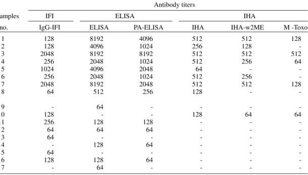
distri-bution of Toxoplasma antibody titers determined
by all these serological tests is demonstrated in Table. We observed titers of 64 to 2,048 for IgG-IFI (GMT = 2.68); titers of 64 to 8,192 for both ELISA and PA-ELISA (GMT = 3.43 and 3.22, re-spectively) and titers of 64 to 512 for IHA (GMT = 2.54).
Based on these results, it was possible to di-vide the positive samples in two different groups: a) a group of positive samples in all three tests (from # 1 to # 8) which presented high titers of
antibodies; and b) a group of positive samples in only one or two tests (from # 9 to # 17) which revealed low titers of antibodies. The positive re-sults of this latter group were mainly related to the IFI and ELISA tests. The GMTs for the high titer group in comparison with the low one were 2.87 and 2.07; 3.67 and 1.98; 3.39 and 1.90; 2.57 and 2.11, respectively for IgG-IFI, ELISA, PA-ELISA and IHA (p < 0.01). In addition, the reactive samples for the IHA with 2ME and M-Toxo tests (GMT= 2.56 and 2.32, respectively) were predomi-nantly included in the group of positive samples with high titers. No significant differences were seen according to sex, age and ancestry.
The correlation coefficient between the titers
provided by ELISA versus PA-ELISA was highly
significant (.98; p < 0.01). On the other hand, the comparison between IHA and IgG-IFI shows a non-significant correlation index of .51 (p > 0.05),
while the comparison between ELISA versus
IgG-IFI and IHA versus ELISA demonstrated
signifi-cant correlation indexes (.69 and .81, respectively; p < 0.01).
Fig. summarizes the results found in this in-vestigation. It was observed 20% (8/17) of posi-tivity in all three tests, 23% (9/17) in only one or
TABLE
Distribution of Toxoplasma antibody titers determined by indirect immunofluorescence test (IgG-IFI), immunoenzymatic test (ELISA and PA-ELISA) and indirect haemagglutination test (IHA, IHA-w2ME, M-Toxo)
among 17 positive samples from 40 serum samples collected from dogs with clinical signs of infectious diseases as examined at Veterinary Hospital of Universidade Federal de Uberlândia, State of Minas Gerais, Brazil
(1990-1993)
Antibody titers
Samples IFI ELISA IHA
no. IgG-IFI ELISA PA-ELISA IHA IHA-w2ME M -Toxo
1 128 8192 4096 512 512 128
2 128 4096 1024 256 128
3 2048 8192 8192 512 512 512
4 256 2048 1024 512 256 64
5 1024 4096 2048 64 -
-6 256 2048 1024 512 256
-7 2048 8192 2048 512 512 128
8 64 512 256 128 -
-9 - 64 - - -
-10 128 - - 128 64 64
11 256 128 128 - -
-12 64 64 64 - -
-13 64 - - - -
-14 - 128 64 - -
-15 64 - - - -
-16 128 128 64 - -
-17 - 64 - - -
788 788 788 788
788 Antibodies Anti-T. gondii in Dogs • Deise AO Silva et al.
two tests, establishing a total of 43% (17/40) of
seropositive samples for T. gondii antibodies, and
57% (23/40) of negativity in all tests.
samples from dogs exhibiting clinical signs sug-gestive of toxoplasmosis.
Our results showed that, even though all se-rum samples came from dogs exhibiting clinical signs of infectious disease compatible with toxo-plasmosis, only 43% of them presented antibodies
against T. gondii. Thus, three groups were
identi-fied: (1) seronegativesamples (57%) - this
find-ing can be explained by the fact that there is an overlapping of clinical signs among toxoplasmo-sis and many infectious diseases in dogs and other animals, so that they can mimic each other (Ahmed et al. 1983). Thus, a negative serological result gen-erally rules out a diagnosis of toxoplasmosis and shows significance, as it may guide the procedures of the clinicians in research for other diseases; (2)
seropositivesamples in all three tests (20%) with
high titers of antibodies and predominance of IgM antibodies detected mainly by the M-Toxo test. This test, developed for the serodiagnosis of acute hu-man toxoplasmosis (Yamamoto et al. 1991), was carried out with the dog serum samples in order to verify its suitability for detection of IgM-specific antibodies in dogs. It was found 4 positive samples by M-Toxo out of 8 positive samples (50%) for IgG antibodies in all three tests, suggesting that these animals might be presenting an active
infec-tion by T. gondii. The 4 M-Toxo non-reactive
samples in this group but with high titers of IgG may represent IgM false-negative results due to competitive interaction with IgG which presents higher avidity; and (3) seropositive samples in only 1 or 2 tests (23%) with low titers of antibodies and mainly related to the IFI and ELISA tests. The re-sults of this group suggest that the samples might belong to animals with chronic infection and the titer and the positivity rates were totally dependent of the sensibility of the employed tests. From the serological methods used in the present study, ELISA and IFI can be strongly recommended. On the other hand, by using IHA, a negative result should be taken carefully because the possibility of T. gondii infection cannot be excluded.
It was shown in the present investigation high correlation among the ELISA and PA-ELISA tests. Since it seems that the use of Protein A - peroxi-dase in the ELISA test for the determination of
Toxoplasma antibodies in dogs has not previously been reported, we concluded that PA-ELISA could also be used in serological surveys in dogs for
de-tection of anti-T. gondii IgG antibodies.
Taken together, the results presented here show that the diagnosis of toxoplasmosis in dog have to be based on combination of serological tests that
are able to detect different anti-T. gondii
antibod-ies and with emphasis in the determination of the titers and the classes of the immunoglobulins.
Percentage of negativity or positivity for Toxoplasma gondii
antibodies as determined by indirecthaemagglutinationtest (IHA), indirectimmunofluorescencetest (IgG-IFI) and immunoenzymatictest (ELISA and PA-ELISA) among 40 se-rum samples collected from dogs with clinical signs of infec-tious diseases as examined at Veterinary Hospital of Universidade Federal de Uberlândia, State of Minas Gerais, Brazil (1990-1993).
DISCUSSION
Many investigations have been conducted on
serological diagnosis of T. gondii infection in dogs.
However, the majority of them have not established a comparative study of the antibody titers and their
possible correlation with active infection by T.
gondii. So far, it has not been possible to make valid comparisons among prevalence of antibody to T. gondii in serum samples of dogs from these various studies because of the differences in sensi-tivity and specificity of the employed serological tests and also the lack of standardization for each test (Dubey 1985).
The variations in incidence of Toxoplasma
se-rum titers could be related to modifications in in-fection rates or to the sensitivity of the different tests used as well as the cut-off established for each test (Ahmed et al. 1983). In this context, it should also be pointed out that the prevalence of animals
with T. gondii antibodies is closely dependent on
the titer regarded as positive. In most studies the usual dilutions of 1:16, 1:64 or 1:100 (for IFI, IHA or ELISA, respectively) were considered as posi-tive (Ahmed et al. 1983, Svoboda 1987, Ugla et al. 1990).
The present study is the first publication con-cerning the comparative analyses of the IFI, IHA and ELISA tests in dogs. Therefore, we evaluate
the titers of anti-T. gondii antibodies determined
by three serological tests conducted with serum
Positive in only 1 or 2 tests (23%) Positive in
all 3 tests
(20%)
Negative
789 789789 789789 Mem Inst Oswaldo Cruz, Rio de Janeiro, Vol. 92(6), Nov./Dec. 1997
REFERENCES
Ahmed BA, Gaafar SM, Weirich WE, Kanitz CL 1983. Relationship of Toxoplasma infections to other dis-eases in dogs. Vet Parasitol 12: 199-203.
Björkman C, Lundén A, Uggla A 1994. Prevalence of antibodies to Neospora caninum and Toxoplasma gondii in Swedish dogs. Acta Vet Scand 35: 445-447.
Camargo ME 1964. Improved technique of indirect im-munofluorescence for serological diagnosis of toxo-plasmosis. Rev Inst Med Trop S Paulo 6: 117-118. Camargo ME, Ferreira AW, Mineo JR, Takiguti CK,
Nakahara OS 1978. Immunoglobulin G and immu-noglobulin M enzyme-linked immunosorbent assays and defined toxoplasmosis serological titers. Infect Immun 21: 35-38.
Dubey JP 1985. Toxoplasmosis in dogs. Canine Pract 12: 7-28.
Dubey JP 1987. Toxoplasmosis. Vet Clin North Am Small Animal Practice 17: 1389-1404.
Guimarães AM, Ribeiro MFB, Lima JD, Cury MC, Spiewak G 1992. Freqüência de anticorpos anti-Toxoplasma gondii em cães de Belo Horizonte, Minas Gerais. Arq Bras Med Vet Zootec 44: 67-68. Hejlicek K, Literak I, Lhotak M 1995. Prevalence of antibodies to Toxoplasma gondii in army dogs in the Czech Republic and Slovak Republic. Vet Medicina 40: 137-140.
Lindsay DS, Dubey JP, Upton SJ, Ridley RK 1990. Se-rological prevalence of Neospora caninum and Toxo-plasma gondii in dogs from Kansas. J Helminthol Soc Wash 57: 86-88.
Lutz W 1967. Statistical methods applied to immuno-logical data, p. 1163-1202. In DM Weir, Handbook of Experimental Immunology, Blackwell, Oxford. Mineo JR, Camargo ME, Ferreira AW 1980.
Enzyme-linked immunosorbent assay for antibodies to Toxo-plasma gondii polysaccharides in human toxoplas-mosis. Infect Immun 27: 283-287.
Shad-Del F, Sarvestani RG, Milani MS 1993. Sero-preva-lence of Toxoplasma infection in human and dog population in Shiraz, Iran. J Appl An Research 3: 83-89.
Svoboda M 1987. Incidence of Toxoplasma gondii anti-bodies in dogs from Brno and its environs. Acta Vet Brno 56: 475-486.
Uggla A, Mattson S, Juntti N 1990. Prevalence of anti-bodies to Toxoplasma gondii in cats, dogs and horses in Sweden. Acta Vet Scand 31: 219-222.
Ulón SN, Marder G 1990. Tasas de infección toxoplasmica en el hombre y su relación con los animales domesticos en la ciudad de Corrientes. Vet Arg 68: 518-522.
White C 1973. Statistical Methods in Serum Surveys, p. 19-32. In JR Paul, C White (eds), Serological Epi-demiology, Academic Press, New York.
Wilson MB, Nakane PK 1978. Antibody conjugated to horse-radish peroxidase, p. 215-224. In W Kanpp, K Holubar, G Wick (eds), Immunofluorescence and related staining technique,. Elsevier North Holland Biomedical Press, Amsterdam.