Impro ve me nt o f the indire ct
he magglutinatio n te st fo r
the de te ctio n o f antibo die s
to
Strep tococcus p yogenes
1Laboratório de Soroepidemiologia, Instituto de Medicina Tropical de São Paulo,
São Paulo, SP, Brasil
2Seção de Imunologia, Instituto Adolfo Lutz, São Paulo, SP, Brasil
3Departamento de Análises Clínicas e Toxicológicas, Faculdade de Ciências
Farmacêuticas, Universidade de São Paulo, São Paulo, SP, Brasil G. Rubinsky-Elefant1,
S. Hoshino-Shimizu2, E.M. Mamizuka3 and M.M.R. Asciutti3
Abstract
An indirect hemagglutination test for a seroepidemiological survey of Streptococcus pyogenes infection was standardized. This is an im-proved modification of the indirect hemagglutination test which uti-lizes an unstable reagent prepared with fresh blood cells. Two types of bacterial antigens represented by extracellular products and purified streptolysin O were assayed, but only the former antigen gave good results. Pretreatment of the bacterial antigen with 0.15 M NaOH and neutralization to pH 5.5, as well as postfixation of sensitized red cells with 0.1% glutaraldehyde at 56oC for 30 min were found to be
essential to give long stability to the reagent in liquid suspension, at least 9 months at 4oC. A total of 564 serum samples with high,
moderate and low anti-streptolysin O antibodies as determined by the neutralization assay were studied by the indirect hemagglutination test using the new reagent. The sensitivity, specificity, efficiency, positive predictive value and negative predictive value of the test in relation to the neutralization assay were 0.950, 0.975, 0.963, 0.973, and 0.955, respectively. The kappa agreement index between the two techniques was high (0.926) and ranked as almost perfect. Antibody levels detected by both techniques also presented a high positive correlation (rs = 0.726). Five reagent batches successively produced proved to be
reproducible. Thus, the improved indirect hemagglutination test seems to be useful for public health laboratories.
Co rre spo nde nce G. Rubinsky-Elefant
Laboratório de Soroepidemiologia Instituto de Medicina Tropical de São Paulo
Av. Enéas C. Aguiar, 470, 4º andar 05403-000 São Paulo, SP Brasil
Fax: 55 (011) 852-3622
Research supported by Laboratório de Investigação Médica, Hospital das Clínicas, Faculdade de Medicina, USP (LIM-48) and CNPq. Publication supported by FAPESP.
Received February 4, 1998 Accepted May 25, 1998
Ke y words
•Streptococcus pyogenes
•Hemagglutination test
•Anti-streptolysin O antibodies
Intro ductio n
The infection caused by Streptococcus
pyogenes (group A, ß hemolytic streptococci)
constitutes a public health problem in sev-eral countries (1), mainly due to nonsuppu-rative sequelae such as acute rheumatic fe-ver (ARF) and acute glomerulonephritis. ARF is considered to be a more relevant sequela and also a severe form of the disease with the
infection, since infective microorganisms cannot be isolated at this stage of the disease. Specific antibodies to bacterial cell com-ponents or extracellular products have been used in serologic tests. The streptolysin O (SLO) neutralization assay (4,5) has long been widely used. Recently, other assays such as latex agglutination, toraysphere ag-glutination and rapid hemagag-glutination, and immunoenzymatic, radiometric and nephelo-metric tests have been introduced. Among them, the immunoenzymatic, latex aggluti-nation and nephelometric tests are preferred for use in routine laboratories (2,6-11).
Practical, reliable, and low cost assays are desirable for public health laboratories. The neutralization assay is a very complex and time-consuming technique (2). The la-tex agglutination test and rapid hemaggluti-nation test were shown to provide nonproducible results (6,12). Other assays re-quire special equipment. The indirect hem-agglutination test is one of the practical and economic tests, first described by Alouf et al. (13) for the diagnosis of streptococcal infections. Nevertheless, the need to use short-lived reagents makes this test inad-equate for routine diagnosis. Thus, in the present study we improved the test by using a stable extracellular antigen of
Streptococ-cus pyogenes, which also provides stable
and reproducible reagent batches for the detection of antibodies to S. pyogenes in population surveys.
Mate rial and Me thods
The study was carried out in two steps. The first step involved the study of the factors influencing the indirect hemagglutination test such as type of antigen, stabilizer agents for red cells, pH, buffer solution, incubation time, temperature, tannic acid concentration, sta-bilizer agents for the reagent, and so on. Different types of reagents were prepared in a pilot study, in which the sensitivity, speci-ficity, stability and reproducibility of the test
were also determined. In the second step, the reagent showing the best diagnostic perfor-mance was selected and evaluated in the study of 546 serum samples from individuals showing high, moderate and low anti-strep-tolysin O antibodies.
Serum samples
A total of 546 serum samples were stud-ied. Blood was collected from outpatients with antibodies to S. pyogenes (N = 332) and clinically healthy individuals (N = 214) at the Serology Laboratory of the University Hospital and at the Clinical Analysis Labo-ratory, both laboratories belonging to the University of São Paulo, and in the Immu-nology Section of the Fleury Laboratory, São Paulo. Serum samples were divided into three groups. The first group comprised 263 serum samples from outpatients showing antibodies to streptolysin O equal or higher than 250 Todd units (TU) as determined by the neutralization assay. The second group consisted of 69 serum samples from patients with unrelated diseases: toxoplasmosis (N = 6), syphilis (N = 14), rubella (N = 2), rheu-matoid arthritis (N = 13), nonspecific in-flammatory processes with high levels of C-reactive proteins (N = 17), lupus erythema-tosus (N = 15) and salmonellosis (N = 2). These patients had no significant anti-SLO levels of antibodies (<250 TU). The third group consisted of 214 serum samples from clinically healthy individuals also showing low levels of anti-SLO antibodies (<250 TU).
Bacte rial e xtrace llular products
Extracellular products (ECP) of S.
pyogenes were obtained in 400 ml of culture
medium according to Alouf and Raynaud (14), with some modifications. Culture me-dium was centrifuged at 9,000 g, at 4o
C, for 10 min. In order to obtain a concentrated antigen, the supernatant was precipitated with 80% ammonium sulfate and kept at 4o
24 h. The sediment was separated by cen-trifugation, resuspended in 40 ml of 0.15 M phosphate-buffered saline solution (PBS), pH 6.8, and dialyzed in the same buffer solution. This extracellular antigen was cen-trifuged again to remove all the insoluble particles and stored at -20o
C, in 5-ml aliquots (13). The extracellular antigen was then ti-trated in the neutralization assay, to deter-mine the hemolytic activity.
Also, a commercially available purified streptolysin O (PSLO) (Biolab S.A., São Paulo, Brazil) was assayed.
Re age nt for the indire ct
he magglutination te st
Type O, Rh negative human red blood cells were treated with formaldehyde accord-ing to Camargo et al. (15). Prior to the sensi-tization of formaldehyde-preserved cells, the ECP was treated with 0.15 M NaOH at 4o
C for 24 h, with pH adjusted to 5.5 with 3 M HCl. The sensitization process of cells with ECP was standardized based on a previously reported method (16) with several modifica-tions. Briefly, a volume of 2.2 ml formalde-hyde-preserved cells (6.5%) was twice washed in 0.15 M NaCl and resuspended in 10 ml of 1:15,000 tannic acid diluted in PBS, pH 6.4, and incubated at 56o
C for 15 min with shaking. Two milliliters ECP (1:40 in PBS, pH 6.4) was added to the mixture and incubated at 37o
C for 60 min with shaking. Then, 1.2 ml glutaraldehyde (1%) was mixed and incubated at 56o
C for 30 min. Finally, 1.2 ml skim milk (Nestlé, Brazil) was added. The latter solution was prepared with 1 g skim milk dissolved in 10 ml 0.15 M NaCl, auto-claved at 121o
C, at 1.5 atm. for 30 min and centrifuged at 6,000 g at 4o
C for 30 min. Sodium azide was added to the supernatant to a final concentration of 0.01% and the prepared reagent was stored at 4o
C.
A reagent was also prepared with puri-fied SLO following the same sensitization process as described above, except that this
antigen was used in its native form without pretreatment with 0.15 M NaOH.
Se rologic te sts
Neutralization assay. All serum samples
were assessed in the neutralization assay (2,4) to confirm the results of neutralization assays previously obtained by the different laboratories mentioned above.
Indirect hemagglutination test. The test
was carried out with the standardized re-agent in V-shaped wells of plastic microplates using 0.1 M NaCl for serum sample dilution, as described (17). Twenty-five microliters of sensitized cell suspension was added to 50 µl of serial double dilutions of serum samples from 1/20 to 1/5,120. The red cell agglutina-tion patterns were scored after incubating the microplates for 90 min at room tempera-ture. Two control sera, one with <250 TU and the other with >625 TU, were always included in the test. The reagent was con-trolled by adding 25 µl of sensitized cell suspension to 50 µl 0.1 M NaCl.
Re producibility and stability of the
indire ct he magglutination re age nt
Several batches of indirect hemaggluti-nation reagent (N = 6) were produced suc-cessively in the study and their reproducibil-ity was checked by a control chart method (18). Each reagent batch was assayed against a panel of 20 serum samples comprising 10 serum samples with positive anti-SLO titers (≥333 TU) and 10 with negative anti-SLO titers (<250 TU). The stability of the reagent was also studied at temperatures of 4o
, 25o
(room temperature) and 37o
C. Tests were performed against 3 control sera (<250, 333 and >625 TU) for a period of 9 months.
Statistical analyse s
re-lation to the neutralization assay as described by Galen and Gambino (19). The 95% confi-dence intervals were also calculated for dif-ferent diagnostic parameters. The kappa in-dex of agreement was determined for the comparison of two tests (20,21). The anti-body levels (Todd units or titer) were trans-formed to logfor statistical analysis (22). The Spearman correlation coefficient (23) was calculated for antibody levels expressed as titers and as Todd units.
Re sults
Factors influe ncing the indire ct
he magglutination te st
Factors influencing the indirect hemag-glutination test were determined in the first step of the present study. A total of about 60 different types of reagents were prepared and assayed because of the many influenc-ing factors investigated.
Four types of antigens were tested: PSLO and ECP in their native forms, and the same antigens submitted to previous treatment with 0.15 M NaOH and then neutralized. The first and fourth types of antigens were more sen-sitive and also gave more reproducible re-sults than the other two. PSLO and ECP were neutralized at different pH values rang-ing from 5.0 to 9.0; however, the best results were obtained when the antigens were kept at pH 5.5.
Red cell sensitization was carried out in two different processes. The first process, known as the direct sensitization process, consisted of incubating the PSLO or ECP antigen directly with formaldehyde-preserved red cells, whereas in the second process red cells were previously treated with tannic acid and then incubated with the antigen. In the first process of direct sensitization, the incubation period ranged from 3 to 24 h at room temperature or at 37o
C, but the results obtained under these conditions were poor. In the second process, which yielded the best
results, tannic acid was tested at concentra-tions ranging from 1:10,000 to 1:30,000 and the concentration of 1:15,000 was chosen because of its sensitivity. Also, tannic acid provided better results when diluted in PBS at pH 6.4 than when diluted in PBS at pH 7.2. Red cells were incubated with tannic acid, at 37o
and 56o
C for periods ranging from 10 to 60 min. Best results were obtained with in-cubation at 56o
C for 10 min or at 37o
C for 50 min. However, incubation at 56o
C for 10 min was preferred because of its practical aspects.
In the sensitization process, tanned red cells were incubated with PSLO and ECP in their native form and after alkaline treat-ment. Buffer solutions were tested at pH ranging from 6.4 to 8.2, and 0.15 M PBS, pH 6.4, was chosen for this process. Different incubation times (10 to 60 min) and tem-peratures (37o
and 56o
C) were assayed for the sensitization process, and the optimal conditions were found to be 37o
C for 50 min. Glutaraldehyde at concentrations rang-ing from 0.04 to 0.8% was tested in order to enhance antigen binding to the red cell sur-face. Glutaraldehyde was incubated with sen-sitized cells at different temperatures (37o
to 100o
C). The glutaraldehyde concentration and the conditions selected to keep the anti-gen steadily fixed onto the cell surface were 0.1% and 56o
C for 30 min, respectively. The reagent prepared with PSLO in its native form gave better results than that pre-pared with PSLO pre-treated with 0.15 M NaOH. Conversely, the reagent prepared with ECP submitted to alkaline treatment gave more sensitive results than that prepared with the same antigen in its native form. Thus, these two types of reagents were pro-duced in large amounts in order to assess their diagnostic features in the second step of the study.
bovine serum albumin (BSA), and sodium thioglycolate were assayed at different con-centrations and pH values but an autoclaved skim milk solution in 0.9% NaCl was found to provide the best stability.
Evaluation of the indire ct
he magglutination te st
The neutralization assay was taken as a reference technique to evaluate the diagnos-tic features of the indirect hemagglutination test with the use of two types of reagents. Serum samples with ≥250 Todd units were considered to be positive and those with lower values were considered to be negative. The serologic performance of the indirect hemagglutination test using two different re-agents was assessed in terms of sensitivity, specificity, efficiency and positive and nega-tive predicnega-tive values. The values obtained in the indirect hemagglutination test with the ECP reagent were high, as shown in Table 1.
Comparison of the se rologic assays
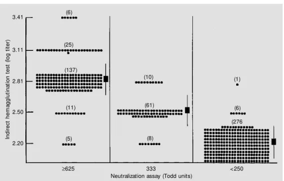
Data obtained by the indirect hemagglu-tination test using the ECP reagent and the PSLO reagent were compared to data ob-tained by the neutralization assay. Serum samples were divided into three groups ac-cording to their antibody levels (TU) as detected by the neutralization assay (Figure 1). The first group had antibody levels ≥625
TU (N = 184), the second had 333 TU (N = 79), and the third <250 TU (N = 283). The indirect hemagglutination test using the ECP reagent showed that about 91% of the se-rum samples from the first group presented titers ≥640 (2.81 in log), 90% of the second group had titers between 320 and 640 (from 2.50 in log to 2.81 in log), and 98% of the third group showed titers <160 (<2.20 in log). The indirect hemagglutination test with the use of the PSLO reagent showed that 83% of the serum samples from the first group presented titers ≥640, 54% of the samples from the second group had titers between 320 and 640, and 54% of the samples from the third group had titers <160.
The geometric mean titers (GMT) and 95% confidence intervals obtained in the indirect hemagglutination test with the use of ECP were 2.84 (2.64-3.04) for the first group of serum samples, 2.51 (2.36-2.66) for the second group, and 2.21 (2.13-2.29) for the third group (Figure 1), and the values obtained in the indirect hemagglutination test with the PSLO reagent were 3.03 (2.73-3.34), 2.50 (2.21-2.80) and 2.43 (2.13-2.74) for the first, second and third groups of serum samples, respectively.
The kappa agreement index between the indirect hemagglutination test and neutral-ization assay was 0.926 (zobtained = 16.85;
zcritical = 3.89, P<0.001), a value ranked as
almost perfect. The indirect
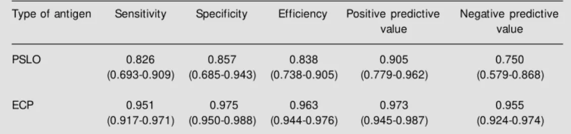
hemaggluti-Table 1 - Diagnostic features of the indirect hemagglutination test using tw o different antigens for the detection of anti-SLO antibodies in the study of 546 serum samples.
PSLO, Purified streptolysin O w ith no pretreatment; ECP, extracellular products of alkali-pretreated S. pyogenes. The 95% confidence interval is indicated w ithin parentheses.
Type of antigen Sensitivity Specificity Efficiency Positive predictive Negative predictive value value PSLO 0.826 0.857 0.838 0.905 0.750
(0.693-0.909) (0.685-0.943) (0.738-0.905) (0.779-0.962) (0.579-0.868) ECP 0.951 0.975 0.963 0.973 0.955
nation test using the PSLO reagent gave, as expected, a lower kappa agreement index, 0.665 (zobtained = 5.64; P<0.001) and ranked
as substantial.
The antibody titers detected by the indi-rect hemagglutination test using the ECP reagent were compared with the correspond-ing Todd units determined by the neutraliza-tion assay, as shown in Figure 2. The Spearman correlation coefficient (rs) was
0.726. On the other hand, the rs obtained in
the indirect hemagglutination test with PSLO was 0.440.
Re producibility and stability of
he magglutination re age nt batche s
In this study 6 reagent batches were successively produced with ECP antigen and tested against a reference panel of 20 serum samples. Standard deviations (SD) were calculated and compared with a stipu-lated control limit corresponding to 0.20 SD. Five reagent batches were approved by this control method because the SD values were 0.184, 0.069, 0.138, 0.140 and 0.155, i.e., lower than the control limit. However, one reagent batch was rejected because its SD was 0.296 i.e., higher than the control limit.
The hemagglutination reagent was stored at 4o
C, at room temperature and at 37o
C. The reagent kept at 4o
C showed no change in reactivity even after 9 months, when the present study was concluded. At room tem-perature, the reagent showed no change in reactivity for a period of 6 months, whereas
In
di
re
ct
h
em
ag
gl
ut
in
at
io
n
te
st
(
lo
g
tit
er
)
3.41
3.11
2.81
2.50
2.20
(6)
(25)
(137)
(11)
(5)
(10)
(61)
(8)
(1)
(6) (276
<250 333
≥625
Neutralization assay (Todd units)
In
di
re
ct
h
em
ag
gl
ut
in
at
io
n
te
st
(
lo
g
tit
er
) 3.5
3.0
2.5
2.0
2.0 2.5 3.0 3.5 Neutralization assay (log Todd units)
1 10 14
1 5
11 130
61 276 8
9 7 2
1 4 Figure 1 - Antibodies detected
by the indirect hemagglutination test in the study of 546 serum samples w ith high (≥625), mod-erate (333) and low (<250) Todd units of streptolysin O anti-bodies as detected by the neu-tralization assay.
Figure 2 - Relationship betw een antibodies detected by the indi-rect hemagglutination test and by the neutralization assay (rs =
0.726).
the reactivity of the reagent stored at 37o
C lasted only 45 days.
D iscussio n
Antibodies to Streptococcus pyogenes
ECP have been reported to occur in strepto-coccal infections (4). Alouf et al. (13) found an excellent correlation between the anti-ECP antibodies detected by the indirect hem-agglutination test and the conventional anti-SLO antibodies detected by the neutraliza-tion assay. In populaneutraliza-tion surveys the indirect hemagglutination test is thought to be suit-able because of its practical and easy execu-tion aspects, requiring no special equipment. In the present study, two types of re-agents, one of them prepared with PSLO antigen without any pretreatment and the other with an alkali-pretreated ECP antigen, gave sensitive results in the standardization step. Nevertheless, a significant difference between these two reagents was observed thereafter when large numbers of serum samples were tested, with a clear superiority of the ECP reagent. This difference may be explained by the fact that the commercially supplied PSLO antigen has some protein contaminants which are not antigenic and which attach to the tanned red cells during reagent preparation. These contaminants, however, did not interfere with the neutral-ization assay. Thus, the diagnostic features of the indirect hemagglutination test using the ECP reagent were shown to be signifi-cantly better than those of the test using the PSLO reagent.
The reagent prepared with alkali-pre-treated ECP can be considered as an im-provement of the reagent previously de-scribed by Alouf et al. (13) who used an unstable and short-lived reagent in liquid solution.
The new reagent differed from the previ-ous one in the following ways: a) tannic acid was used instead of glutaraldehyde to bind
antigen to the red cells; b) the antigen was treated with 0.15 M NaOH which conferred stability, requiring no protease inhibitors; c) the time for red cell sensitization was re-duced to 50 min instead of the previous 24 h; d) antigens coating red cells were postfixed with 1% glutaraldehyde at 37o
C for 30 min for long reagent stability. The previous re-agent was found to be stable only for 15 days at 4o
C, in contrast to the new reagent which lasted at least 9 months at 4o
C, 6 months at room temperature and 6 weeks at 37o
C. The processes to keep a reagent in liquid solu-tions without changing its reactivity are usu-ally manufacturers patents or secret formu-lation. e) Serum dilutions with hypotonic saline solution (0.1 M NaCl) gave better results than the isotonic saline solution (0.15 M NaCl) used for the previous reagent, con-firming the findings of Yamamoto et al. (17) for the toxoplasma system; f) V-shaped plas-tic microplates showed clear-cut results within 90 min compared to U-shaped plates which gave results within 2 or 3 h. These conditions for the preparation of the ECP reagent were also optimal for obtaining the PSLO reagent.
cutoff for the NA was based on the value reported by Pereira et al. (27) which is simi-lar to that adopted in the United States (28). The correlation coefficient found between antibody titers obtained by the indirect hem-agglutination test and Todd units obtained by the neutralization assay was high (r = 0.726), as illustrated in Figure 2, showing that titers and Todd units are closely related. This value was similar to those observed for the commercial slide agglutination test (Streptozyme) (29), and for the immunoen-zymatic ELISA (9,30), both assays in com-parison to the neutralization assay.
The sensitivity obtained for the indirect hemagglutination test with the use of PSLO was significantly lower (0.826) than that of the indirect hemagglutination test with the use of the ECP reagent, as also was its speci-ficity (0.857). Therefore, the indirect hem-agglutination test with the use of PSLO in this test was considered unsuitable for diag-nostic purposes.
The sensitivity (0.978) and specificity (0.689) of the indirect hemagglutination test obtained by Alouf et al. (13) were calculated based on their reported data. The original indirect hemagglutination test had a sensi-tivity similar to that obtained by us (0.951) for the same test with the use of the new reagent. The specificity of the original test, however, was significantly lower in com-parison to the improved indirect hemaggluti-nation test (0.975). The difference in speci-ficity may be explained by the fact that those authors used native S. pyogenes extracellu-lar products which probably crossreacted
with other unrelated infections, whereas in the present study alkaline treatment of the same antigen resulted in high specificity, probably because many small nonspecific fragments derived from this treatment did not attach to the red cell surface.
Also, the sensitivity of the indirect hem-agglutination test using the new reagent was slightly higher than that of the latex aggluti-nation test (7), but much higher than that of immunoenzymatic ELISA (30).
In turn, the specificity of the indirect hemagglutination test with the new reagent was superior to those of the latex agglutina-tion test and of ELISA.
The new reagent will also be tested against sera from patients with defined diagnoses of acute rheumatic fever, acute glomerulone-phritis and pyoderma. The aspects related to the kinetics of antibody production in such patients during the acute and chronic stages of the disease will also be investigated. The improvement of the indirect hemagglutina-tion test with the use of a stable reagent seems useful for epidemiological serum sur-veys in developing countries.
Ackno wle dgme nts
Re fe re nce s
1. Rosa EC, Rizzo M C, Giavina-Bianchi-Jr PF, Forte WCN & M imica IM (1988). Imuno-patologia da febre reumática. Ciências M édicas, 16: 21-30.
2. Rubinsky Elefant G (1996). Estreptococ-cias. In: Ferreira AW & Ávila SLM (Edi-t ors), Diagnóst ico Laborat orial das Principais Doenças Infecciosas e Auto-Im unes. Guanabara Koogan, Rio de Janeiro, 93-100.
3. WHO CVD (1992). Unit and Principal In-vestigators. WHO programme for the pre-vention of rheumatic fever/rheumatic heart disease in 16 developing countries: a report from phase I (1986-1990). Bulle-tin of the World Health Organization, 70: 213-218.
4. Todd EW (1932). Antihaemolysin titres in haemolytic streptococcal infections and their significance in rheumatic fever. Brit-ish Journal of Pathology, 13: 248-259. 5. Randall EA & Rantz LA (1949). Stable,
reduced, desiccated streptolysin “ O” .
Proceedings of the Society for Experimen-tal Biology and M edicine, 70: 414-416. 6. Curtis GDW, Kraak WAG & M itchell RG
(1988). Comparison of latex and haemol-ysin tests for determination of anti-streptolysin O (ASO) antibodies. Journal of Clinical Pathology, 41: 1331-1333.
7. Gerber M A, Caparas LS & Randolph M F (1990). Evaluation of a new latex aggluti-nation test for detection of streptolysin O antibodies. Journal of Clinical M icrobiol-ogy, 28: 413-415.
8. Heymer B, Schleifer KH, Read S, Zabriskie JB & Krause RM (1976). Detection of anti-bodies to bacterial cell w all peptidoglycan in human sera. Journal of Immunology, 117: 23-26.
9. Reitano M , Pisano M A, Eriquez LA & D’Amato RF (1986). Enzyme-linked im-munosorbent assay for detection of strep-tolysin O antibodies. Journal of Clinical M icrobiology, 23: 62-65.
10. Barbosa SFC, Nakamura PM & Hoshino-Shimizu S (1996). Detection of antibody isotypes to streptolysin O by dot ELISA.
Brazilian Journal of M edical and Biological Research, 29: 763-767.
11. Kodama T, Ichiyama S, M orishita Y, Fukatsu T, Shimokata K & Nakashima N (1997). Determination of anti-streptolysin O antibody titer by a new passive aggluti-nation method using sensitized toray-sphere particles. Journal of Clinical M icro-biology, 35: 839-842.
12. Gerber M A, Wright LL & Randolph M F (1987). Streptozyme test for antibodies to group A streptococcal antigens. Pediatric Infectious Disease Journal, 6: 36-40. 13. Alouf JE, Saint M artin J, Eyquem A,
Geoffrey C, Jacquemot C & Duphot M (1978). Titrage des anticorps sériques humains anti-exoprotéines du strepto-coque du groupe A par microhémaggluti-nation en plaque: corrélation avec les taux de l’streptolysine O et de divers anti-enzymes. Annales de M icrobiologie, 129A: 447-472.
14. Alouf JE & Raynaud M (1973). Purification and some properties of streptolysin O.
Biochimie, 55: 1187-1193.
15. Camargo M E, Hoshino S & Siqueira GRV (1973). Hemagglutination w ith preserved, sensitized cells, a practical test for routine serological diagnosis of American trypan-osomiasis. Revista do Instituto de M edi-cina Tropical de São Paulo, 15: 81-85.
16. Nagasse-Sugahara TK, Hoshino-Shimizu S, Pagliarini RC & Celeste BJ (1996). Im-provement of the slide hemagglutination test for rapid Chagas’ disease screening in epidemiological surveys. Brazilian Jour-nal of M edical and Biological Research,
29: 623-628.
17. Yam am oto YI, Hoshino-Shim izu S & Camargo M E (1991). A novel IgM -indirect hemagglutination test for the serodiagno-sis of acute toxoplasmoserodiagno-sis. Journal of Clinical and Laboratory Analysis, 5: 127-132.
18. Hoshino-Shimizu S, Nagasse-Sugahara TK, Castilho EA, Camargo M E & Shimizu T (1986). A control chart method for evalu-ating hemagglutination reagent used in Chagas’ disease diagnosis. Pan American Health OrganizationBulletin, 20: 170-178. 19. Galen RS & Gambino SR (1975). Beyond Normality: the Predictive Value and
Effi-ciency of M edical Diagnosis. John Willey & Sons, Inc., New York.
20. Fleiss JL (1973). Statistical M ethods in Rates and Proportions. John Wiley & Sons, Inc., New York.
21. M acLure M & Willet DC (1987). M isinter-pretation and misuse of the kappa statis-tic. American Journal of Epidemiology, 126: 161-169.
22. White C (1973). Statistical methods in se-rum surveys. In: Paul JR & White C (Edi-tors), Serological Epidemiology. Academ-ic Press, New York, 9-32.
23. Siegel S (1979). Estatística Não Paramé-trica para a Ciência do Comportamento.
M cGraw -Hill do Brasil, São Paulo, 228-240.
24. Federico WA, Fava Netto C, Amato Neto V & Debes AC (1967). Título de anti-estreptolisina O no soro de indivíduos normais da cidade de São Paulo. O Hospi-tal, 72: 269-278.
25. Watanabe N, Kobayashi M , Arimura A & Oshima M (1981). Follow up study of ASO, ADN-B and ASK levels in children w ith rheumatic fever. Japanese Circula-tion Journal, 45: 1379-1381.
26. Braida M , Gaido E, Panarisi P & Fiorio C (1986). In tema di diagnostica sierologica delle malattie streptococciche. Ulteriori dati sul confronto tradue metodologie (Streptozyme e ASLO). M inerva M edica, 77: 1679-1688.
27. Pereira JAA, Plotkow iski M CM , Suassuna A & Suassuna I (1982). Faringite estrepto-cócica em população de escolares no Rio de Janeiro. Revista da Associação M édica Brasileira, 28: 45-48.
28. Blum G & Ellner PD (1970). Evaluation of a rapid slide test as a sceening procedure for antistreptolysin O. American Journal of Clinical Pathology, 53: 936-937. 29. Tolliver PR & Roe M H (1979).
Compari-son of tw o slide tests for detection of group A streptococcal antibodies. Ameri-can Journal of Clinical Pathology, 72: 218-221.
30. Rykner G (1980). Titrage immunoenzymol-ogique des antistreptolysines (ELISA).
Contact us for specific services not listed above
Banco de Células do Rio de Janeiro - BCRJ Programa Avançado de Biologia Celular Aplicado à Medicina - PABCAM Hospital Universitário Clementino Fraga Filho - HUCFF Universidade Federal do Rio de Janeiro - UFRJ Sala 4A9, Caixa Postal 68021 CEP 21941-970 Rio de Janeiro, RJ Tel: (021)564-2010 Ramal 249 Tel/Fax: (021)590-8736
Rio de Janeiro Cell Bank
•
Deposit and Distribution of Cell Lines•
Cell Cryopreservation•
Development and Distribution of Human and Animal Primary Cell Cultures•
Development of New Monoclonal Antibody Secreting Hybridomas•
Monoclonal Antibodies, in Cell Culture Supernatants or Purified•
Identification of Microbiological Contaminants•
Decontamination of Cell Lines•
Cytotoxicity Tests•
Characterization of Cell Lines•
Training in in vitro Culture of Human and Animal Cells•
Consultation Assistance in Cell CultureContact us for specific services not listed above
Banco de Células do Rio de Janeiro - BCRJPrograma Avançado de Biologia Celular aplicado à Medicina - PABCAM
Hospital Universitário Clementino Fraga Filho - HUCFF
Universidade Federal do Rio de Janeiro - UFRJ
Sala 4A9, Caixa Postal 68021
CEP 21941-970 Rio de Janeiro, RJ
Tel: (021)564-2010 Ramal 249
Tel/Fax: (021)590-8736