High occurrence of
Fusobacterium nucleatum
and
Clostridium difficile
in the intestinal microbiota of colorectal carcinoma patients
Márcia H. Fukugaiti
1, Aline Ignacio
1, Miriam R. Fernandes
1, Ulysses Ribeiro Júnior
2,
Viviane Nakano
1, Mario J. Avila-Campos
11
Departamento de Microbiologia, Universidade de São Paulo, São Paulo, SP, Brazil. 2
Instituto do Câncer do Estado de São Paulo, São Paulo, SP, Brazil.
Submitted: August 5, 2014; Approved: February 16, 2015.
Abstract
Colorectal carcinoma is considered the fourth leading cause of cancer deaths worldwide. Several mi-croorganisms have been associated with carcinogenesis, includingEnterococcusspp.,Helicobacter pylori, enterotoxigenicBacteroides fragilis, pathogenicE. colistrains and oralFusobacterium. Here we qualitatively and quantitatively evaluated the presence of oral and intestinal microorganisms in the fecal microbiota of colorectal cancer patients and healthy controls. Seventeen patients (between 49 and 70 years-old) visiting the Cancer Institute of the Sao Paulo State were selected, 7 of whom were diagnosed with colorectal carcinoma. Bacterial detection was performed by qRT-PCR. Al-though all of the tested bacteria were detected in the majority of the fecal samples, quantitative differ-ences between the Cancer Group and healthy controls were detected only forF. nucleatumandC. difficile. The three tested oral microorganisms were frequently observed, suggesting a need for fur-thers studies into a potential role for these bacteria during colorectal carcinoma pathogenesis. Despite the small number of patients included in this study, we were able to detect significantly more F. nucleatumandC. difficilein the Cancer Group patients compared to healthy controls, suggesting a possible role of these bacteria in colon carcinogenesis. This finding should be considered when screening for colorectal cancer.
Key words:oral and intestinal bacteria, colorectal cancer, intestinal microbiota.
Introduction
The human intestinal microbiota is a complex ecolog-ical environment that harbors up to 100 trillion bacteria. The collective genome of these intestinal bacteria contains at least 100 times as many genes as there are in the human genome (Ahmedet al., 2007).
It is estimated that the intestinal microbiota comprises 40,000 or more different microbial species (Castellarinet al., 2012), which account for ~50% of the fecal volume (De Cruzet al., 2012). It is know that the intestinal microbiota collaborate with the host in provision of additional meta-bolic capabilities, protection against pathogens, modula-tion of the immune system and gastrointestinal develop-ment (Franket al., 2007).
The microbial ecology of the human intestine can be affected by the host environment and dietary habits. Al-though these effects are not yet completely understood, it has been hypothesized that country-specific dietary habits affect the microbiota development and subsequently host health(Franket al., 2007).
The human intestinal microbiota has been analyzed using various molecular techniques, such as microarray analysis, quantitative real-time polymerase chain reaction (qRT-PCR), fluorescencein situhybridization (FISH) and metagenomic sequencing as reviewed by Ivanov and Lit-tman (2011). qRT-PCR is advantageous because of its specificity and suitability for quantifying population size (or prevalence) of specific bacterial groups/species. In ad-dition, as a high throughput method qRT-PCR can be used
DOI: http://dx.doi.org/10.1590/S1517-838246420140665
Send correspondence to M.J. Avila-Campos. Departamento de Microbiologia, Universidade de São Paulo, Av. Prof. Lineu Prestes 1374, 05508-900 São Paulo, SP, Brazil. E-mail: [email protected].
to detect specific pathogens in large numbers of clinical samples.
Colorectal carcinoma is considered the fourth leading cause of cancer deaths, estimated to be responsible for ap-proximately 610,000 deaths per year worldwide (WHO, 2011). Although inflammation is a well-established risk factor (McLeanet al., 2011), the cause of colorectal carci-noma remains unclear.
Colorectal carcinoma is typically caused by earlier-stage adenomatous lesions or polyps, and analyses for the presence of biological markers (e.g., microbial species) will likely be important for further understanding the devel-opment of this inflammatory process. Several risk factors of colon cancer have been identified and include older age (> 50 years-old), personal history of colorectal cancer or polyps, inflammatory intestinal conditions, genetic predis-position, family history, low-fiber and high-fat diet, seden-tary lifestyle, diabetes, obesity, smoking, alcohol and can-cer radiation therapy (Ponnusamyet al., 2011).
In colorectal cancer, several microorganisms have been associated with carcinogenesis, including
Enterococcus spp., Helicobacter pylori, enterotoxigenic
Bacteroides fragilisand pathogenicE. colistrains (Collins
et al., 2010). Oral and intestinal bacteria can alter the intes-tinal environment andin vivostudies aimed at determining the presence/absence of specific organisms under different host environments and dietary habits, as well as their co-occurrence with specific risk factors and biological markers (e.g., diabetes, obesity, smoking, alcohol, radiation ther-apy, adenomatous lesions), would likely provide novel in-sights into the microbiota-carcinoma relationship.
Oral microorganisms are capable of producing infec-tious diseases, including endocarditis, acute appendicitis, gastrointestinal diseases, lung and brain abscesses and periodontal diseases (Nakanoet al., 2007). These anaerobic bacteria are found in subgingival biofilms and have a well-established association with periodontitis(Signat et al., 2011). Among these, Fusobacterium nucleatum is most frequently detected in the oral cavity (presumably because of its role as bridge between early and later oral colonizers) and is the most common bacteria in both healthy and dis-eased oral cavities (Castellarinet al., 2012). In addition to
F. nucleatum, other periodontal bacteria (e.g.,
Porphyromonas gingivalisandPrevotella intermedia) are involved in human and animal periodontal diseases and have pro-inflammatory properties (Castellarinet al., 2012).
Recent studies have detected a prevalence of oral fusobacteria (mainlyF. nucleatum) in tissues from colo-rectal cancer patients and shown that strains isolated from inflamed biopsy tissue of intestinal disease patients display a more invasive phenotype (Strausset al., 2011). Since oral and intestinal microorganisms have been implicated in gas-trointestinal disorders, here we qualitatively and quantita-tively evaluated the presence of these microorganisms in
the fecal microbiota of colorectal cancer patients and healthy controls.
Material and Methods
Cohort and sample collection
Seventeen patients (13 male, 4 female) between 49 and 70 years-old (mean age: 60 years) visiting the Cancer Institute of the Sao Paulo State (Sao Paulo, SP, Brazil) were included in the study. Among these, 7 patients (5 male, 2 fe-male) displaying polyps, tumors and an inflamed area were diagnosed with colorectal carcinoma (by colonoscopy). The remaining 10 patients (8 male, 2 female) did not pres-ent polyps, tumors, abnormal areas or inflammation and were considered healthy. Fecal samples were collected 2 d before colonoscopy. Patients who had taken antibiotics or with any systemic infection were excluded. All patients were asked to participate in this study and provided written informed consent. This study was approved by the Ethic Committee of the Biomedical Science Institute at Univer-sity of Sao Paulo (CEPSH-1165).
Bacterial quantitative determination
Bacterial DNA from feces was obtained using a com-mercial QIAmp DNA Stool Mini Kit (QIAGEN, Hilden, Germany) according to the manufacturer’s instructions. DNA was stored at -80 °C until use.
The quantitative determination of oral and intestinal microorganisms (F. nucleatum, P. gingivalis, P. intermedia,Clostridium difficile,Clostridium perfringens,
B. fragilis, Bacteroides vulgatus, Parabacteroides distasonis,Lactobacillus spp., Bifidobacterium spp., and
Escherichia coli) was performed by qRT-PCR (SybrGreen detection system). DNA amplification was performed in fi-nal volumes of 20 mL, containing 10 mL of 2X Go Taq
qPCR Master Mix (Promega), 5mM of each primer and ul-tra-pure water (Table 1) using a Rotor Gene 6000 instru-ment (Corbett Life Science, Mort lake, New South Wales, Australia). Cycling parameters were as follows: 95 °C for 10 min (initial denaturation); 40 cycles of 95 °C for 15 s and a primer pair-specific annealing temperature (see Table 1) for 60 s. A melting curve was used to evaluate the presence of primers-dimers. All primer sequenceswere analyzed
us-ing the NetPrimer Analysis Software
(http://www.premierbiosft.com/netprimer). The specifi-cities of the primers were predicted by comparison to all available sequences in the BLAST database (www.ncbi.nlm.nih.gov/BLAST).
coliATCC 25922. A no DNA reaction was used as a nega-tive control.
Statistical Analyses
The Fischer’s exact test was used to evaluate the in-fluence of sex. Unpairedt-tests were used to assess differ-ences in the bacterial quantification results between the Cancer and Healthy Groups and to test for differences across age groups. A p-value of < 0.05 was considered sta-tistically significant.
Results
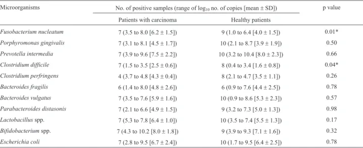
Patients with cancer were significantly older than pa-tients without cancer (65.4±1.1vs.54.8±1.3 years-old, p < 0.0001). Log10values [mean±standard deviation (SD)] for the copy number per gram of feces were calculated for each microorganism. The number of positive samples and the target copy numbers of each microorganism are shown in Table 2. Most of the evaluated microorganisms were de-tected in both the Cancer and Healthy Groups.
Statistically significant differences in the qRT-PCR detection limits ofF. nucleatumandC. difficilewere de-tected between the Cancer Group and Healthy Group. For
F. nucleatumthe detection limits were log103.5 and 1.0 copies for the Cancer and Healthy Group respectively (p = 0.01). ForC. difficilethe detection limits were log101.5 (Cancer Group) and log100.4 (Healthy Group) (p = 0.04).
Clostridium difficile was more prevalent than C. perfringens in cancer patients. We also detected signifi-cantly moreC. perfringensin the Cancer Group than in the Healthy Group (p = 0.04).
Bacteroides fragiliswas present in both the Cancer (log10 1.4 to log10 8.0) and Healthy Group (log10 0.9 to log10 7.6). B. vulgatus was present in all fecal samples, ranging from log103.5 to log107.6 in the Cancer Group and from log10 0.9 to log10 8.6 in the Healthy Group. P.
distasoniswas detected in all 7 of the cancer patients and in 9/10 healthy patients, with copy numbers ranging from log102.1 to 6.6 and log103.2 to 7.3, respectively.
Lactobacillus(log105.3 to 7.8) andBifidobacterium (log104.3 to 10.2) species were detected in all 7 cancer pa-tients. Lactobacillus was detected in 10/10, and
Bifidobacteriumin 9/10, of the healthy patients, with copy numbers ranging from log103.5 to 7.4 and log103.9 to 9.3, respectively.E. coliwas detected in all evaluated patients with values ranging from log102.8 to 9.5 (Cancer Group) and log101.7 to 9.5 (Healthy Group). We detected no signif-icant difference in theE. colicopy number between groups. Table 1- Species-specific oligonucleotides used to the bacterial quantitative detection.
Microorganisms Oligonucleotides Tm (ºC) Amplicon size (bp) References
5’®3’
Fusobacterium nucleatum F: CTT AGG AAT GAG ACA GAG ATG 56 120 Periasamyet al.(2009)
R: TGA TGG TAA CAT ACG AAA GG
Porphyromonas gingivalis F: ACC TTA CCC GGG ATT GAA ATG 60 83 Nonnenmacheret al.(2005)
R: CAA CCA TGC AGC ACC TAC ATA GAA
Prevotella intermedia F: CGT GGA CCA AAG ATT CAT CGG TGGA 55 259 Okamotoet al.(1999)
R: CCG CTT TAC TCC CCA ACA AA
Clostridium difficile F: ATT AGG AGG AAC ACC AGT TG 56 307 Kanget al.(2010)
R: AGG AGA TGT CAT TGG GAT GT
Clostridium perfringens F: TCA TCA TTC AAC CAA AGG AGC AAT CC 60 105 Siragusa and Wise (2007)
R: CCT TGG TAG GCC GTT ACC C
Bacteroides fragilis F: TCR GGA AGA AAG CTT GCT 56 162 Tonget al.(2011)
R: CAT CCT TTA CCG GAA TCC T
Bacteroides vulgatus F: GCA TCA TGA GTC CGC ATG TTC 60 287 Wanget al.(1996)
R: TCC ATA CCC GAC TTT ATT CCT T
Parabacteroides distasonis F: GTC GGA CTA ATA CCG CAT GAA 60 273 Wanget al.(1996)
R: TTA CGA TCC ATA GAA CCT TCA T
Lactobacillusspp. F: AGC AGT AGG GAA TCT TCC A 60 380 Ponnusamyet al.(2011)
R: ATT YCA CCG CTA CAC ATG
Bifidobacteriumspp. F: GCG TGC TTA ACA CAT GCA AGT C 60 125 Ponnusamyet al.(2011)
R: CAC CCG TTT CCA GGA GCT ATT
Escherichia coli F: AGA AGC TTG CTC TTT GCT GA 57 120 Leeet al.(2010)
Discussion
Intestinal diseases (e.g., inflammatory bowel disease and necrotizing enterocolitis) have been associated with a disequilibrium of the gastrointestinal microbiota and this has been addressed by several ecological studies applying culture-dependent and -independent methods. Several in-testinal microorganisms are capable of inducing the inflam-mation of gastrointestinal tissue. Interestingly, oral fuso-bacteria can migrate to extra-oral sites where they can cause inflammatory infections and have been found in high numbers in colorectal cancer (Han and Wang, 2013).
Fusobacterium nucleatumis the most commonly ob-served microorganism in the subgingival biofilm and is in-volved in periodontal diseases. This microorganism is con-sidered an important pro-inflammatory factor in the oral cavity (Signat et al., 2011). AlthoughF. nucleatum has been recently detected in colorectal carcinoma, its involve-ment in tumorigenesis remains to be determined (Castel-larinet al., 2012).F. nucleatumis highly capable of colo-nizing the human intestinal tract and it will therefore be important to verify whether the presence ofF. nucleatumin immune-compromised sites (e.g., colorectal cancer) repre-sents opportunistic infections.
Porphyromonas gingivalisandPrevotella intermedia
belong to oral microbiota and have been associated with several periodontal disease types (Socransky and Haffajee, 2005). Here we were able to detect P. gingivalisandP. intermediain the fecal samples of patients from both the Cancer and Healthy Group. These bacteria are fastidious under atmospheric conditions, however,P. gingivaliscan produce various fimbriae types that are associated with ad-herence to different cell surfaces (Sojaret al., 2002). In ad-dition, it is known thatP. gingivalisandP. intermediaare capable of several adaptive regulatory and metabolic
activi-ties that contribute to their pathogenicity (Sojar et al., 2002).
In a study by De Cruzet al.(2012), 6.8% of patients that had undergone colorectal cancer surgery were diag-nosed as havingC. difficile-associated colitis.Clostridium
species (e.g.,C. difficileandC. perfringens) in unbalanced ecosystem can produce intestinal inflammatory diseases in humans and animals. Here we report thatC. difficileis sig-nificantly abundant in both cancer patients and healthy con-trols (p = 0.04).
Bacteroides fragilis,B. vulgatusandP. distasonisare considered important commensal bacteria in the intestinal resident microbiota of humans and animals. Some species ofB. fragilisare able to produce an enterotoxin and these have been detected among strains isolated from colon can-cer patients, normal feces and extra-intestinal infections. Due to the lowB. fragiliscopy number found here, the pres-ence of enterotoxigenicB. fragiliswas not evaluated.
Species of Lactobacillus and Bifidobacterium are also commensal bacteria and represent less than 10% of the human oral and intestinal microbiota. These bacteria are considered the most numerous probiotics, and it is sug-gested that their contribution to the intestinal microbiota might be dependent on age and diet (Zoetendalet al., 2006).
Lactobacillusspp. are widely used as probiotics and there is an assumed interaction with the host via the binding of its extracellular pili to human mucus. However, the molecular details of the probiotics signaling mechanisms are not yet understood and it remains to be established whether the ef-fect is direct (e.g., through metabolites or structural compo-nents modulating the immune responses of the host) or indirect (via alteration of the intestinal microbiota) (Kan-kainenet al., 2009).
Species ofBifidobacteriumrepresent around 3% of the fecal microbiota (approximately 9.4 x 10⁹cells/g of fe-Table 2- Qualitative and quantitative analysis of oral and intestinal microorganisms from fecal samples.
Microorganisms No. of positive samples (range of log10no. of copies [mean±SD]) p value
Patients with carcinoma Healthy patients
Fusobacterium nucleatum 7 (3.5 to 8.0 [6.2±1.5]) 9 (1.0 to 6.4 [4.0±1.5]) 0.01*
Porphyromonas gingivalis 7 (3.1 to 8.1 [4.5±1.7]) 10 (2.1 to 8.7 [3.9±1.9]) 0.50
Prevotella intermedia 7 (3.9 to 9.6 [7.5±2.2]) 10 (3.2 to 10.4 [8.0±2.3]) 0.66
Clostridium difficile 7 (1.5 to 3.5 [2.5±0.6]) 8 (0.4 to 3.4 [1.6±0.8]) 0.04*
Clostridium perfringens 4 (3.7 to 4.8 [4.3±0.4]) 8 (2.1 to 4.7 [3.5±1.1]) 0.26
Bacteroides fragilis 6 (1.4 to 8.0 [4.8±2.6]) 6 (0.9 to 7.6 [4.4±2.5]) 0.78
Bacteroides vulgatus 7 (3.5 to 7.6 [5.9±1.6]) 10 (0.9 to 8.6 [5.3±2.3]) 0.57
Parabacteroides distasonis 7 (2.1 to 6.6 [4.9±1.5]) 9 (3.2 to 7.3 [5.0±1.3]) 0.98
Lactobacillusspp. 7 (5.3 to 7.8 [6.4±1.0]) 10 (3.5 to 7.4 [5.5±1.3]) 0.17
Bifidobacteriumspp. 7 (4.3 to 10.2 [8.0±1.8]) 9 (3.9 to 9.3 [7.1±1.6]) 0.32
Escherichia coli 7 (2.8 to 9.5 [6.7±2.4]) 10 (1.7 to 9.5 [6.4±2.5]) 0.78
ces) and are more prominent in the large intestine compared to the terminal ileum. Whereas Lactobacillus are more abundant in the distal intestine (proximal region of colon) than in the terminal ileum (Ahmedet al., 2007).
As a commensal microorganism of the human in-testinal microbiota, we expectedE. colito be present in all clinical samples. Although associations between mu-cosa-adherentE. coliand colorectal cancer have been re-ported (Arthuret al., 2012), we detected no significant difference in E. coli abundance between the patient groups.
Since carcinogenesis is a lengthy and multifactorial process, factors such as diet, habit, ethnicity and environ-mental exposure might participate in the cancer process (Arthuret al., 2012). Surprisingly, we detected high occur-rences of the three evaluated oral microorganisms in our patients. Based on these findings, future studies (i.e., obser-vational and longitudinal studies) should focus on potential roles for these bacteria (especiallyF. nucleatum) during colorectal carcinoma pathogenesis.
Despite the small number of patients evaluated in this study, we detected statistically significant differences in the abundance ofF. nucleatumandC. difficilebetween healthy and cancer patients, suggesting a possible role of these bacteria in colon carcinogenesis. This finding should be considered when designing screens for colo-rectal cancer.
Acknowledgments
This study was supported by Fundação de Amparo à Pesquisa do Estado de São Paulo (FAPESP Grant 10/52417-4) and CAPES-PNPD (2472/09-0).
References
Ahmed S, MacFarlane GT, Fite Aet al. (2007) Mucosa-asso-ciated bacterial diversity in relation to human terminal ileum and colonic biopsy samples. App Environ Microbiol 73:7435-7442.
Arthur JC, Perez-Chanona E, Muhlbauer Met al.(2012) Intestinal inflammation targets cancer-inducing activity of the micro-biota.Science 338:120-123.
Castellarin M, Warren RL, Freeman JD et al. (2012) Fusobacterium nucleatuminfection is prevalent in human colorectal carcinoma. Genoma Res 22:299-306.
Collins D, Hogan AM, Winter DC (2010) Microbial and viral pathogens in colorectal cancer. Lancet Oncol 12:504-512.
De Cruz P, Prideaux L, Wagner JET AL. (2012) Characterization of the gastrointestinal microbiota in health and inflamma-tory bowel disease. Inflamm Bowel Dis 18:758-777.
Frank DN, St Amand AL, Feldman RAet al.(2007) Molecu-lar-phylogenetic characterization of microbial community imbalances in human inflammatory bowel diseases. Proc Natl Acad Sci USA 104:13780-1385.
Han YW, Wang X (2013) Mobile microbiome: Oral bacteria in extra-oral infections and inflammation. Cell Host Microbe 14:195-206.
Ivanov II, Littman DR (2011) Modulation of immune homeostasis by commensal bacteria. Curr Opin Microbiol 14:106-114. Kang S, Stuart E, Denman SEet al.(2010) Dysbiosis of fecal
microbiota in Crohn’s disease patients as revealed by a cus-tom phylogenetic microarray. Inflamm Bowel Dis 16:2034-2042.
Kankainen M, Paulin L, Tynkkynen Set al.(2009) Comparative genomic analysis ofLactobacillus rhamnosus GGreveals pili containing a human-mucus binding protein. Proc Natl Acad Sci USA 106:17193-17198.
Lee DH, Bae JE, Lee JHet al.(2010) Quantitative detection of re-sidualEscherichia colihost cell DNA by Real-time PCR. J Microbiol Biotechnol 20:1463-1470.
McLean MH, Murray GI, Stewart KNet al.(2011) The inflamma-tory microenvironment in colorectal neoplasia. PloS One 6:15366.
Nakano K, Inaba H, Nomura Ret al.(2007) Detection and sero-type distribution ofAggregatibacter actinomycetemcomitans in cardiovascular specimens from Japanese patients. Oral Microbiol Immunol 22:136-139.
Nonnenmacher C, Dalpke A, Rochon Jet al.(2005) Real-time polymerase chain reaction for detection and quantification of bacteria in periodontal patients. J Periodontol 6:1542-1549.
Okamoto M, Maeda N, Kondo Ket al.(1999) Hemolytic and hemagglutinating activities of Prevotella intermedia and Prevotella nigrescens. FEMS Microbiol Lett 178:299-304. Periasamy S, Chalmers NI, Du-Thumm Let al. (2009).
Fuso-bacterium nucleatum ATCC 10953 requires Actinomyces naeslundii ATCC 43146 for growth on saliva in a three-species community that includesStreptococcus oralis.Appl Environ Microbiol 75:3250-3257.
Ponnusamy K, Choi JN, Kim Jet al.(2011) Microbial community and metabolomic comparison of irritable bowel syndrome faeces. J Med Microbiol 60:817-827.
Signat B, Roques C, Poulet Pet al.(2011) Role ofFusobacterium nucleatumis periodontal health and disease. Curr Issues Mol Biol 13:25-35.
Siragusa GR, Wise MG (2007) Quantitative analysis of the intes-tinal bacterial community in one- to three-week-old com-mercially reared broiler chickens fed conventional or antibi-otic-free vegetable-based diets. J Appl Microbiol 102:1138-1149.
Socransky SS, Haffajee AD (2005) Periodontal microbial ecol-ogy. Periodontol 2000 38:135-187.
Sojar HT, Sharma A, Genco RJ (2002)Porphyromonas gingivalis fimbriae bind to cytokeratin of epithelial cells. Infect Immun 70:96-101.
Strauss J, Kaplan GG, Beck PLet al.(2011) Invasive potential of gut mucosa-derive Fusobacterium nucleatum positively correlates with IBD status of the host. Inflamm Bowel Dis 17:1971-1978.
fragilisgroup and related organisms in human wound sam-ples. Anaerobe 17:64-68.
Wang RF, Cao W, Cerniglia CE (1996) PCR Detection and quantitation of predominant anaerobic bacteria in human and animal fecal samples. App Envir Microbiol 4:1242-1247.
World Health Organization (2011) Fact Sheet no. 297. World Health Organization, Geneva, Switzerland.
http://www.who.int/mediacentre/factsheep/fs297/en/. Available at: Accessed December, 2011.
Zoetendal EG, Vaughan EE, de Vos WM (2006) A microbial world with us. Mol Microbiol 59:1639-1650.
Associate Editor: Marina Baquerizo