THE EXPLORATION OF THE ANTIBACTERIAL MECHANISM OF FE3+ AGAINST BACTERIA
Hong-qi Sun, Xue-mei Lu, Pei-ji Gao*
State Key Laboratory of Microbial Technology, Shandong University, Jinan 250100, China.
Submitted: June 06, 2009; Approved: June 21, 2010.
ABSTRACT
This study demonstrated that the bacteria could adsorb Fe3+ and reduce Fe3+ to Fe2+. Iron had significant
bacteriostatic effects, which were directly proportional to the iron concentration and under the influence of
pH and chelator. It presumed that the inhibition of Fe3+ acts through the formation of hydroxyl free
radicals.
Key words: antibacterial activity, chelator, hydroxyl radical, ferric ion, siderophore
Iron is a nutritionally essential trace element and has
important functions in the metabolic processes of aerobic
organisms, such as photosynthesis, respiration, oxygen
transport, gene regulation and DNA biosynthesis (1, 3). Under
physiological conditions, iron mainly exists in two readily
reversible redox states, the reduced Fe2+ form and the oxidized
Fe3+ form. Although abundant in nature, iron tends to form
highly insoluble hydroxides in the aerobic neutral pH
environment. The concentration of free ferric ion in solution at
biological pH is probably 10-9-10-18 mol l-1 (2, 5, 9), a
concentration too low to allow growth by aerobic
microorganisms. Microorganisms have evolved a range of
strategies to acquire iron. The major strategies include
production and utilization of siderophores, utilization of host
iron proteins such as transferrin, and lactoferrin, and reduction
of Fe3+ to Fe2+ with subsequent transport of Fe2+ (4). On the
other hand, iron is a devastating metal. Fe2+ reacts with H2O2 to
form a hydroxyl radical (6). HO· is known to be a highly
reactive, indiscriminate oxidizing agent, which can damage
proteins and nuclear acids. Thus, aerobic organisms have a
dilemma: they need a scavenging system that is effective in
accumulating iron, without allowing too much to accumulate.
Much attention has been given to the response of bacteria to
iron-limited conditions, but not to how the bacteria respond to
adequate or excess iron supply. The growth of bacteria usually
requires 10-6 mol l-1 of iron, and the proper amount of iron can
stimulate the growth of bacteria, but under excessive
concentrations, the microorganism's growth will be inhibited.
In this article, we studied the adsorption and reduction of 2
Gram-negative bacteria (Escherichia coli CVCC 249 and
Ralstonia solanacearum) and 2 Gram-positive bacteria (Staphylococcus aureus ATCC 25923 and Bacillus subtilis) to Fe3+ under excess Fe3+ conditions. The antibacterial effects of
Fe3+ were investigated and we conducted a preliminary
analysis of the inhibition mechanism of Fe3+.
Each bacterial strain was inoculated into 100 ml LB broth
and incubated at 37°C with shaking at 170 rpm for 12 h. The
culture was centrifuged and washed twice with sterile
physiological saline, then the pellet was resuspended in
appropriate amount of sterile physiological saline and the
turbidity of the samples was measured at 600 nm using a
UNICO 2000 UV-Vis spectrophotometer, made the turbidity
values were 2.0. A sample volume of 5 ml of bacteria
suspension was centrifuged and the cell was resuspended into 5
ml of FeCl3 solution (1 mmol l-1, containing 2.5 mmol l-1 HCl)
and then incubated at 37°C, with shaking at 170 rpm for 2 h.
Similarly, the 5 ml bacteria suspension containing 5 ml FeCl3
solution (1, 0.5 and 0.25 mmol l-1, containing 2.5 mmol l-1 HCl,
respectively) was incubated at room temperature with shaking
for 0-45 min at every 5 min interval and then it was centrifuged
the supernatant was collected. The ferrous and ferric ion
concentrations in the supernatant were measured using 1,
10-phenanthroline spectrophotometry at 510 nm and 364 nm,
respectively (8, 12). A volume of 1 ml hydroxylamine
hydrochloride (w/v 5%) was added to 1 ml of supernatant,
incubated 15 min, and then mixed with 1 ml 1,
10-phenanthroline (m/v, 0.25%), and 1 ml sodium acetate buffer
(0.5 mol l-1, pH 4.5), and then added to deionized water to
make a total volume of 5 ml. The mixture was further
incubated at room temperature for about 30 min, after which
the absorbance at 510 nm was measured to determine the total
iron content in the supernatant. The hydroxylamine
hydrochloride was replaced with the same amount of deionized
water and the absorbance at 510 nm and 364 nm were
measured to characterize the Fe2+ and Fe3+ ion content,
respectively. A solution of 1 mmol l-1 FeCl3 was used as a
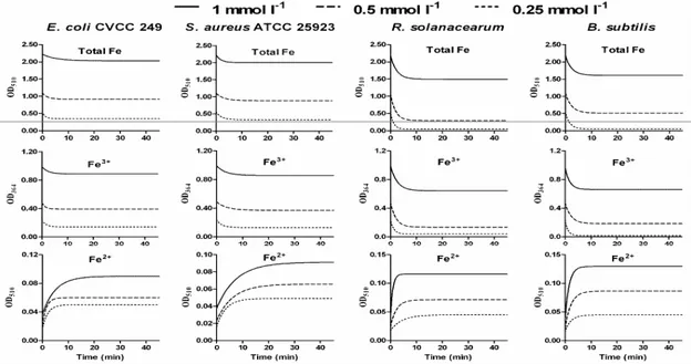
control. The results showed that all four bacteria could adsorb
Fe3+. R. solanacearum had the strongest adsorptive capacity for Fe3+ at about 50%, followed by B. subtilis and S. aureus ATCC 25923 which retained about 30% while the absorption capacity
of E. coli CVCC 249 for Fe3+ was the weakest at close to 15%. As can be seen from Figure 1, the iron absorption of the 4
bacteria reached equilibrium in 5-10 min and thus, the
adsorption was an extremely rapid process. The four bacteria
had similar absorption dynamics curves of total iron and Fe3+.
For E. coli CVCC 249, S. aureus ATCC 25923 and B. subtilis
bacteria, the Fe3+ adsorption could achieve saturation when the
Fe3+ concentration was 0.25 mmol l-1, but for R. solanacearum, saturation could only be reached when the concentration
increased to 0.5 mmol l-1. This indicated that R. solanacearum
had the strongest Fe3+ adsorptive capacity. The results also
indicated that the decrease of the total iron and Fe3+ content
was accompanied by the generation of Fe2+. The dynamics
curve of the Fe2+ production was similar for the 4 bacteria.
Moreover, the Fe2+ production of R. solanacearum and B. subtilis was higher than E. coli CVCC 249 and S. aureus
ATCC 25923, which corresponded directly with their Fe3+
adsorptive capacity.
To investigate the antibacterial effects of high
concentrations of ferric iron, the appropriate amount of FeCl3
was sterilized using ultraviolet radiation and then it was
combined with sodium hydrogen phosphate - citric acid buffer
solutions (pH 4.0, 5.0, 6.0, and 7.0, prepared using 10 mmol l-1
sodium hydrogen phosphate and 5 mmol l-1 citric acid,
separately and then sterilized at 121°C for 30 min.) or sterile
water to compound the FeCl3 solutions separately. It was
prepared just before use and diluted to the necessary
concentrations. The bacterial culture was appropriately diluted
with sterile saline and then mixed with different concentrations
and pHs of FeCl3 solution at room temperature for 10, 20, 30,
and 40 min, respectively. A volume of 100 l of the solution
was spread onto a LB plate. Plates were incubated at 37°C
overnight, and colonies on the plates were counted. Samples
were plated in triplicate and sterile saline was used as a
negative control. The results illustrates that the different
concentrations of FeCl3 aqueous solutions (without buffer) had
definite antibacterial activities. The bactericidal capacity of
Fe3+ increased with the increasing concentration of Fe3+.
Additionally, the inhibitory effect was an extremely rapid
process. In 10 min, 70% of E. coli CVCC 249 bacteria were inhibited by 1 mmol l-1 Fe3+, and 90% of S. aureus ATCC
25923 were inhibited at the same concentration. Both R. solanacearum and B. subtilis were completely inhibited when the Fe3+ concentration was only 250 mol l-1 and 62.5 mol l-1,
respectively. The antibacterial results of four pH Fe3+ solutions
on the growth of the bacteria. The four different pH buffer
solutions did not obviously inhibited E. coli CVCC 249 and S. aureus ATCC 25923. However, for R. solanacearum and B. subtilis, they had varying degrees of inhibition while a solution with a pH value of 4.0 could suppress their growth completely.
For solutions with pH values of 4.0, 5.0, and 6.0, 1 mmol l-1
Fe3+ could suppress the growth of E. coli CVCC 249, while at 0.5 mmol l-1 Fe3+ no inhibitory effects were observed. For the
pH 7.0 solution, only the 2 mmol l-1 Fe3+ displayed inhibitory
effects. Under all pH conditions, only 2 mmol l-1 Fe3+ could
obviously inhibit the growth of S. aureus ATCC 25923. For solutions with pH values of 5.0 and 6.0, 500 mol l-1 and 250
mol l-1 Fe3+ had obvious inhibitory effects on the growth of R. solanacearum and the B. subtilis, respectively. When the pH value was 7.0, the inhibitory effects weakened. By comparison
of these results, it was determined that the antibacterial effects
of the buffered Fe3+ solutions were weaker than those of the
Fe3+ aqueous solutions. Thus, it is necessary that higher
concentrations of FeCl3 were needed in buffered solutions in
order to achieve the same antibacterial efficiency. During the
preparation of the fresh FeCl3 solution, the hydrolysis state of
ferric ion changes continuously. Depending on the pH of the
sodium hydrogen phosphate - citric acid buffer solution, the
Fe3+ will be in different hydrolysis forms which have
correspondingly different relative antibacterial capacities. At
pH 7, the concentration of free ferric ion is 1.4 × 10⁻⁹ mol l-1 in
the aqueous solution and the main species is Fe(OH)2+. In the
presence of 40 mmol l-1 phosphate, the concentration of free
ferric ion is decreased to 3.3 × 10⁻¹² mol l-1 because of the
formation of ferric phosphate (Chipperfield and Ratledge
2000). Depending on the different pH conditions, the
hydrolysis of Fe3+ and the formation of ferric phosphate
complexes results in a decline in the free Fe3+ content, which
make the antibacterial activity of the iron weaker. The
sensitivities of the various bacteria to the ferric iron were
different, but the bacteriostasis dynamics curves were the same.
This indicates that the antibacterial effect of Fe3+ is likely a
non-selective mechanism.
The FeCl3 solution was mixed with three different chelator
solutions, acetohydroxamic acid (ICN Biomedicals), 2,
3-dihydroxybenzoic acid (Acros Organics) or a siderophore
which produced by Aspergillus niger An76 (11) for 2 min, and then added into a bacterial suspension at room temperature for
10 min, respectively. In addition, the FeCl3 solution was added
into the bacterial suspension at room temperature for 5 min and
then mixed with the above three chelator solutions for 5 min,
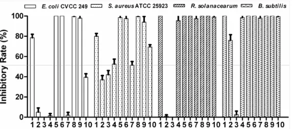
respectively. Results of the comparison between the
antibacterial activity of Fe3+ and chelators are shown in Figure
2. Solutions of Fe3+ alone displayed remarkable bacteriostasis.
min treatment time, and 83 mol l-1 Fe3+ could completely
suppress the growth of R. solanacearum and B. subtilis. All chelators had no obvious inhibitory effects on E. coli CVCC 249, but did inhibit S. aureus ATCC 25923. 2, 3-Dihydroxybenzoic acid could strongly inhibit the growth of B. subtilis. Acetohydroxamic acid showed a certain extent inhibitory effect for S. aureus ATCC 25923, but for other three bacteria did not obvious inhibitory action. Aspergillus niger
An76 siderophore can completely inhibit R. solanacearum and
B. subtilis growth. When 2, 3-dihydroxybenzoic acid or acetohydroxamic acid was combined with Fe3+ together, it was
determined that both of the chelators could enhance the Fe3+
inhibition of E. coli CVCC 249 and S. aureus ATCC 25923 regardless of whether they were added before or after the Fe3+
solutions were mixed with the bacterial suspension. But the
An76 siderophore could reduce the Fe3+ inhibition of E. coli
CVCC 249 and S. aureus ATCC 25923.
Figure 2. Comparison of the antibacterial effects of Fe3+ and chelators. Notes: 1: FeCl3; 2: 2, 3-dihydroxybenzoic acid; 3:
acetohydroxamic acid; 4: A. niger An76 siderophore; 5, 6, 7: First, FeCl3 with 2, 3-dihydroxybenzoic acid or acetohydroxamic
acid or A. niger An76 siderophore were mixed for 2 min, respectively; 8, 9, 10: After FeCl3 treatment for 5 min, it was mixed with
2, 3-dihydroxybenzoic acid or acetohydroxamic acid or A. niger An76 siderophore for 5 min, respectively. The final concentrations of FeCl3, 2, 3-dihydroxybenzoic acid, and acetohydroxamic acid were 1 mmol l-1 for E. coli CVCC 249 and S. aureus ATCC 25923, and those were 83 mol l-1 for R. solanacearum and B. subtilis. The final concentrations of A. niger An76 siderophore were 1 mg/ml for E. coli CVCC 249 and S. aureus ATCC 25923, and those were 100 g/ml for R. solanacearum and
B. subtilis.
Acetohydroxamic acid and 2, 3-dihydroxybenzoic acid can
form complexes with Fe3+, which cause the hydrolysis of ferric
ion to be suppressed. One molecule of Fe3+ can bind to the 2,
3-dihydroxybenzoic acid between the acid group and a
neighboring hydroxyl group and form an iron complex that
subsequently reduces an additional Fe3+ (13). The 2,
3-dihydroxybenzoic acid – Fe3+ complex can catalyze the
generation of hydroxyl free radicals which reach a maximum at
about a 1:1 ratio (10). The Fenton reagent, which is a complex
of hydrogen peroxide and an iron catalyst, has a strong
sterilization action, which has been proven to work through
HO·. In our study, 4 bacteria were able to rapidly reduce Fe3+
to Fe2+. Moreover, H2O2 is a normal metabolite in aerobic and
facultative aerobic organisms (7). Our previous research
demonstrated that the siderohpre produced by Aspergillus niger
radicals (11). Therefore, it is presumed that pure Fe3+ does not
have the antibacterial effect alone, but instead, it becomes
effective as an inhibitor after the first reduction of Fe3+ to Fe2+,
and further oxidation with oxidized factors, such as superoxide
radical and hydrogen peroxide, with the ultimate formation of
hydroxyl free radicals which have a strong non-selective
sterilization ability.
The formation of drug resistant microorganisms remains a
difficult medical problem. Non-selective sterilization would be
a useful technique to combat drug resistance and the
information provided herein should provide knowledge
towards making a disinfectant using iron chelators and iron.
However, the interaction of iron ions with chelating agents and
their inhibitory mechanism still needs more in-depth research.
ACKNOWLEDGEMENTS
This work was supported by the Natural Science
Foundation of Shandong Province (Grant No. Y2004D07).
REFERENCES
1. Braun, V. (1997) Surface signaling: novel transcription initiation mechanism starting from the cell surface. Arch. Microbiol. 167, 325-331.
2. Chipperfield, J.R.; Ratledge, C. (2000) Salicylic acid is not a bacterial siderophore: a theoretical study. Biometals. 13, 165-168.
3. Crichton, R.R. (1991) Inorganic biochemistry of iron metabolism. Ellis Horwood, West Sussex.
4. Guerinot, M.L. (1994) Microbial iron transport. Anun. Rev. Microbiol. 48, 743-772.
5. Haas, H. (2003) Molecular genetics of fungal siderophore biosynthesis and uptake: the role of siderophores in iron uptake and storage. Appl. Microbiol. Biotechnol. 62, 316-330.
6. Halliwell, B.; Gutteridge, J.M.C. (1989) Free Radical in Biology and Medicine. 2nd edn. Clarendon, Oxford.
7. Howard, D.H. (1999) Acquisition, transport, and storage of iron by pathogenic fungi. Clin. Microbiol. Rev. 12 (3), 394-404.
8. Institute of Health, Chinese Academy of Sciences (IHCAS) (1983) Analytical method for water quality. People’s Medical, Beijing. 9. Neilands, J.B. (1995) Siderophore: Structure and function of microbial
iron transport compounds. J. Biol. Chem. 270, 26723-26726.
10. Qian, Y.; Goodell, B.; Felix, C.C. (2002) The effect of low molecular weight chelators on iron chelation and free radical generation as studied by ESR measurement. Chemosphere. 48, 21-28.
11. SUN, H.; ZHANG, W.; LU, X.; Gao, P. (2008) Siderophore production from 27 filamentous fungal strains and a novel siderophore with potential biocontrol applications from Aspergillus niger An76. J. Life. Sci. 2(1), 19-26.
12. Tesfaldet, Z.O.; Van Staden, J.F.; Stefan, R.I. (2004) Sequential injection spectrophotometric determination of iron as Fe (II) in multi-vitamin preparations using 1,10-phenanthroline as complexing agent. Talanta. 64, 1189-1195.