Recebido para publicação em 28/09/2014 Aceito para publicação em 12/02/2015
Antibacterial activity and chemical compounds of leaves and branches of
Protium
hebetatum
CONRADO, G.G.1; SIMPLICIO, F.G2*; COSTA, K.R.C3; REHDER, V.L.G1; ESPINAR, M.F.4; SOUZA, G.O.2; SAMPAIO, P.T.B.5
1Divisão de Química Orgânica e Farmacêutica, Centro Pluridisciplinar de Pesquisas Químicas, Biológicas e Agrícolas-CPQBA, Rua Alexandre Cazelatto, 999, Vila Betel, CEP 13081-970, Paulínia-SP, gabrielly_conrado@ hotmail.com, [email protected]; 2Laboratório de Produtos Naturais, Universidade Federal do Amazonas, Rua Alexandre Amorim, 330, Aparecida, CEP 69103-00, Amazonas-AM, fgsfarmaceutica@gmail. com, [email protected]; 3Laboratório de Microbiologia, Universidade Federal do Amazonas, Rua Afonso Pena, 1053, Centro, CEP 69020-160, Amazonas-AM, [email protected]; 4Laboratório de Produtos Naturais, Instituto Nacional de Pesquisas da Amazônia, Av. André Araújo, 2.936, Petrópolis, CEP 69067-375, Amazonas-AM, [email protected]; 5Laboratório de Propagação de Plantas, Instituto Nacional de Pesquisas da Amazônia; Av. Efigênio Sales, 2239, Aleixo, CEP 69067-375, Amazonas-AM, sampaio@inpa. gov.br. *[email protected].
ABSTRACT: The extracts and fractions of leaves and branches of Protium hebetatum D. C. Daly (Burseraceae) were investigated for their antibacterial activity and chemical composition. The methanol extract of branches (EMG) was considered active against the Escherichia coli and the Proteus vulgaris, showing an inhibition zone of 13 mm, and was selected for bioassay-guided phytochemical fractionation. From the technique of broth microdilution, the extract was considered a moderate inhibitor against Staphylococcus aureus, Pseudomonas aeruginosa and Enterococcus faecalis, with a minimum inhibitory concentration (MIC) of 1 mg/mL. The dichloromethane fraction was considered a moderate inhibitor against S. aureus (MIC of 1 mg/mL) and a potent inhibitor against E. faecalis (MIC of 0.5 mg/mL). F1, F2, F5 and F6 from chromatographic column of dichloromethane fraction were considered moderate inhibitors against S. aureus (MIC of 1 mg/mL). Through analysis by a gas chromatography mass spectrometry, eighteen compounds were identified, from which thirteen (isoeugenol, p-vinylguaiacol, metoxyeugenol, coumarin, 5-hydroxy-scopoletin, 4,7-dihydroxy-6-metoxicromam-2-one, 4[(1E]-3-hydroxy-1-propenyl)-2-methoxyphenol, piperonal, scoparon, o-guaiacol, spathulenol, seringol
and antiarol) are unprecedented in these species. We also identified the triterpenes α-amyrin and β-amyrin, the steroids stigmasterol and sitosterol and the coumarin scopoletin, which was closely linked to the antibacterial activity of the samples.
Keywords: Protium hebetatum; antibacterial activity, coumarins, Staphylococcus aureus, Enterococcus faecalis.
RESUMO: Atividade antibacteriana e compostos químicos de folhas e galhos de Protium hebetatum. Extratos e frações de folhas e galhos de Protium hebetatum D. C. Daly (Burseraceae) foram investigados quanto sua atividade antibacteriana e composição química. O extrato metanólico dos galhos (EMG) foi considerado ativo contra Escherichia coli e Proteus vulgaris, apresentando um halo de inibição de 13 mm, sendo selecionado para um
fracionamento fitoquímico biomonitorado. A partir da técnica de microdiluição em caldo o EMG
foi considerado um inibidor moderado contra Staphylococcus aureus, Pseudomonas aeruginosa e Enterococcus faecalis, apresentando uma concentração inibitória mínima (CIM) de 1mg/mL. A fração diclorometânica foi considerada inibidora moderada contra S. aureus (CIM de 1 mg/ mL) e inibidora potente contra E. faecalis (CIM de 0,5 mg/mL). F1, F2, F5 e F6 provenientes da fração diclorometânica foram consideradas inibidoras moderadas contra S. aureus (CIM
de 1 mg/mL). Através da análise por cromatografia gasosa acoplada a espectrometria de massa, foram identificados dezoitos compostos, dos quais treze (isoeugenol, p-vinilguaiacol,
INTRODUCTION
Among the numerous species of plants used in popular medicine in Brazil, the species of the Protium genus are noteworthy, as this is the largest and most heterogeneous of the Burseraceae family, with about 150 species, 80% of which occur in the North of country, where a large portion of the Amazon rainforest is located (Siani et al., 2004). Species of the Burseraceae family are characterized by their resin exudate, popularly known as “breus”, widely used in local popular medicine for its
anti-inflammatory, analgesic, expectorant, wound-healing
and insect repellent properties (Bandeira et al., 2002; Maia et al., 2000), besides being used as fixative in artistic inks and in the manufacture of cosmetics, toiletries and perfumes (Ramos et al., 2000).
The extensive popular use of these species has promoted several scientific studies, which have intensified in recent years, leading to the
identification of several interest pharmacological
substances (Costa et al., 2012), as well as evidence of various therapeutic activities. Based on the ethnomedicinal use, it was found that ethyl ether extract of the resin of Protium kleinii showed
excellent results its potent anti-inflammatory activity
in topical use (Otuki et al., 2005). Pharmacologic
studies with triterpenes α and β-amyrin isolated
from extracts of the leaves and resin of species P. heptaphyllum demonstrated analgesic (Holanda Pinto et al., 2008), anti-inflammatory (Susunaga et al., 2001), antidepressant (Aragão et al., 2006), gastroprotective (Oliveira and Amaral, 2004) and cytotoxic (Taylor et al., 2012) activities. Another study with P. glabrescens resin demonstrated potent antimalarial activity for the species (Deharo etal., 2001).
In terms of chemical constitution, the literature reports the presence of triterpenes in resins of P. grandifolium and P. subserratum, and in bark and stem extracts of P. paniculatum and P. hebetatum, respectively, and the presence of steroids and coumarins in extract of P. hebetatum. Overall, triterpenes represent the most frequently occurring class of natural substances in the Protium genus (Costa et al., 2012; Silva et al., 2009; Zoghbi et al., 1993). Nevertheless, their antibacterial potential
has been poorly investigated. A study of the essential oils extracted from the leaves, fruits, stems and bark of P. confusum showed that the essential oil extracted from the leaves demonstrated the best antibacterial activity against Staphylococcusaureus and Mycobacterium smegmatis (Santana et al., 2009). On the other hand, Protium hebetatum D. C. Daly, in which triterpenes and coumarins were
previously identified, has not yet been investigated
with this approach.
It is known than the continuous emergence of new mechanisms of bacterial resistance has generated a rush of pharmaceutical industries for the production and launch of new antimicrobial compounds that act by a different action mechanism
drugs in use (Bérdy, 2012). Natural products and
derived drugs have been crucial for the discovery of anti-infection drugs, and many of the antibacterial drugs currently in clinical use are natural products, or were designed using natural products as templates (Mishra and Tiwari, 2011).
Thus, considering the urgency of the search for new drugs for the treatment of bacterial infections, and the recognized antimicrobial activity of various plant extracts, this study conducted a bioassay-guided phytochemical study of antibacterial activity of extracts and fractions obtained from leaves and branches of the species Protium hebetatum against bacteria of medical interest, seeking to identify substances with antibacterial activity, as alternatives to the present therapeutic arsenal.
MATERIALS AND METHODS Chemicals
Hexane, ethyl acetate, dichloromethane (Nuclear), methanol (Vetec) and equipment were provided of the Natural Products Laboratory of the Universidade Federal do Amazonas (UFAM). All other chemicals used in this experiment were purchased from the local chemical suppliers in Manaus.
Microorganism
Standard strains Enterococcus faecalis metoxieugenol, cumarina, 5-hidroxi-escopoletina, 4,7-dihidroxi-6-metoxicromam-2-ona,
4[(1E]-3-hidroxi-1-propenil)-2-methoxifenol, piperonal, escoparona, o-guaiacol, espatulenol, seringol
e antiarol) foram identificados pela primeira vez nesta espécie. Foram também identificados os triterpenos α-amirina e β-amirina, os esteroides estigmasterol e sitosterol e a cumarina escopoletina, que estão intimamente ligados à atividade antibacteriana da espécie.
CCCD-E002, Escherichia coli E004-CCCD, Klebsiella pneumoniae CCCD-K003, Salmonella enterica S001-CCCD, Staphylococcus aureus ATCC 25923, Proteus vulgaris CCCD-P002, Pseudomonas aeruginosa P004 CCCD were commercially obtained in lyophilized form from the company Didactic SP. The culture media was provided of the Microbiology Laboratory of the UFAM.
Plant material
The collection of vegetal samples was authorized by the Instituto Chico Mendes de Conservação da Biodiversidade (ICMBio), chaired by the Ministry of Environment of Federative Republic of Brazil, under registration number
41553-1. Leaves and branches of five individuals of Protium hebetatum were collected in the Reserva Florestal Adolpho Ducke Manaus – AM (forestry reserve), in the following code and coordinates: 1 (S 02°55’97’’ W 59°58’57’’, ALT 78 m), 2 (S 2°55’93’’ W59°58’53’’, ALT 65 m), 3 (S 02°55’96’’ W 59°58’48’’, ALT 93 m), 4 (S 2°55’96’’ W 59°58’48’’, ALT 59 m), 5 (S 02°55’97’’
W 59°58’47’’, ALT 72 m), and taxonomically identified
by Dr. Mike Hopkins of the Herbarium of the Instituto Nacional de Pesquisas da Amazonia – INPA, where a voucher specimen was deposited for each individual under numbers 251968, 251969, 251970, 251971 e 251972, respectively.
Preparation of extracts
Leaves and branches (nodes and internodes) were dried at room temperature for 7 days followed by a period of 24 hours at 40ºC in an oven (WHO, 2003), then pulverized using a mechanical grinder equipped with a 0.5 mm sieve. The extracts were obtained by maceration using 480 g leaves and 638 g of branches and portions of 500 mL of hexane (total of 7 L), ethyl acetate (total of 6 L) and methanol (total of 8 L), in cycles of 15 min, in an ultrasound bath (Ultrasonic Cleaner Unique ®), sequentially. The new portions of solvents were added to residue of the previous extraction and the polarity of the solvents was altered until the volumes described above were achieved. The solvents were eliminated in a rotary evaporator (IKA ® RV10 rotaevaporator basic) with a pressure of 500 mmHg, at 45°C and 110 rpm, yielding the respective extracts, which were kept under refrigeration (8 ºC) until the time of use (Simões et al., 2007).
Fractionation from the methanolic extract of the branches of P. hebetatum
Fractionation of the methanolic extract of the branches was performed by liquid-liquid partition (Collins et al., 2006). For this purpose, 35 g of the extract was solubilized in 500 ml of methanol:water 1:1 and extracted with 400 mL
of hexane, dichloromethane and ethyl acetate, sequentially. The fractions were dried in a rotary evaporator (IKA® RV10 rotaevaporator basic),
providing the following fractions: FHMEB, FDMEB and FEAMEB. The solvent residual fraction, hydromethanol (FHMEB) was eliminated using a spray dryer apparatus. In the second stage of fractionation, 480 mg of FDMEB was fractionated by column chromatography (Collins et al., 2006) using a glass column of (32 cm high and 2 cm diameter as support, and 40.31 g of silica gel 60 (70-230 mesh, VETEC) as stationary phase. The column was initially eluated with dichloromethane and ethyl acetate (60:40 v/v), with a gradual increase in polarity toward 100% methanol. The obtained fractions were analyzed by thin layer chromatography (TLC) using silica gel 60 (ALUGRAM® Xtra SIL G, Macherey-Nagel) and grouped according to their similarities, totaling 10 subfractions.
Preparative Thin Layer Chromatography (PTLC)
The separation of active substances from samples obtained in item 2.5, in the Bioautography test was performed by preparative thin layer chromatography (PTLC). Using micro-syringes, 0.5 mL of a solution at 10 mg/mL of each sample were applied to a plate of silica gel 60 of 8 x 10 cm (ALUGRAM® Xtra SIL G, Macherey-Nagel), which was eluted with hexane and ethyl acetate 50:50 v/v. A portion of 1 cm of the plate was cut and revealed with sulfuric anisaldehyde solution, followed by heating in an oven at 100°C to visualize and calculate the retention factors of the spots. The regions corresponding to the spots in the untreated portion of the plate were then cut into small pieces and transferred to glass vials. The substances in silica were extracted with ethyl acetate with the aid of an ultrasound bath (Ultrasonic Cleaner Unique®). The
solutions were then filtered with cotton and dried in a
rotary evaporator (IKA® RV10 basic rotaevaporator) and sent for gas chromatography analysis.
Gas chromatography coupled to mass spectrometry (GC-MS) analyzes
The identification of active substances was performed on a gas chromatograph Agilent, HP-6890 model equipped with a mass selective
detector (HP-5975 model, Agilent) using a finjector = 250° C; column = 110° C; heating rate of 5° C.min-1;∙ temperature to 280° C (26 min); detector = 300° C. Helium was used as carrier gas at a flow rate of 1
mL min-1. Mass selective detector was operated at
Qualitative Test of antibacterial activity - Agar Diffusion
The strains were initially hydrated in Brain Heart Infusion Broth (BHIB - Himedia) and incubated at 37°C for 24 h for reactivation. A small fraction of growth colonies was withdrawn and diluted in sterile saline solution (0.9% NaCl) to obtain a standard inoculum of turbidity on a scale 0.5 to Mac Farland,
equivalent to a final concentration of 1.0 x 10⁸ CFU/
mL (NCCLS, 2003). Antibacterial tests of extracts and fractions of the P. hebetatum obtained by liquid-liquid partition were performed in triplicate by the agar diffusion method, according to the protocol proposed by the Clinical and Laboratory Standards Institute (CLSI, 2009) with slight modifications. The microorganisms were inoculated in a laminar
flow hood, where for each 25 mL of media
Mueller-Hinton Agar (AMH - Himedia) molten, 500µL of
standard inoculum was added. After solidification
of the agar, a circular cavity of 6 mm diameter were made, at points equidistant from the plate, to which was added 20 µL of the extracts and partitions at a concentration of 4 mg/mL per well. Gentamicin at a concentration of 10 µg/mL was used as positive control and dimethylsulfoxide (DMSO) as solvent of the samples and negative control. The plates were incubated at 37°C for 24 h. After the incubation period the antibacterial activity was determined by measuring the inhibition zone (mm) and analyzed as follows (Alves et al., 2000): inactive (<9mm), little active (9-12mm), active (13-18mm) and very active (> 18mm).
Determination of Minimum Inhibitory Concentration (MIC) - Broth Microdilution
The MIC was determined only for samples that showed activity in the well diffusion method and the fractions obtained from the FDEMG. The assays were performed by the micro-dilution (96 wells per plate) according to standard M7-A6 Manual of Clinical and Laboratory Standards Institute (CLSI,
2006) with slight modifications. The inoculates were
standardized according to the 0.5 McFarland scale
and subjected to dilution in Mueller-Hinton Broth (HCM - Himedia) to achieve a final concentration of
1.5 x 10⁴ CFU/mL. Next, 100µL of culture medium
with the bacterial inoculum and 100µL of sample in serial concentrations (4 mg/mL, 2 mg/mL, 1 mg/mL, 0.500 mg/mL, 0.250 mg/mL 0.125 mg/mL, 0.06 mg/ mL and 0.03 mg/mL) were added to each well of the microplate. Wells were also prepared with growth control micro-organism, sterility of the medium, and positive control (gentamicin 10 µg/mL). The plates
were incubated at 37°C for 24h for later verification
of microbial growth. After the incubation period, 20 µL of resazurin developer (10 mg/mL) was added to each well of the microplate. The microplates are
reincubated for a period of 3h and analyzed for color change of the cultures, with blue indicating the absence of bacterial growth, and pink indicating the presence of bacterial growth (Palomino et al., 2002). The minimum inhibitory concentration was revealed by the lowest concentration that promoted growth inhibition, evidenced by the permanence of the original color, classifying samples with MIC up to 0.5 mg/mL as potent inhibitor samples, those with MIC of between 0.6 and 1.5 mg/mL as moderate inhibitors, and those with MIC above 1.6 mg/mL as weak inhibitors (Aligiannis et al., 2001).
Bioautography
To determine what substances were responsible for the antibacterial activity presented by F1, a screening test called Bioautography (Choma and Grzelak, 2011) was performed only against Staphylococcus aureus. Initially 30 µL of the F1 was applied, with the aid of micropipettes, to silica gel 60 CCM® Xtra Alugram SIL G/UV254 chromatogram
plates (5.5 x 3cm) and eluted with hexane and ethyl acetate 50:50 v/v. After 30 minutes, the duly dried chromatograms were placed in sterile Petri plates (90 x 15 mm), to which were added 15 mL of AMH medium containing 300 µL of the standard inoculum of S. aureus, suitably adjusted to a scale of 0.5 which
is equivalent to the MacFarland final concentration
of 1.0 x 10⁸ CFU/mL and 600 µL of an aqueous
developer solution 2,3,5-triphenyltetrazolium chloride (TTC) at a concentration of 20 mg/mL. After
medium solidification, Petri plates were incubated
at 37°C for 18h. After the incubation period, the inhibition zone (colorless points) were visualized against a red background on the plate surface, indicating the presence of antibacterial substances (Duarte et al., 2005).
RESULTS AND DISCUSSION
Antibacterial Activity of Protium hebetatum We analyzed the antibacterial potential of the extracts hexane of the leaves (HEL, 1.38 % yield), ethyl acetate of the leaves (EaEL, 1.84 % yield), methanolic of the leaves (MEL, 11.09 % yield), hexane of the branches (HEB, 0.97 % yield), ethyl acetate of the branches (EaEB, 1.14 % yield) and methanolic of the branches (MEB, 6.07 % yield) using the agar diffusion technique against seven bacterial strains responsible for respiratory, gastrointestinal, urinary and skin infections, and that
more frequently acquire antibiotic resistance (Bérdy,
confusum presented activity against Staphylococcus aureus and Mycobacterium smegmatis (Santana et al., 2009). MEB was also active against Klebsiella pneumoniae, Enterococcus faecalis and Salmonella enterica, presenting inhibition zones of 11 mm in diameter. However, MEB was inactive for S. aureus and Pseudomonas aeruginosa. The other extracts were inactive against all bacteria tested (Table 1).
For the bioguided fractionation, we performed a liquid-liquid partition with MEB dissolved in methanol and water using hexane, dichloromethane and ethyl acetate, sequentially, yielding the hexane (FHMEB, 21.9 % yield), dichloromethane (FDMEB, 1.43 % yield), ethyl acetate (FEAMEB, 26.2 % yield) and hydromethanol (FHyMEB, 42.55 % yield) fractions, respectively. With these fractions, we performed a new antibacterial screening in the agar diffusion technique against same bacterial strains, against which EMG was tested. Among the fractions analyzed, the FDMEB showed activity against S. aureus, presenting inhibition zones with diameter of 13 mm and little activity against E. faecalis, presenting inhibition zones of 11.3 mm and inactivity for the remaining bacterial strains. All other fractions were inactive against all the bacteria tested (Table 2).
Fractionation of FDMEB by chromatographic column yielded 10 fractions, from which we determined the Minimum Inhibitory Concentration (MIC) against the bacteria used in the agar diffusion test. The MIC was also determined for MEB and FDMEB. MEB showed moderate inhibition for S. aureus, E. faecalis and P. aeruginosa, presenting MIC of 1000 µg/mL, and weak inhibition for K. pneumoniae, S. enterica, E. coli and P. vulgaris, with MIC of 2000 µg/mL (Table 2).
As noted, MEB proved to be a moderate inhibitor against P. aeruginosa and S. aureus when tested by the microdilution method, but was inactive in the agar diffusion test. The agar diffusion technique is a qualitative test based on the diffusion of the samples in the medium. Therefore, it is possible that the lack of antibacterial activity against these bacteria is due to a lack of diffusivity of the extract. In this context, it is proposed that this variation may be
justified by the chemical composition of the samples,
since higher molecular weight molecules tend to be more soluble and dispersible in liquid medium than in solid medium (Valgas et al., 2007).
More than half of the fractions (F3, F4 and F7 - F10) presented as weak inhibitors, with MIC
≥ 2000 µg/mL. However, FDMEB was a moderate
inhibitor against S. aureus, presenting MIC of 1000 µg/mL, and a potent inhibitor against E. faecalis, presenting MIC of 500 µg/mL. Fractions F1, F2, F5 and F6 also proved to be moderate inhibitors against S. aureus, with MIC of 1000 µg/mL. However, although the MICs determined in this study are not
as significant as for other species reported in the scientific literature, they are unpublished results on
the biological potential of Protium hebetatum, adding
relevant information to the scientific knowledge of
the species.
Of all the active fractions, we selected only F1 for the follow up research, as it demonstrated
an interesting profile of separation of substances
in the analysis by thin layer chromatography (TLC), which is important for further fractionation. On the bioautography of F1, the sample showed an inhibition zone around all the compounds present on the TLC plate, indicating that the same is rich
(-) no inhibition zone was observed. DMSO a - dimethylsulfoxide.
TABLE 1. Antibacterial activity of extracts and fractions of Protium hebetatum in the agar diffusion test
SAMPLESa
MICROORGANISMS
S. aureus E. faecalis K. pneumoniae S. enterica P .
aeruginosa E. coli P. vulgaris
HEL - - - - - -
-EAEL - - - - - -
-MEL - - - - - -
-HEB - - - - - -
-EAEB - - - - - -
-MEB - 11 mm 11 mm 11 mm - 13 mm 13 mm
FHMEB - - -
-FDMEB 13 mm 11.3 mm - - - -
-FEAMEB - - -
-FHyMEB - - -
-GENTAMICIN 17 mm 17 mm 17.5 mm 20 mm 27 mm 29 mm 15 mm
-in bioactive substances. This also suggests that the antibacterial activity observed in MEB and its fractions are not due to a single active substance,
but to a joint action of the compounds in the mixture,
given that that the crude extract was quantitatively more active than the samples obtained from its fractionation, as indicated by the results of the agar diffusion test. Figure 1 shows the result of the bioautography of F1.
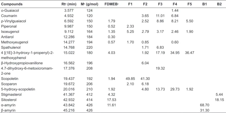
The pink spot (retention factor of 0.67) and the purple spot (retention factor of 0.64), both highlighted by brackets in Figure 2A, had good separation profile. Furthermore, it was active when stained with TCC (Fig. 2B). We performed the separation of these spots by preparative thin layer chromatography (PTLC), a simple, rapid and inexpensive technique that is frequently used to isolate substances with minimal yields. This technique led to two fractions, named B1 and B2, which along with fractions FDMEB, F1, F2, F3, F4 and F5, were analyzed by gas chromatography coupled with mass spectrometry (GC-MS) to identify the compounds.
Chemical Composition of Protium hebetatum We analyzed the active samples in the screening of antibacterial activity by the GC-MS method and obtained mass spectra for each signal in the chromatogram, comparing these with the data from the NIST Mass Spectral Search Program version 2.0, available in the equipment used (Figure 2). We identified eighteen compounds, thirteen of which (isoeugenol, p-vinylguaiacol, methoxyeugenol, coumarin, 5-hydroxy-scopoletin, 4,7-dihydroxy-6-metoxicromam-2-one,
4-[(1E]-3-TABLE 2. MIC of the active fractions of Protium hebetatum
SAMPLES
MIC (µg/mL)
MICROORGANISMS
S. aureus E. faecalis K. pneumoniae S. enterica P. aeruginosa E. coli P. vulgaris
MEB 1000 1000 2000 2000 1000 2000 2000
FDMEB 1000 500 - - - -
-F1 1000 2000 - - - -
-F2 1000 2000 - - - -
-F3 2000 1000 - - - -
-F4 2000 1000 - - - -
-F5 1000 1000 - - - -
-F6 1000 1000 - - - -
-F7 2000 2000 - - - -
-F8 2000 2000 - - - -
-F9 >2000 >2000 - - - -
-F10 >2000 >2000 - - - -
-GENTAMYCIN 10 10 10 10 10 10 10
FIGURE 1. F1 antibacterial agents identified by the Bioautography method. (A) Plate revealed with sulfuric anisaldehyde solution; (B) Plate revealed with 2,3,5-triphenyltetrazolium chloride (TTC). Eluent: hexane: ethyl acetate 1:1.
TABLE 3. Compounds identified from the FDMEB and subsequent fractions
Compounds Rta (min) Mb (g/mol) FDMEBc F1 F2 F3 F4 F5 B1 B2
o-Guaiacol 3.577 124 1.44
Coumarin 4.932 120 3.65 11.01 6.84
ρ-Vinylguaiacol 6.592 150 1.79 2.52 8.86 8.21 5.50
Piperonal 9.987 150 0.52 2.33
Isoeugenol 9.112 164 1.35 5.25 2.79 3.17 2.46 1.90
Antiarol 12.286 184 0.30
Methoxyeugenol 14.277 194 0.57 1.70 0.85 0.60
Spathulenol 14.768 220 1.71 6.83
4 [(1E]-3-hydroxy-1-propenyl)-2-methoxyphenol
15.022 180 4.03 1.92 17.19 34.95 36.47
β-Hydroxypropiovanillone 16.562 196 6.04
4.7-dihydroxy-6-metoxicromam-2-one
17.376 208 19.32
Scopoletin 19.437 192 1.94 49.85 41.30
Scoparon 19.672 206 2.10 6.18
5-hydroxy-scopoletin 20.016 210 1.92 4.80 13.73 29.73 1.92
Stigmasterol 41.367 412 4.32 5.44
Sitosterol 42.932 414 17.53 18.15
α-amyrin 43.842 426 11.61 68.70
β-amyrin 45.216 426 31.30
FIGURE 2. Chromatograms (GC-MS) of the active fractions obtained from MEB oh the P. hebetatum. A-Chromatogram of the FDMEB, (1) sitosterol, (2) α- amyrin; B – Chromatogramof the F1, (3) scopoletin; C – Chromatogram of the B1, (2) α- amyrin, (4) β- amyrin; D – Chromatogram of the B2, (1) sitosterol, (5) stimasterol. Other chromatograms and spectral data are available in the supplementary material.
The compounds identified in sample B1 correspond to α-amyrin and β-amyrin triterpenes, and the compounds identified in sample B2 correspond to
stigmasterol and sitosterol steroids (Figure 2).
Among the compounds identified, isoeugenol
(Santin et al., 2011), guaiacol, spathulenol (Cooper et al., 2013), α- and β-amyrin (Coloma et al., 2011) and scopoletin (Musa et al., 2011) have known antibacterial activity. There were higher percentage areas of coumarin, 5-hydroxy-scopoletin, 4,7-dihydroxy-6-metoxicromam-2-one and scopoletin, in the samples, and it is suggested that the antibacterial activity presented by MEB is attributed to this class of coumarins. This also means that Protium hebetatum is a potential source of coumarins, which have a wide range of industrial applications, including in the pharmaceutical industry.
This is a pioneering work on the evaluation of the antibacterial activity of Protium hebetatum. Its activity against Staphylococcus aureus and
Enterococcus faecalis is noteworthy, due to the impact of these microorganisms on public health, and its involvement in the etiology of various gastrointestinal disorders with high prevalence. Our results,
particularly those relating to the identification of substances, motivate further scientific investigations
of Protium hebetatum, focusing, for example, on
its analgesic and anti-inflammatory activities due
the presence of coumarins and triterpenes, as well as prompting studies with other species of genus Protium, which are still poorly studied. The GC-MS analyses indicate that the species is rich in substances of industrial interest, such as isoeugenol, guaiacol, and scopoletin, which are widely used in the pharmaceutical, food, toiletries, cosmetics and agricultural industries.
SUPPLEMENTARY MATERIAL
Chromatograms (GC-MS) of the fractions obtained from MEB on P. hebetatum.
FIGURE 3. Cromatogramof the F2, (3) scopoletin
ACKNOWLEDGMENTS
This research was supported by a grant from FAPEAM (AM, Brazil).
REFERENCES
ALIGIANNIS, N; KALPOTZAKIS, E; MITAKU, S; CHINOU, I.B. Composition and antimicrobial activity of the essential oils of two Origanum species. Journal of Agricultural and Food Chemistry, v.40, p.4168-4170, 2001.
ALVES, T.M.A.; SILVA, A.F.; BRANDÃO, M.; GRANDI, T.S.M.; SMÂNIA, E.F.; SMÂNIA, J.R.A.; ZANI, C.L. Biological screening of Brazilian medicinal plants.
Memória Instituto Oswaldo Cruz, v. 95, p.367-373,
2000.
ARAGÃO, G.F.; CARNEIRO, L.M.V.; JUNIOR, A.P.F.; VIEIRA, L.C.; BANDEIRA, P.N.; LEMOS, T.L.G.; VIANA, G.S.B. A possible mechanism for anxiolytic and antidepressant effects of alpha- and beta-amyrin from
Protium heptaphyllum (Aubl) March. Pharmacology
Biochemistry&Behavior, v.85, p.827-834, 2007. BANDEIRA, P.N.; PESSOA, O.D.L.; TREVISAN, M.T.S.;
LEMOS, T.L.G. Metabólitos secundários de Protium
heptaphyllum March. Química Nova, v.25,
p.1078-1080, 2002.
BÉRDY, J. Thoughts and facts about antibiotics: Where we are now and where we are heading. Journal ofAntibiotics, v.65, p.385-395, 2012.
CHOMA I.M.; GRZELAK E.M. Bioautography detection in thin-layer chromatography. Journal of Chromatography A. v.1218, p.2684–2691, 2011.
CLSI. Manual Clinical and Laboratory Standards Institute. Methods for dilution antimicrobial susceptibility tests for bacteria that grow aerobically;
approved standards- 6th ed. Document M7-A6 performance standards for antimicrobial susceptibility testing. Clinical and Laboratory Standards Institute, Wayne, PA, 2006.
CLSI. Performance Standards for Antimicrobial Disk
Susceptibility Tests; 88 Approved Standard—Eighth
FIGURE 5. Cromatogramof the F4, (8) ρ-Vinylguaiacol, (6) 4 [(1E]-3-hydroxy-1-propenyl)-2-methoxyphenol, (9) 5-hydroxy-scopoletin
Edition. CLSI document M2-A8 (ISBN 1-56238-485-6). CCLS, 940 West Valley Road, Suite 1400, Wayne, Pennsylvania 19087-1898 USA, 2009.
C O L L I N S , C . H . ; B R A G A , G . L . ; B O N ATO , P. S .
Fundamentos de cromatografia. Campinas: Editora da Unicamp, 456p, 2006.
COLOMA, A.G.; BALBOA, C.L.; REINA, O.S.M.; FRAGA, B.M. Triterpene based plant defenses. Phytochemistry,
v.10, p.245-260, 2011.
COOPER, R.A. Inhibition of biofilms by glucose oxidase,
lactoperoxidase and guaiacol: the active antibacterial component in an enzyme alginogel. International
Wound Journal, v.37, p.373-376, 2013.
COSTA, T.O.G.; ALMEIDA, R.A.; KOOLEN, H.H.F.; SILVA, F.M.A.; PINTO, A.C. Constituintes químicos do caule de
Protium hebetatum (Burseraceae). Acta Amazônica,
v.42, p.557-560, 2012.
DEHARO, E.; BOURDY, G.; QUENEVO, C.; MUNOZ, V.; RUIZ, G.; SAUVAINA, M. A search for natural bioactive compounds in Bolivia through a multidiscipilinary approach. Part V. Evolution of antimalarial activity of plants used by Tacana Indians. Journal of
Ethnopharmacology, v.77, p.91-98, 2001.
DUARTE, M.C.T.; FIGUEIRA, G.M.; SARTORATTO, A.; REHDER, V.L.G.; DELARMELINA, C. Anti-Candida
activity of Brazilian medicinal plants. Journal of
Ethnopharmacology, v.97, p.305-311, 2005.
HOLANDA PINTO, S.A.; PINTO, L.M.; CUNHA, G.M.; CHAVES, M.H.; SANTOS, F.A.; RAO, V.S.
Anti-inflammatory effect of alpha, beta-amyrin, a pentacyclic
triterpene from Protium heptaphyllum in rat model of acute periodontitis. Inflammopharmacology v.16, p.48-52, 2008.
MAIA, R.M.; BARBOSA, P.R.; CRUZ, F.G.; ROQUE, N.F.; FASCIO, M. Triterpenos da resina de Protium
heptaphyllum March (Burseraceae): Caracterização
em misturas binárias. QuímicaNova, v.23, p. 623-626, 2000.
MISHRA, B.B.; TIWARI, V.K. Natural products: An evolving role in future drug Discovery. European
Journal of Medicinal Chemistry, v.46, p.4769-4807,
2011.
MUSA, M.A.; COOPERWOOD, J.S.; KHAN, M.O. A review of coumarin derivatives in pharmacotherapy of breast cancer. Current Medicinal Chemistry, v.15, p.2664-2679, 2008.
NCCLS- National Committee for Clinical Laboratory S t a n d a r d s – P a d r o n i z a ç ã o d o s Te s t e s d e
Sensibilidade a Antimicrobianos por Disco-difusão:
Norma Aprovada – 8ed., v. 23, 2003.
OLIVEIRA, N.A.; AMARAL, I.L. Florística e fitossociologia
de uma floresta de vertente na Amazônia Central, Amazonas, Brasil. Acta Amazônica, v.34, p.21-34, 2004.
OTUKI, M.F.; VIEIRA-LIMA, F.V.; MALHEIROS, A.;
YUNES, R.A.; CALIXTO, J.B. Topical antiinflammatory
effects of the ether extract from Protium kleinii and alpha-amyrin pentacyclic triterpene. European Journal
of Pharmacology, v.507, p.253-259, 2005.
PALOMINO; J.C.; MARTIN, A.; CAMACHO, M.; GUERRA, H.; SWINGS, J.; PORTAELS, F. Resazurin Microtiter Assay Plate: simple and inexpensive method for detection of drug resistance in Mycobacterium tuberculosis.
Antimicrobial Agents and Chemotherapy, v.46,
p.2720-2722, 2002.
RAMOS, M.F.S.; SIANI, A.C.; TAPPIN, M.R.R.; GUIMARÃES, A.C.; RIBEIRO, J.E.L.S. Essential oils from oleoresins of Protium spp. of the Amazon region.
Flavour and Fragrance Journal, v.150, p.383-387,
2000.
SANTANA, A.I.; VILA, R.; ESPINOSA, A.; OLMEDO, D.; GUPTA, M.P.; CAÑIGUERAL, S. Composition and biological activity of essential oils from Protium
confusum. Natural Products Communication, v.4,
p.1401-1406, 2009.
SANTIN, J.R.; LEMOS, M.; KLEIN-JÚNIOR, L.C.; MACHADO, I.D.; COSTA, P.; DE OLIVEIRA, A.P.; TILIA, C.; DE SOUZA, J.P.; DE SOUZA, J.P.B.; BASTOS, J.K.; DE ANDRADE, S.F. Gastroprotective activity of
essential oil of the Syzygium aromaticum and its major
component eugenol in different animal models. Naunyn
-Schmiedeberg’s Archives of Pharmacology, v.38,
p.149-158, 2011.
SIANI, A.C.; GARRIDO, I.S.; MONTEIRO, S.S.; CARVALHO, E.S.; RAMOS, M.F.S. Protium icicariba
as a sourse of volatile essences. Biochemical
Systematics and Ecology v.32, p.477-489, 2004.
SILVA, J.R.A.S.; ZOGHBI, M.G.B.; PINTO, A.C.; GODOY, R.L.O.; AMARAL, A.C.F. Analysis of the hexane extracts from seven oleoresins of Protium species. Journal Essential Oil Research, v.21, p.305-308, 2009. SIMÕES, C. M. O. (Org.). Farmacognosia: da planta ao
medicamento. 6ed. Porto Alegre: Editora da UFRGS,
2007.
SUSUNAGA, G.S.; SIANE, A.C.; PIZZOLATTI, M.G.; YUNES, R.A.; MONAACHE, F.D. Triterpenes from the resin of Protiumheptaphyllum. Fitoterapia, v.72, p.709-711, 2001.
TAYLOR, P.; ARSENAK, M.; ABAD, M.J.; FERNÁNDEZ, A.; MILANO, B.; GONTO, R.; RUIZ, M.C.; FRAILE, S.; TAYLOR, S.; ESTRADA, O.; MICHELANGELI, F. Screening of Venezuelan medicinal plant extracts for cytostatic and cytotoxic activity against tumor cell lines.
Phytotherapy Research, v.27, p.530-539, 2012.
VALGAS, C.; SOUZA, S.M.; SMÂNIA, E.F.A.; JÚNIOR, A.S. Screening methods to determine antibacterial activity of natural products. Brazilian Journal Microbiology, v.38, p.369-380, 2007.
ZOGHBI, M.G.B.; SIQUEIRA, J.B.G.; WOLTER, E.L.A.; JÚNIOR, O.L.P. Constituintes químicos de Protium
paniculatum (BURSERACEAE). Acta Amazônica v.23,
p.187-189, 1993.


![FIGURE 6. Cromatogram of the F5, (6) 4 [(1E]-3-hydroxy-1-propenyl)-2-methoxyphenol, (9) 5-hydroxy-scopoletin](https://thumb-eu.123doks.com/thumbv2/123dok_br/15855531.661473/9.918.221.738.81.347/figure-cromatogram-f-hydroxy-propenyl-methoxyphenol-hydroxy-scopoletin.webp)
