Taxonomic revision of
Galeocharax
(Characiformes: Characidae: Characinae)
Victor Giovannetti
1, Mônica Toledo-Piza
1and Naércio A. Menezes
2The taxonomy of Galeocharax, a genus of freshwater fishes distributed in most South American cis-Andean river systems, except the rio Negro, rio São Francisco and rio Xingu basins and the eastern drainages of Brazil, is herein revised. A total of 1229 specimens were examined from which 680 had meristic and morphometric data taken. Osteological and morphological features were also examined through radiographs, scanning electron microscopy and in cleared and stained specimens. Three of the four species previously considered as valid are herein recognized: Galeocharax humeralis from rio Paraguay and lower rio Paraná basins; Galeocharax goeldii, from rio Madeira basin, with records of geographical distribution expanded to the río Madre de Dios, río Beni, rio Mamoré and rio Guaporé basins and Galeocharax gulo, which is widespread throughout rio Amazonas, río Orinoco, rio Tocantins, and upper rio Paraná basins. Galeocharax knerii is herein considered a junior synonym of Galeocharax gulo. A key to species of Galeocharax is presented.
Keywords: Key of identification, Ostariophysi, Systematics, Widespread species.
A taxonomia de Galeocharax, um gênero de peixes de água doce distribuído nos principais sistemas hídricos da América do Sul cisandina, com exceção da bacia do rio Negro, do rio São Francisco, do rio Xingu e das drenagens litorâneas do leste, é aqui revisada. Foram examinados 1229 exemplares, dos quais 680 tiveram dados merísticos e morfométricos tomados. Características osteológicas e morfológicas também foram examinadas por meio de radiografias, microscopia eletrônica de varredura e exemplares diafanizados. Três das quatro espécies previamente consideradas válidas são reconhecidas: Galeocharax humeralis das bacias do rio Paraguai e baixo rio Paraná; Galeocharax goeldii da bacia do rio Madeira, com registros de distribuição geográfica ampliados para as bacias dos rios Madre de Dios, Beni, Mamoré e Guaporé, e Galeocharax gulo amplamente distribuída nas bacias dos rios Amazonas, Orinoco, Tocantins e alto rio Paraná. Galeocharax knerii foi considerada sinônimo júnior de Galeocharax gulo. Uma chave de identificação para as espécies de Galeocharax é apresentada.
Palavras-chave: Chave de identificação, Espécies amplamente distribuídas, Ostariophysi, Sistemática.
1Departamento de Zoologia, Instituto de Biociências, Universidade de São Paulo, Rua do Matão, Travessa 14, n° 101, 05508-090 São Paulo, SP, Brazil. (VG) [email protected] (corresponding author), (MTP) [email protected]
2Museu de Zoologia, Universidade de São Paulo. P. O. Box 42494, 04218-970 São Paulo, SP, Brazil. [email protected] Introduction
Galeocharax Fowler, 1910 is a genus of freshwater fishes that reach up to 25 cm in length. They are popularly known as “saicanga”, “peixe-cigarra”, “peixe-cadela”, and “dentudo” in the Brazilian territory (Graça, Pavanelli, 2007; Langeani, Rêgo, 2014) and as “dentón” and “dientón alargado” in Peru and Venezuela (Taphorn, 1992; Ortega et al., 2012). Species of the genus are found in most of the main river systems of the cis-Andean South America with the exception of the rio São Francisco, rio Negro, rio Xingu basins, and the eastern drainages of Brazil.
They are carnivorous, feeding on small fishes and invertebrates (Ribeiro Neto et al., 1998; Gandini et al., 2012). They reproduce throughout the year and there is no record of parental care or migratory behavior (Magalhães et al., 2004). The presence of the parasitic isopoda (Braga
cigarra, Cymothoidae), popularly known as “cigarrinha”, in the oral cavity of specimens of Galeocharax in the Paraná basin is fairly common (Brandão et al., 2013), justifying the popular name peixe cigarra.
The latest taxonomic revision of the genus was carried out by Menezes (1976) who recognized three valid species: Galeocharax humeralis (Valenciennes, 1834) from the rio Paraguai and the lower rio Paraná basins; Galeocharax gulo (Cope, 1870) from the rio Amazonas and the rio Tocantins basins and Galeocharax knerii (Steindachner, 1879) from the upper rio Paraná basin. At that time, Menezes considered Galeocharax goeldii (Fowler, 1913) from the rio Madeira basin, as a synonym of G. gulo, but more recently, after the examination of additional specimens, the species was recognized as valid (Menezes, 2007a).
More recent revisions of Acestrocephalus and Cynopotamus resulted in the descriptions of new species (Menezes 1987, 2006, 2007b). Moreover, Taphorn (1992: 185) suggested that specimens of Galeocharax from the río Apure, in the río Orinoco drainage, Venezuela, could represent an undescribed species.
The number of specimens of Galeocharax available in ichthyological collections increased considerably in the past decades, showing that the genus is more widely distributed than previously recorded. Therefore, the aim of the present study was to undertake a taxonomic study of the genus including the highest number of specimens as possible in order to determine how many and which species can be recognized as valid, to reevaluate their diagnostic features and to estimate their geographic distribution.
Material and Methods
A total of 1229 specimens deposited in several ichthyological collections and covering the entire area of distribution of Galeocharax were examined, from which 680 specimens had meristic and morphometric data taken. Morphometric data were taken point to point with a digital caliper with 0.1 mm precision, on the left side of the specimen, whenever possible. Nineteen different measurements were made on each specimen and they are presented in the tables and text as percentages of standard length (SL), with the exception of measurements of subunits of the head, which were expressed as percentages of head length (HL).
Data of specimens from which sex could be determined based on the examination of gonads were initially analyzed separately to check for possible sexual dimorphism, but are presented together in the tables and text since no differences were detected. Variation of the number of teeth on the posterior row of the dentary is only partially associated with growth, and was represented by scatter plots. The same association was observed for number of maxillary teeth.
Measurements follow Menezes (1969), with the exception of: head length, which was measured from the tip of the snout to the posterior bony margin of the opercle at the ventral tip of the opercle concavity. The following measurements were included: upper jaw length: distance
between the tip of the snout and posterior margin of the maxilla. Dorsal-fin length: distance between the base of the first ray and the distal tip of the longest unbranched ray. Dorsal-fin base length: distance between the bases of the anterior- and posteriormost dorsal-fin rays. Anal-fin length: distance between the base of the first visible unbranched ray and the distal tip of the longest unbranched ray. Anal-fin base length: distance between the bases of the anterior- and posteriormost anal-fin rays. Prepectoral distance: distance between the tip of the snout and the base of the first pectoral-fin ray. Caudal-peduncle length: distance between the base of the posteriormost anal-fin ray and the posterior margin of the hypural plate.
Meristic data were taken from the left side of specimens whenever possible. Scales, fin-rays and teeth were counted with the aid of a stereomicroscope. The number of unbranched rays is expressed with roman numerals and the number of branched rays with arabic numerals. For a better visualization of the teeth, a solution of methylene blue in 70% ethanol was applied to the specimens. The stain is temporary and fades away a few minutes after returning the specimen to 70% ethanol. Counts of vertebrae and supraneurals were made on radiographs taken from a limited number of specimens and examined in the RadiAnt DICOM viewer (32-bit) software.
Counts follow Menezes (1969) with the exception of number of scales below the lateral line that was counted as the number of horizontal rows of non-perforated scales between the lateral line and the base of the first pelvic-fin ray. This way of counting showed to be more precise than that made at the vertical through the first anal fin ray, since the scales in this latter region tended to be slightly disorganized. The following counts were added: number of scales around the caudal peduncle; number of scales in the anal-fin sheath: number of horizontal rows on the sheath of scales that covers the base of the anal-fin rays; number of pelvic-fin rays; number of teeth on the outer row of the dentary; number of teeth on the inner row of the dentary; number of teeth on the outer row of the premaxilla; number of teeth on the inner row of the premaxilla; total number of vertebrae; number of abdominal vertebrae: including the four anteriormost vertebrae of the Webberian apparatus until the posteriormost vertebra without a haemal spine and number of caudal vertebrae (from the first vertebra with a haemal spine to the compound ural centrum, which was counted as one). Counts of unbranched anal-fin rays, principal and procurrent caudal-fin rays were made only on cleared and stained specimens. In the descriptions, meristic values are followed by their respective frequency in parentheses, except those presented on tables.
Meristic and morphometric data were summarized using descriptive statistics with the help of MYSTAT 12 software.
images made in a Zeiss DSM940 electronic microscope. Since no diagnostic differences were observed, details of those structures are shown for a single species.
Specimens were cleared and stained following the procedure of Taylor, Van Dyke (1985).
Specimens from the same locality were initially compared and if differences on the examined characteristics were not detected they were grouped. This approach was carried out successively for localities near to each other, then to localities on the same river basins until all the specimens were analyzed and preliminary groups were defined.
In the discussion of geographic distribution, the Amazon basin was defined excluding the rio Tocantins (see Goulding et al., 2003a, for a discussion). Maps of geographic distribution were made using Google Earth and Quantum GIS Wroclaw softwares.
Institutional abbreviations are as listed in Fricke, Eschmeyer (2016) with the addition of UFRO-I for Coleção Ictiológica of Universidade Federal de Rondônia, Porto Velho.
Results
Galeocharax Fowler, 1910
Galeocharax Fowler, 1910: 790 [type species Cynopotamus gulo Cope, 1870: 565, by original designation]. -Géry,
Vu-Tân-Tuê, 1963b: 239 [as a synonym of Acestrocephalus,
subgenus of Cynopotamus]. -Géry, 1972: 27 [as a synonym of Acestrocephalus, subgenus of Cynopotamus].
Type species. Cynopotamus gulo Cope, 1870, original designation.
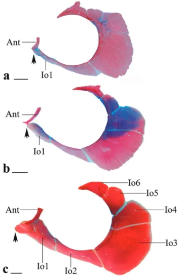
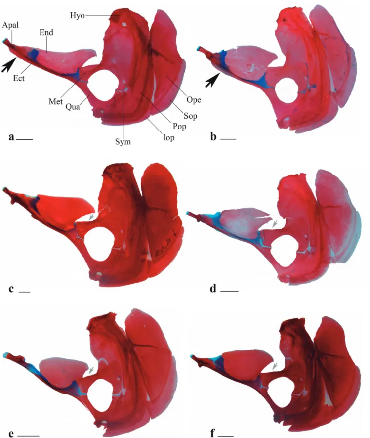
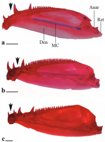
Diagnosis. Galeocharax differs from all genera of the Characinae (sensu Mattox, Toledo-Piza, 2012) except Cynopotamus and Acestrocephalus, in the presence of spinoid scales. It can be distinguished from members of the latter two genera by the presence of a lateral bony projection on the nasal (vs. projection absent in Acestrocephalus and Cynopotamus, Fig. 1); by having infraorbital 1 anteriorly elongated, reaching the premaxilla and extending anteriorly beyond the antorbital (vs. infraorbital 1 not as anteriorly elongated falling short of the anterior margin of the antorbital in Acestrocephalus and Cynopotamus, Fig. 2); and by the absence of a bony crest on the medial surface of the ectopterygoid (vs. bony crest present in Acestrocephalus and Cynopotamus, Fig. 3). Galeocharax also differs from Cynopotamus in the presence of 6-13 small conical teeth posterior to the dentary canines (vs. small conical teeth posterior to the dentary canines absent, Fig. 4); and in the lack of a pronounced notch on the posteroventral margin of the cleithrum (vs. notch on posteroventral margin of the cleithrum present, Fig. 5).
Fig. 1. Nasal bone of a. Cynopotamus xinguano, MZUSP 94196, 97.9 mm SL, scale bar 500 µm; b. Acestrocephalus stigmatus, MZUSP 94216, 85.0 mm SL, scale bar 500 µm, and c. Galeocharax gulo, MZUSP 62839, 127.2 mm SL, scale bar 1 mm. Right side, ventral view, anterior tip oriented downwards. Arrow points to lateral bony projection.
Fig. 4. Lower jaw of a. Cynopotamus xinguano, MZUSP 94196, 97.9 mm SL; b. Acestrocephalus stigmatus, MZUSP 94216, 85.0 mm SL and c. Galeocharax gulo, MZUSP 62839, 127.2 mm SL. Right side, medial view. Black arrows points to region posterior to dentary canines. Anar - anguloarticular; MC - Meckel’s cartilage; Den - dentary; Ret - retroarticular. Scale bars 2 mm.
Remarks. The nominal species of Galeocharax were all described between 1834 and 1913, but the genus name was proposed only in 1910 (Fowler, 1910). Before the proposal of the genus, the species were allocated in several distinct genera such as Charax Scopoli, Hydrocyon Cuvier (= Hydrocynus Cuvier), Cynopotamus Valenciennes, Xiphorhamphus Müller, Troschel (= Acestrorhynchus Eigenmann, Kennedy), Anacyrtus Günther (= Charax), Eucynopotamus Fowler (= Roeboides Günther) and Cyrtocharax Fowler (= Cynopotamus). The name Galeocharax was proposed in order to resolve a nomenclatural confusion originated by publications of Fowler (1901, 1904, 1910) and Eigenmann (1903, 1907, 1910) that was later detailed by Géry, Vu-Tân-Tuê (1963a). It should be noted that Fowler’s proposition was not followed by a discussion about the species included neither presented a diagnosis of Galeocharax. Most of the publications citing Galeocharax, subsequent to the proposition of the genus name consisted of species lists that did not include any discussion or characterization of the genus or the species included.
Menezes (1976) recognized a natural group comprising Cynopotamus, Acestrocephalus and Galeocharax, the Cynopotaminae on the basis of morphological characters and differentiated Galeocharax from Acestrocephalus and Cynopotamus based on the three characters mentioned above in the Diagnosis.
Galeocharax, Acestrocephalus and Cynopotamus have also been considered phylogenetically related in studies subsequent to that of Menezes (1976), however hypotheses of relationships among the three genera differ among authors (Lucena, 1998; Mirande, 2010; and Mattox, Toledo-Piza, 2012, based on morphological data, and Oliveira et al., 2011, based on molecular data). Mattox,
Piza (2012) corroborated the hypothesis of monophyly of Galeocharax and proposed additional synapomorphies for the genus: 1) the presence of scales covering, at least part of the caudal fin lobes on the median region of the fin; 2) the ventral portion of the levator arcus palatini muscle extending until the anterior margin of the metapterygoid; and 3) the anteroventral margin of the cleithrum with a well developed and anteriorly oriented projection.
Key to the species of Galeocharax
1. Perforated lateral line scales 77-87 ...Galeocharax gulo. Perforated lateral line scales 88-105 ... 2. 2. 1-3 Small conical teeth between the third and fourth
dentary canines ...Galeocharax humeralis. Small conical teeth between the third and fourth dentary
canines absent ...Galeocharax goeldii.
Galeocharax goeldii (Fowler, 1913)
Fig. 6
Anacyrtus humeralis. -Perugia, 1897:26 [listed; Reyes, río Beni,
Bolívia; misidentification].
Charax goeldii Fowler, 1913:568 [original description; type
locality: tributary of rio Madeira, near Porto Velho, Brazil]. -Schultz, 1950:62 [synonym of Cyrtocharax amazonum
(Günther) = Cynopotamusamazonum]. -Menezes, 1976:44 [as
a synonym of Galeocharax gulo (Cope)]. -Lucena, Menezes,
2003:203 [as a synonym of Galeocharax gulo (Cope)]. Cyrtocharax goeldii. -Fowler, 1950:311 [listed].
Cynopotamus (Acestrocephalus) goeldii. -Géry, Vu-Tân-Tuê,
1963b:240 [diagnosis in key]. -Géry, 1972:25 [description; diagnosis in key]; -Géry, 1977:306 [listed].
Galeocharax gulo. -Barthem et al., 2003:80 [río Madre de Dios
basin].
Galeocharax kneri. -Barthem et al., 2003:80 [río Madre de Dios
basin]. -Goulding et al., 2003b:143 [río Madre de Dios basin]. Galeocharax goeldii. -Menezes, 2007a:21 [redescription, species
removed from synonymy]. -Rapp Py-Daniel, 2007:111 [as
Galeocharax sp., rio Madeira basin]. -Lima et al., 2013:253
[listed, rio Madeira basin].
Diagnosis. Galeocharax goeldii differs from G. humeralis in the absence of small conical teeth between the third and fourth dentary canines (Fig. 7a) (vs. 1-3 small conical teeth, Fig. 7c), and from G. gulo in the higher number of perforated lateral line scales (88-97 vs. 77-87, Tab. 1). Although there is overlap in the number of scales above and below the lateral line between G. goeldii and G. gulo, the former species shows a tendency towards higher values of scales above (17-20, median 18 vs. 14-19, median 16, in G. gulo, Tab. 2) and below (15-17, median 16 vs. 11-16, median 13 in G. gulo, Tab. 3) the lateral line, respectively.
Tab. 2. Frequency distribution of number of scale rows between the dorsal-fin origin and lateral line of species of Galeocharax.
14 15 16 17 18 19 20 Total
G. goeldii 13 20 18 4 55
G. gulo 12 131 141 83 24 3 394
G. humeralis 9 46 38 9 102
Tab. 3. Frequency distribution of number of scale rows between the pelvic-fin origin and lateral line of species of Galeocharax.
11 12 13 14 15 16 17 Total
G. goeldii 13 30 15 58
G. gulo 10 138 158 54 44 7 411
G. humeralis 11 55 36 102
Description. Morphometric data of Galeocharax goeldii presented on Tab. 4. Largest specimen examined 213.3 mm SL. Body elongated, laterally compressed and moderately deep, largest body depth at vertical through dorsal-fin origin. Dorsal profile of head slightly concave from tip of snout to posterior tip of supraoccipital spine. Dorsal profile of body slightly convex from posterior tip of supraoccipital spine to base of first dorsal-fin ray, posteroventrally slanted to slightly convex along dorsal-fin base. Dorsal profile from base of last dorsal-fin ray to caudal peduncle straight, slightly concave along caudal peduncle. Ventral profile of head and body convex from tip of lower jaw to anal-fin origin, posterodorsally slanted along anal-fin base, straight to slightly concave along caudal peduncle. Profile of posterior bony margin of opercle slightly concave or straight, rarely convex.
Tab. 1. Frequency distribution of perforated lateral-line scales of species of Galeocharax.
77 78 79 80 81 82 83 84 85 86 87 88 89 90 91 92 93 94 95 96 97 98 99 100 101 102 103 104 105 Total
G. goeldii 3 8 14 5 7 11 7 4 4 5 68
G. gulo 3 18 52 72 57 70 63 48 23 13 8 427
G. humeralis 5 8 3 8 12 15 20 12 6 6 7 4 3 1 1 1 112
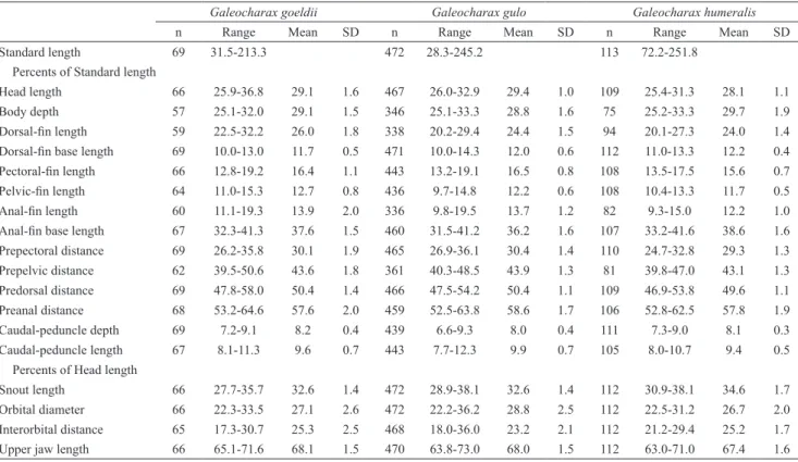
Tab. 4. Morphometric data of species of Galeocharax. SD = standard deviation, Standard length in mm.
Galeocharax goeldii Galeocharax gulo Galeocharax humeralis
n Range Mean SD n Range Mean SD n Range Mean SD
Standard length 69 31.5-213.3 472 28.3-245.2 113 72.2-251.8
Percents of Standard length
Head length 66 25.9-36.8 29.1 1.6 467 26.0-32.9 29.4 1.0 109 25.4-31.3 28.1 1.1
Body depth 57 25.1-32.0 29.1 1.5 346 25.1-33.3 28.8 1.6 75 25.2-33.3 29.7 1.9
Dorsal-fin length 59 22.5-32.2 26.0 1.8 338 20.2-29.4 24.4 1.5 94 20.1-27.3 24.0 1.4
Dorsal-fin base length 69 10.0-13.0 11.7 0.5 471 10.0-14.3 12.0 0.6 112 11.0-13.3 12.2 0.4
Pectoral-fin length 66 12.8-19.2 16.4 1.1 443 13.2-19.1 16.5 0.8 108 13.5-17.5 15.6 0.7
Pelvic-fin length 64 11.0-15.3 12.7 0.8 436 9.7-14.8 12.2 0.6 108 10.4-13.3 11.7 0.5
Anal-fin length 60 11.1-19.3 13.9 2.0 336 9.8-19.5 13.7 1.2 82 9.3-15.0 12.2 1.0
Anal-fin base length 67 32.3-41.3 37.6 1.5 460 31.5-41.2 36.2 1.6 107 33.2-41.6 38.6 1.6
Prepectoral distance 69 26.2-35.8 30.1 1.9 465 26.9-36.1 30.4 1.4 110 24.7-32.8 29.3 1.3 Prepelvic distance 62 39.5-50.6 43.6 1.8 361 40.3-48.5 43.9 1.3 81 39.8-47.0 43.1 1.3
Predorsal distance 69 47.8-58.0 50.4 1.4 466 47.5-54.2 50.4 1.1 109 46.9-53.8 49.6 1.1 Preanal distance 68 53.2-64.6 57.6 2.0 459 52.5-63.8 58.6 1.7 106 52.8-62.5 57.8 1.9 Caudal-peduncle depth 69 7.2-9.1 8.2 0.4 439 6.6-9.3 8.0 0.4 111 7.3-9.0 8.1 0.3
Caudal-peduncle length 67 8.1-11.3 9.6 0.7 443 7.7-12.3 9.9 0.7 105 8.0-10.7 9.4 0.5
Percents of Head length
Snout length 66 27.7-35.7 32.6 1.4 472 28.9-38.1 32.6 1.4 112 30.9-38.1 34.6 1.7
Orbital diameter 66 22.3-33.5 27.1 2.6 472 22.2-36.2 28.8 2.5 112 22.5-31.2 26.7 2.0
Interorbital distance 65 17.3-30.7 25.3 2.5 468 18.0-36.0 23.2 2.1 112 21.2-29.4 25.2 1.7
Upper jaw length 66 65.1-71.6 68.1 1.5 470 63.8-73.0 68.0 1.5 112 63.0-71.0 67.4 1.6
Dorsal-fin origin situated anterior to half of SL, base of last dorsal-fin ray posterior to vertical through anal-fin origin. Proximal-medial radial of first dorsal-anal-fin pterygiophore inserted posterior to neural spine of ninth vertebra. Dorsal-fin rays ii,9. Profile of distal margin of dorsal fin straight in most specimens. Anal-fin origin
situated at approximately half of SL. Proximal-medial radial of first anal-fin pterygiophore situated posterior to haemal spine of 17th vertebra (1). Anal-fin rays iv (1), 40
at posterior third. Pectoral-fin rays i, 13 (1), 14 (28), 15 (31), or 16 (10); longest ray reaching vertical through pelvic-fin origin when fin is depressed. Profile of posterior margin of pectoral fin almost straight, with four or five proximal rays shorter than others. Specimens of at least 33.7 mm SL still retain larval rayless pectoral fin. Pelvic-fin origin situated anterior to vertical through dorsal-Pelvic-fin origin; pelvic-fin rays i,7; longest ray reaching vertical through anterior border of urogenital opening when fin is depressed, but failing to reach anal-fin origin. Profile of posterior margin of pelvic fin slightly convex, fan-like aspect when extended. Caudal fin forked, principal rays i,9,8,i (1) dorsal procurrent rays 16 (1), ventral procurrent rays 13(1); lower lobe slightly longer and deeper than upper lobe. Adipose fin present, its base anterior to vertical through base of last anal-fin ray.
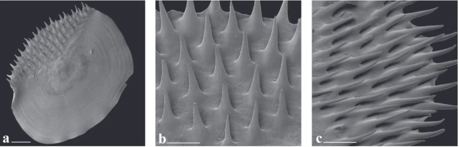
Spinoid scales (Fig. 8) distributed along entire body, except on region of axilla of paired fins and on region posterior to last ray of dorsal and anal fins where cycloid scales are present. Axillary scale present in pelvic fin. Anal-fin scale sheath with three horizontal scale rows over base of anteriormost anal-fin rays, covering one-fifth of length of anterior fin rays; number of horizontal scale rows gradually decreasing posteriorly to one scale row on posteriormost rays. Base of caudal-fin rays covered by scales.
Mouth terminal, obliquely oriented relative to horizontal body axis. Upper jaw slightly more anteriorly projected than lower jaw. Posterior margin of maxilla extending beyond vertical through posterior margin of orbit. Infraorbitals 1 to 6 present, supraorbital absent, posterior margin of infraorbital 3 not reaching preopercle, posterior margin of infraorbital 4 reaching preopercle in some specimens. Teeth conical. Premaxilla with two teeth rows, inner row with two teeth oriented to interior of mouth, outer row with 8 (4), 9 (28), or 10 (39) teeth,
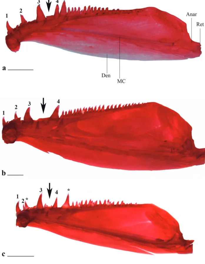
medialmost and lateralmost teeth canines. Maxilla with 38 (8), 39 (10), 40 (12), 41 (12), 42 (8), 43 (4), 44 (6), 45 (2), 46 (3), 47 (2), or 48 (2) conical teeth distributed along entire ventral margin. Dentary with two teeth rows. Inner row with 7 (14), 8 (29), 9 (16), 10 (2), 11 (1), 12 (2), or 13 (2) posteriorly oriented teeth. Outer row with anterior group formed by four canine teeth, third tooth larger, first and fourth usually of similar size, some specimens with fourth tooth intermediate in size between third and first teeth, second tooth always smallest and more external. Space between third and fourth canines occupied by lateral premaxillary canine when mouth is closed. Posterior group of outer teeth with 23 (1) small conical teeth forming row adjacent and continuous to fourth canine.
Lateral line complete. Lateral-line tube present between bases of middle caudal-fin rays. Lateral-line scales 88-97 (68) (Tab. 1), laterosensory canal on each scale with one posteroventral ramification; number of horizontal scale rows between dorsal-fin origin and lateral line 17-20 (55) (Tab. 2); number of horizontal scale rows between lateral line and pelvic-fin origin 15-17 (58) (Tab. 3); number of horizontal scale rows around caudal peduncle 23 (3), 24 (3), 25 (8), 26 (8), 27 (15), or 28 (6).
Total number of vertebrae 41 (11) or 42 (2), abdominal vertebrae 16 (12) or 17 (1), caudal vertebrae 25 (12) or 26 (1). Supraneurals 4 (12) obliquely oriented, proximal tip more anterior than distal tip, anteriormost supraneural lying anterior to neural spine of fifth vertebra, posteriormost lying between neural spines of seventh and eighth vertebrae.
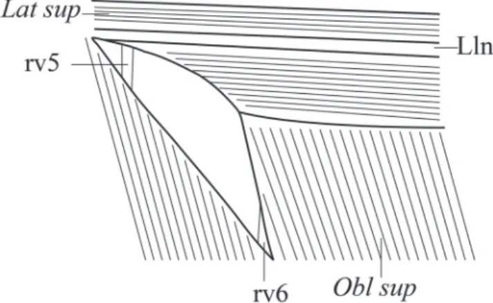
Pseudotympanum present, bordered anteroventraly by obliquus superioris muscle, posteriorly by obliquus superioris muscle and by rib associated with sixth vertebra, posterodorsaly by lateralis superficialis muscle and lateral line nerve. Rib associated with fifth vertebra visible anteriorly, but not bordering muscle hiatus anteriorly (Fig. 9).
Fig. 9. Schematic drawing of the fibers of muscles that limit the pseudotympanum in Galeocharax. rv5 - rib of vertebra 5; rv6 - rib of vertebra 6; Lat sup - lateralis superficialis muscle; Lln - lateral line nerve; Obl sup - obliquus superioris muscle.
Coloration. Overall body coloration yellowish, dorsal region darker than ventral region due to presence of chromatophores distributed on posterior field of dorsal scales. Dorsal median dark band from supraoccipital spine to dorsal-fin origin, not distinguishable posterior to dorsal fin base.
Dorsal region of head darker until approximately horizontal line through dorsal margin of orbit. Few specimens with scattered chromatophores on infraorbitals, mainly on 4, 5 and 6. Skin bordering mouth more pigmented on premaxilla, maxilla and dentary, mainly on base of canine teeth of lower jaw. Lateral surface of maxilla with sparse chromatophores anteriorly to approximately half of bone length.
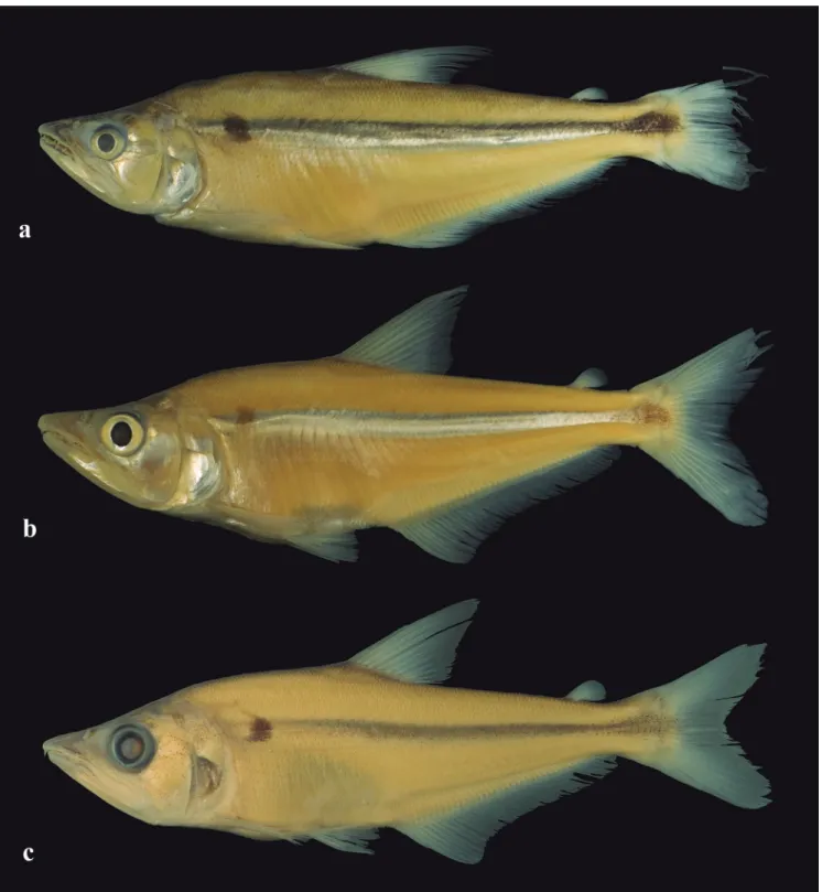
Oval shaped blotch on humeral region, largest diameter along vertical axis. Few specimens with crescent shaped humeral blotch with tips of crescent pointed anteriorly (Fig. 6b).
Silvery midlateral longitudinal band on body, dorsal to lateral line, ventral margin of band reaching lateral line in most specimens. Silvery midlateral longitudinal band extending from posterior margin of supracleithrum to posterior margin of hypural plate, narrower at anterior- and posteriormost regions in most specimens, broader at region of vertical through dorsal-fin base. Silvery midlateral longitudinal band widening from caudal peduncle to posterior margin of hypural plate in some specimens, and with ventral margin extending beyond lateral line on region of caudal peduncle.
Dark midlateral longitudinal band dorsal to lateral line extending from humeral region, to approximately half length of median caudal-fin rays, narrower at anterior- and posteriormost regions, broader at region of vertical through dorsal-fin. Some specimens with dark midlateral longitudinal band evident from posterior margin of supracleithrum to median caudal-fin rays. Dark midlateral longitudinal band forming a diamond shaped blotch over caudal peduncle and base of median caudal-fin rays. Dark midlateral longitudinal band not as wide as silvery midlateral longitudinal band except on caudal peduncle region. Dorsal margin of two longitudinal bands usually overlap.
Specimens smaller than 48.0 mm SL with lines of chromatophores ventral to lateral line, following myosepta on lateral surface of body, region dorsal of anal-fin base and anal-fin rays.
Pectoral and pelvic fins hyaline. Dorsal fin with lines of chromatophores along rays and scattered chromatophores on interradial membrane. Adipose fin pale, weakly pigmented except for line of chromatophores on anterodorsal margin.
Chromatophores forming arched vertical line through base of caudal fin rays; chromatophores more deeply concentrated underneath scales, arched vertical line interrupted in some specimens, with only dorsal and ventral limits evident (Fig. 6b).
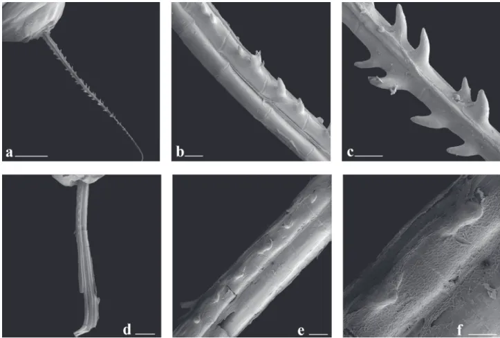
Sexual dimorphism. Males of Galeocharax goeldii have bony hooks on pelvic- and anal-fin rays. Bony hooks on anal-fin ray located on lateral surface of posterior branch of branched rays; bony hooks present on last unbranched ray in few specimens. One or two, rarely three hooks per ray segment, hooks dorsoposteriorly oriented (Figs. 10a-c). Bony hooks present on 10 to 23 anteriormost rays. Bony hooks on pelvic fin located on ventral surface of lepidotrichia slightly posteriorly displaced (one specimen with hooks on dorsal surface of fin ray). One or two, rarely three hooks per segment pointing towards base of ray (Fig. 11). Hooks present on four or five lateralmost branched rays.
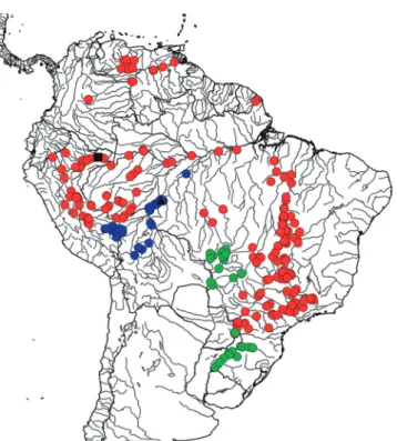
Geographical distribution. Galeocharax goeldii occurs in the rio Madeira, río Madre de Dios, río Beni, rio Mamoré, and rio Guaporé drainages (Fig. 12).
Remarks. Galeocharax goeldii was originally described as Charax goeldii by Fowler (1913) based on a single specimen, 56.0 mm SL, collected in a tributary of the rio Madeira, near Porto Velho, RO, Brazil. Later, Menezes (1976) considered the species as a synonym of Galeocharax gulo, based on data presented by Géry (1972), but after examining the holotype of Charax goeldii and additional material from the rio Madeira basin, Menezes (2007a) recognized the species as valid in Galeocharax.
As part of the present study, specimens examined from the río Madre de Dios, río Beni, rio Mamoré, and rio Guaporé drainages showed that G. goeldii is more widely distributed within the rio Madeira basin.
Fig. 10. Scanning electron microscope images of the second branched anal-fin ray of Galeocharax gulo, male, MZUSP 40901, 110.6 mm SL a.-c. and female, MZUSP 10479, 179.7 mm SLd.-f.. Proximal tip to upper left corner in a.-c., to top in d. and to upper right corner in e.-f. a. posterior view, scale bar 2 mm; b. Detail of middle portion, lateral view, left side, scale bar 200 µm; c. Detail of middle portion, posterior view, scale bar 200 µm; d. lateral view, right side, scale bar 1 mm; e. Detail of middle portion, lateral view, right side, scale bar 200 µm, and f. Detail of middle portion, lateral view, right side, scale bar 100 µm.
Fig. 12. Map of northern and central South America showing the geographical distribution of species of Galeocharax. G. goeldii (blue circles), the black triangle indicates its type locality, G. gulo (red circles), the black square indicates its type locality, and G. humeralis (green circles).
No variation in meristic and morphometric data associated with any geographic pattern was observed in specimens throughout the distribution range of the species, and the slight discrepancies in those counts of the holotype provided by Menezes (2007a) seem most probably related to the relative poor condition of the specimen associated to its small size.
Material examined. Bolívia: Beni: MNHH 1989-1441, 1, 124.7
mm SL; USNM 301901, 1, 190.0 mm SL; USNM 305364, 1, 173.9 mm SL; USNM 305367, 10, 97.1-213.3 mm SL. La Paz:
MZUSP 35951, 1, 144.6 mm. Brazil: Amazonas: INPA 24598, 1,
109.7 mm SL. Rondônia: INPA 20916, 1, 151.8 mm SL; MZUSP
92432, 5, 20.3-38.6 mm SL; MZUSP 92433, 8, 19.5-48.2 mm SL; MZUSP 92434, 1, 108.0 mm SL; MZUSP 92435, 4, 28.9-94.6 mm SL; MZUSP 92436, 24, 26.1-89.7 mm SL; UFRO-I 722, 1, 33.5 mm SL; UFRO-I 2246, 1, 54.5 mm SL; UFRO-I 2566, 1, 50.8 mm SL; UFRO-I 5468, 2, 1 c&s, 77.9-81.2 mm SL; UFRO-I 6854, 1, 121.2 mm SL; UFRO-I 6846, 1, 123.2 mm SL; UFRO-I 9705, 1, 120.8 mm SL; UFRO-I 9668, 1, 160.8 mm SL; UFRO-I 12941, 1, 17.3 mm SL; UFRO-I 13077, 2, 96.9-147.5 mm SL; UFRO-I 13090, 2, 101.6-105.7 mm SL; UFRO-I 14233, 2, 150.3-153.0 mm SL; UFRO-I 15986, 1, 59.3 mm SL; UFRO-I 16104, 1, 32.3 mm SL; UFRO-I 17970, 2, 132.5-144.9 mm SL; UFRO-I 19051, 1, 120.1 mm SL; UFRO-I 19368, 1, 85.2 mm SL; UFRO-I 20611, 1, 67.9 mm SL; UFRO-I 20612, 1, 68.7 mm SL; UFRO-I 20613, 2, 32.3-49.6 mm SL; UFRO-I 20631, 1, 39.4
Tab. 5. Meristic data of the types of the nominal species of Galeocharax presented by previous authors. A= Valenciennes (1849); B = Géry & Vu-Tân-Tuê (1963a); C = Cope (1870); D and G = Géry (1972); E = Steindachner (1879); F = Fowler (1913); H = Menezes (2007a).
G. humeralis G. gulo G. knerii G. goeldii
A B C D E F G H
Lateral line scales 115 97-98 - 80-83 79-90 81 + 6 86 87
Scale rows between dorsal-fin origin and lateral line - 1 14 14 14-18 18 17 18
Scale rows between lateral line and pelvic-fin origin - - - - 13-14 18 -
-Scale rows between lateral line and anal-fin origin - 15 - 16 - 19 16 18
Scale rows around caudal peduncle - - - 26
Scale rows on anal-fin sheath - - -
-Dorsal-fin rays 11 ii,9 12 - - ii,9 - ii,9
Pectoral-fin rays - i,14-15 - - - i,15 - i,15
Pelvic-fin rays - - - i,7 - i,7
Anal-fin rays 44 iv,41 41 iv,40 iv,40-45 iv,40,i iv,40 iv,41
Caudal-fin rays - - - i,9,8,i
Teeth on maxilla - 43-46 - 31-36 - - 36-39 40
Teeth on pre-maxilla outer row - 10 2(canine) 9-10 2(canine) 2(canine) 9 12
Teeth on pre-maxilla inner row - 4 - 3 - - 2 2
Teeth on dentary outer row - 4 4 4 4 3(canine) 5 4
Teeth on dentary inner row - 9 - 8 - - 8 7
Teeth on dentary posterior row - 23 - 22 - - 10 18
Abdominal vertebrae - - -
-Caudal vertebrae - - -
mm SL; UFRO-I 20632, 3, 21.4-29.5 mm SL; UFRO-I 20633, 2, 20.5-31.8 mm SL; UFRO-I 20634, 3, 22.8-29.9 mm SL; UFRO-I 20635, 2, 23.1-24.3 mm SL; UFRO-I 20636, 2, 81.3-83.6 mm SL; UFRO-I 20639, 1, 90.9 mm SL. Peru: Madre de Dios: ANSP
143577, 2, 98.3-114.5 mm SL; MUSM 131, 1, 106.9 mm SL; MUSM 4931, 1, 109.4 mm SL; MUSM 7578, 1, 103.3 mm SL; MUSM 8154, 1, 105.8 mm SL; MUSM 8155, 1, 104.7 mm SL; MUSM 8790, 1, 143.4 mm SL; MUSM 8883, 1, 139.3 mm SL; MUSM 9180, 1, 115.5 mm SL; MUSM 9228, 1, 132.1 mm SL; MUSM 9564, 1, 125.4 mm SL; MUSM 19413, 1, 115.0 mm SL; MUSM 36890, 1, 106.7 mm SL; MUSM 36918, 2, 105.6-133.0 mm SL; MUSM 36958, 1, 98.7 mm SL.
Galeocharax gulo (Cope, 1870)
Fig. 13
Cynopotamus gulo Cope, 1870:565 [original description; type
locality Pebas, Peru]. -Eigenmann, Eigenmann, 1891:58 [listed]. -Fowler, 1907:459 [description]. -Fowler, 1940:268 [description, río Ucayali basin]. -Eigenmann, Allen, 1942:259 [listed, río Marañon basin]. -Schultz, 1950:69 [synonym of
Cynopotamus molossus Kner (= Roestes molossus)].
Anacyrtus (Cynopotamus) knerii Steindachner, 1879:65 [original
description; part, only specimens from Oriçanga; type locality Oriçanga, rio Mogi-Guaçu]. -Schultz, 1950:68 [synonym
of Cynopotamus humeralis (Valenciennes) (= Galeocharax humeralis)]. -Aramburu, 1953:299 [synonym of Cynopotamus humeralis (Valenciennes) (= Galeocharax humeralis)].
-Godoy, 1975:235 [as a synonym of Cynopotamus humeralis
(Valenciennes) (= Galeocharax humeralis)].
Anacyrtus knerii. -Boulenger, 1887:282 [listed; Canelos, Ecuador,
río Pastaza basin]; -Boulenger, 1898:426 [listed; rio Juruá]. -Eigenmann, Eigenmann, 1891:57 [listed; Canelos, Ecuador, río Pastaza basin].
Cynopotamus knerii. -Garman, 1890:13 [listed; Tabatinga,
upper rio Paraná basin]. -Eigenmann, Eigenmann, 1891:58 [listed, part, only Iriçanga (= Oriçanga) and Tabatinga, upper rio Paraná basin]. -Eigenmann, Norris, 1900:359 [listed; Piracicaba, São Paulo, upper rio Paraná basin]. -Eigenmann, Allen, 1942:259 [río Itaya, Iquitos, río Amazanas basin]. -Fowler, 1945:158 [listed; distribution: part, only Iquitos, río Amazonas basin]. -Fowler, 1950:309 [listed; distribution: part, except Bolivia and rio Paraguai].
Cynopotamus humeralis. -Kner, 1860:49 [part; only specimens
from Oriçanga, upper rio Paraná basin]. -Garman, 1890:13 [listed; part, only specimens from Saô Paulo (São Paulo) and Goyaz (Goiás), upper rio Paraná basin]. -Eigenmann, Eigenmann, 1891:58 [listed; distribution: part, only Sao Paolo (São Paulo) and Goyaz (Goiás), upper rio Paraná basin]. -Godoy, 1975:235 [Mogi Guassu = Mogi Guaçu, upper rio Paraná basin, description].
Anacyrtus humeralis. -Pellegrin, 1899:157 [río Apure, Venezuela]. Eucynopotamus gulo. -Eigenmann, 1907:770 [listed].
-Eigenmann, 1910:445 [listed, Pebas, Peru, río Amazonas basin].
Eucynopotamus knerii. -Eigenmann, 1907:770 [listed]. -Eigenmann,
1910:445 [listed; distribution: part, except from La Plata].
Cynopotamus knerii. -Pellegrin, 1909:150 [listed; Tonnantins
(Tocantins)].
Eucynopotamus humeralis. -Eigenmann, 1910:445 [listed; part,
Goyaz (Goiás), upper rio Paraná basin]. -La Monte, 1935:8 [listed; rio Purus].
Galeocharax gulo. -Fowler, 1910:790 [listed]. -Fowler, 1950:314
[listed; part, except rio Paraguai]. -Menezes, 1976:43 [synonymy, description, distribution]. -Ortega, Vari, 1986:7 [listed; Peru]. -Lowe-McConnell, 1991:68 [rio das Mortes, rio Araguaia basin]. -Taphorn, 1992:185 [río Apure, Venezuela]. -Silvano et al., 2001:72 [río Juruá]. -Lucena, Menezes, 2003:203
[listed, distribution]. -Santos et al., 2004:57 [diagnosis, lower
rio Tocantins]. -Lasso et al., 2004:120 [Ventuari, Meta, Arauca, río Apure and río Orinoco]. -Mojica et al., 2005:199 [río
Amazonas, Letícia, Colombia]. -Bogotá-Gregory, Maldonado-Ocampo, 2006:72 [río Amazonas, Colombia]. -Galvis et al.,
2006:201 [Letícia, Colombia, río Amazonas basin]. -Ortega
et al., 2006:104 [río Putumayo, Peru]. -Buckup et al., 2007:
35 [listed, distribution]. -Menezes, 2007a:24 [correction of erroneous locality record for rio São Francisco].
-Maldonado-Ocampo et al., 2008:178 [río Amazonas basin, Colombia].
-Pelicice et al., 2009:35 [environment impact of river dams,
rio Tocantins basin]. -Freitas et al., 2009:47 [reservoir ecology
rio Tocantins basin]. -Neuberger et al., 2009:61 [reproductive
activity in reservoir rio Tocantins basin]. -Monteiro et al.,
2009:82 [diet variation in reservoir rio Tocantins basin].
-Mérona et al., 2010:196 [listed, lower rio Tocantins basin].
-Ortega et al., 2012:36 [listed, Peru, río Amazonas basin]. Galeocharax knerii. -Fowler, 1910:790 [listed]. -Menezes,
1976:45 [synonymy, restriction of type locality to Oriçanga, rio Mogi-Guaçu, São Paulo, description, distribution]. -Lucena, Menezes, 2003:203 [listed, distribution]. -Milko et al., 2003:43 [rio Paranapanema basin]. -Júlio Jr. et al., 2003:79
[listed, upper rio Paraná flood plains]. -Magalhães et al., 2004
[reproduction in reservoir, upper rio Paraná basin]. -Petry et al.,
2003:5 [listed, upper rio Paraná flood plains]. -Gomiero, Braga, 2006:58 [rio Corumbataí basin]. -Shibatta, Dias, 2006:57 [listed]. -Buckup et al., 2007:35 [listed, distribution]. -Graça,
Pavanelli, 2007:90 [upper rio Paraná flood plains]. -Langeani
et al., 2007:184 [listed, upper rio Paraná basin]. -Geahl,
2007:153 [rio Tibagi, upper rio Paraná basin]. -Apone et al.,
2008:102 [rio Quilombo, upper rio Paraná basin]. -Brandão et al., 2009:454 [parasites; rio Paranapanema, upper rio Paraná
basin]. -Zeinad, Prado, 2012:130 [sport fishing, distribution:
part, except rio Paraguai]. -Gandini et al., 2012:58 [feeding
habits, rio Grande]. -Langeani, Rêgo, 2014:90 [rio Araguari basin].
Eucynopstamus gulo. -Pearson, 1937:92 [Tingo de Pauca, Peru, río
Marañon basin].
Cynopotamus (Acestrocephalus) gulo. -Géry, Vu-Tân-Tuê,
1963b:240 [diagnosis in key]. -Géry, 1972:29 [diagnosis in key]. -Géry, Vu-Tân-Tuê, 1977:306 [listed].
Cynopotamus (Acestrocephalus) knerii. -Géry, Vu-Tân-Tuê,
(Valenciennes)]. -Géry, 1972:29 [as a synonym of Cynopotamus humeralis (Valenciennes)]. - Géry, 1977:306 [listed].
Galeocharax aff. gulo. -Planquette et al., 1996:193, 220
[identification key; rio Oyapock basin, French Guiana]. -Le Bail
et al., 2012:300 [rio Oyapock basin; French Guiana]. Galeocharax kneri. -Vaz et al., 2000:70 [listed, rio Grande basin].
Diagnosis. Galeocharax gulo differs from its congeners in having fewer perforated lateral line scales (77-87 vs. 90-105 in G. humeralis and 88-97 in G. goeldii, Tab. 1). Galeocharax gulo further differs from G. humeralis in the absence of small conical teeth between the third and fourth dentary canines (Fig. 7b) (vs. 1-3 small conical teeth, Fig. 7c).
Although there is overlap in the number of scales above and below the lateral line between G. gulo and its congeners, G. gulo shows a tendency towards lower values of number of scales above (14-19, median 16 vs. 17-20, median 18 in G. humeralis and G. goeldii, Tab. 2) and below (11-16, median 13 vs. 14-16, median 15 in G. humeralis and 15-17, median 16 in G. goeldii, Tab. 3) the lateral line, respectively.
Description. Morphometric data of Galeocharax gulo presented on Tab. 4. Largest specimen examined 245.2 mm SL. Body elongated, laterally compressed and moderately deep, largest body depth at vertical through dorsal-fin origin. Dorsal profile of head slightly concave from tip of snout to posterior tip of supraoccipital spine. Dorsal profile of body slightly convex from posterior tip of supraoccipital spine to base of first dorsal-fin ray, posteroventraly slanted to slightly convex along dorsal-fin base. Dorsal profile from base of last dorsal-dorsal-fin ray to caudal peduncle straight, slightly concave along caudal peduncle. Ventral profile of head and body convex from tip of lower jaw to anal-fin origin, posterodorsaly slanted along anal-fin base, straight to slightly concave along caudal peduncle. Profile of posterior bony margin of opercle usually concave, rarely straight or convex.
Dorsal-fin origin situated anterior to half of SL, base of last dorsal-fin ray posterior to vertical through anal-fin origin. Proximal medial radial of first dorsal-fin pterygiophore inserted posterior to neural spine of ninth vertebra. Dorsal-fin rays ii (466) or iii (3), 8 (2), 9 (459), 10 (7), or 11 (1). Profile of distal margin of dorsal fin slightly concave on most specimens. Anal-fin origin situated at approximately half of SL, proximal medial radial of first anal-fin pterygiophore situated posterior to haemal spine of 17th (4) or 18th (4) vertebra. Anal-fin rays
v (5), 33 (2), 34 (5), 35 (21), 36 (31), 37 (64), 38 (42), 39 (76), 40 (63), 41 (77), 42 (38), 43 (20), 44 (3), 45 (1), or 46 (1). Anterior lobe of anal fin poorly developed, profile of distal margin of anal fin concave at anterior two thirds; slightly convex at posterior third. Pectoral-fin rays i, 13 (7), 14 (164), 15 (232), 16 (62), or 17 (1); longest ray reaching pelvic-fin origin when fin is depressed. Profile of posterior margin of pectoral fin straight, four or five proximal rays shorter than others. Specimens of at least 34.0 mm SL still retain larval rayless pectoral fin. Pelvic-fin origin situated anterior to dorsal-Pelvic-fin origin; pelvic-fin rays i,7; longest ray failing to reach anterior border of urogenital opening when fin is depressed in most examined specimens, not reaching anal-fin origin. Profile of posterior margin of pelvic fin slightly convex, fan-like aspect when extended. Caudal fin forked, principal rays i,9,8,i (5), dorsal procurrent rays 14 (2), 15 (1), or 16 (2), ventral procurrent rays 12 (1) or 13 (4); lower lobe longer and deeper than upper lobe. Adipose fin present, base situated anterior to vertical through base of last anal-fin ray.
Spinoid scales (Fig. 8) distributed along entire body, except on region of axilla of paired fins and region posterior to last ray of dorsal and anal fins where cycloid scales are present. Axillary scale present in pelvic fin. Anal-fin scale sheath with three, generaly two horizontal scale rows over base of anteriormost anal-fin rays, covering one-fifth of length of anterior fin rays; number of horizontal scale rows gradually decreasing posteriorly to one or no scale row over posteriormost rays. Base of caudal-fin rays covered by scales.
Mouth terminal, obliquely oriented relative to horizontal body axis. Upper jaw slightly more anteriorly projected than lower jaw. Posterior margin of maxilla generally not surpassing vertical through posterior margin of orbit. Infraorbitals 1 to 6 present, supraorbital absent, posterior margin of infraorbital 3 generally not reaching preopercle, posterior margin of infraorbital 4 not reaching preopercle in most examined specimens. Teeth conical. Premaxilla with two teeth rows, inner row with two teeth oriented to interior of mouth, outer row with 7 (1), 8 (43), 9 (180), 10 (201), or 11 (18) teeth, medialmost and lateralmost teeth canine. Maxilla with 35 (3), 36 (4), 37 (6), 38 (8), 39 (8), 40 (14), 41 (28), 42 (34), 43 (30), 44 (50), 45 (50), 46 (52), 47 (40), 48 (33), 49 (31), 50 (17), 51 (25), 52 (12), 53 (9), 54 (7), or 55 (1) conical teeth distributed along entire ventral margin. Dentary with two teeth rows. Inner row with 6 (2), 7 (110), 8 (219), 9 (58), or 10 (10) posteriorly oriented teeth. Outer row with anterior group formed by four canine teeth, third larger, first and fourth usually of similar size, some specimens with fourth tooth intermediate in size between third and first teeth, second tooth always smallest and more external. Space between third and fourth canines occupied by lateral premaxillary canine when mouth is closed. Posterior group of outer row with 21 (2), 23 (2), 24 (1), 25 (3), 26 (7), 27 (11), 28 (10), 29 (5), 30 (7), 31 (4), 32 (2), 33 (5), or 34 (1) small conical teeth forming row adjacent and continuous to fourth canine.
Lateral line complete. Lateral-line tube present between bases of middle caudal-fin rays. Lateral line scales 77-87 (427) (Tab. 1), laterosensory canal on each scale with one posteroventral ramification; number of horizontal scale rows between dorsal-fin origin and lateral line 14-19 (394) (Tab. 2); number of horizontal scale rows between lateral line and pelvic-fin origin 11-16 (411) (Tab. 3); number of horizontal scale rows around the caudal peduncle 19 (7), 20 (32), 21 (48), 22 (77), 23 (81), 24 (41), 25 (14), or 26 (5).
Pseudotympanum present, bordered anteroventraly by obliquus superioris muscle, posteriorly by obliquus superioris muscle and by rib associated with sixth vertebra, posterodorsaly by lateralis superficialis and by lateral line nerve. Rib associated with fifth vertebra visible anteriorly, but not bordering the muscle hiatus anteriorly (Fig. 9).
Coloration. Overall body coloration yellowish, dorsal region darker than ventral region due to presence of chromatophores distributed on posterior field of dorsal scales. Dorsal median dark band from supraoccipital spine to dorsal-fin origin on most specimens, not distinguishable posterior to dorsal-fin base.
Dorsal region of head darker until approximately horizontal line through dorsal margin of orbit. Few specimens with scattered chromatophores on infraorbitals, mainly on 4, 5 and 6. Skin bordering mouth more pigmented on premaxilla, maxilla, and dentary, mainly on base of canine teeth of lower jaw. Lateral surface of maxilla with sparse chromatophores anteriorly to, approximately half of bone length.
Oval shaped blotch on humeral region, largest diameter along vertical axis. A few specimens with crescent shaped humeral blotch with tips of crescent pointed anteriorly (Fig. 13c).
Silvery midlateral longitudinal band on body, dorsal to lateral line, ventral margin of band reaching lateral line in most specimens. Silvery midlateral longitudinal band extending from posterior margin of supracleithrum to posterior margin of hypural plate, narrower at anterior- and posteriormost regions in most specimens, broader at region of vertical through dorsal-fin base. Silvery midlateral longitudinal band widening from caudal peduncle to posterior margin of hypural plate in some specimens, and with ventral margin extending beyond lateral line on region of caudal peduncle.
Dark midlateral longitudinal band dorsal to lateral line extending from posterior margin of supracleithrum to approximately half length of median caudal-fin rays, narrower at anterior- and posteriormost regions, broader at region of vertical through dorsal-fin base. Dark midlateral longitudinal band forming a diamond shaped blotch over caudal peduncle and base of median caudal-fin rays. Dark midlateral longitudinal band not as wide as silvery midlateral longitudinal band except on caudal peduncle region. Dorsal margin of two longitudinal bands usually overlap.
Specimens smaller than 104.0 mm SL with lines of chromatophores ventral to lateral line, following myosepta on lateral surface of body, region dorsal of anal-fin base and anal-fin rays.
Pectoral and pelvic fins hyaline. Dorsal fin with lines of chromatophores along rays and scattered chromatophores on interradial membrane. Adipose fin pale.
Chromatophores forming arched vertical line through base of caudal-fin rays; chromatophores more deeply concentrated underneath scales; arched vertical line
interrupted in some specimens, with only dorsal and ventral limits evident (Fig. 13c).
Sexual dimorphism. Males of Galeocharax gulo have bony hooks on pelvic- and anal-fin rays. Bony hooks on anal-fin rays located on lateral surface of posterior branch of branched rays; bony hooks present on last unbranched rays in a few specimens. One or two, rarely three hooks per ray segment, hooks dorsoposteriorly oriented (Figs. 10a-c). Bony hooks present on 8 to 28 anteriormost rays. Bony hooks on pelvic fin located on ventral surface of lepidotrichia slightly posteriorly displaced. One or two, rarely three hooks per segment pointing towards base of ray (Fig. 11). Hooks present on one to five lateralmost rays.
Eight relatively large sized females (e.g., MZUSP 10479, 179,0 mm SL) also have bony hooks on anal fin, however less developed, fewer in number and distributed on fewer rays compared to males (Figs. 10d-f).
Geographical distribution. Galeocharax gulo is widely distributed in the Amazon basin including most of its tributaries except the rio Negro and rio Xingu. It also occurs in the río Orinoco, rio Oyapok, rio Araguaia, rio Tocantins, and the upper rio Paraná basins (Fig. 12).
Geographic variation. Although there is overlap in the range of variation of all meristic and morphometric characters (Tabs. 6-7) throughout the geographic distribution of the species, specimens from the upper rio Paraná and the rio Tocantins basins are more similar to each other, than they are to specimens from the rio Amazonas (excluding specimens from the rio Tapajós basin) and the río Orinoco basins, regarding mainly the number of scales above the lateral line (Tab. 8), number of scales below the lateral line (Tab. 9) and number of maxillary teeth (Tab. 10).
Specimens from the upper rio Paraná basin show lower values of interorbital distance compared to specimens from the rio Amazonas basin (Fig. 14). However, when specimens from other drainages are included in the comparison (rio Tocantins and rio Orinoco basis) such difference becomes less evident (Fig. 15). Similarly, specimens from the rio Tocantins basin show higher values of orbital diameter compared to specimens from the rio Amazonas basin (Fig. 16). However, when specimens from the upper rio Paraná and río Orinoco basins are included in the comparison, their values are intermediate between those from specimens from the rio Tocantins and rio Amazonas basins (Fig. 17).
Tab. 6. Morphometric data of specimens of Galeocharax gulo from rio Amazonas, upper rio Paraná and rio Tocantins basins. SD = standard deviation. Standard length in mm.
Galeocharax gulo
Amazonas Upper Paraná Tocantins
n Range Mean SD n Range Mean SD n Range Mean SD
Standard length 97 28.3 - 176.92 198 53.8 - 245.2 135 44.1 - 152.2
Percents of Standard length
Head length 95 26.7 - 32.9 29.3 1.1 196 26.0 - 31.2 29.1 0.8 134 27.8 - 32.5 30.2 0.8
Body depth 73 25.5 - 33.3 29.8 1.6 136 25.2 - 33.2 28.4 1.7 104 25.1 - 31.6 28.4 1.3
Dorsal-fin length 70 22.7 - 28.6 25.6 1.1 128 20.2 - 27.2 23.6 1.3 110 20.6 - 27.3 24.2 1.2
Dorsal-fin base length 97 10.5 - 13.4 12.0 0.5 197 10.0 - 13.8 11.8 0.6 135 10.8 - 14.3 12.2 0.5
Pectoral-fin length 93 13.2 - 18.8 16.7 0.8 185 14.2 - 18.0 16.1 0.5 126 14.7 - 19.1 16.7 0.8
Pelvic-fin length 94 11.3 - 14.8 12.6 0.6 186 9.7 - 13.2 12.0 0.5 118 9.8 - 13.9 12.1 0.6
Anal-fin length 60 11.2 - 16.1 13.4 1.2 149 11.1 - 15.9 13.4 0.9 98 11.6 - 17.4 14.1 1.1
Anal-fin base length 92 33.5 - 40.4 37.5 1.1 194 31.5 - 39.0 35.6 1.2 132 32.1 - 38.0 35.4 1.0 Prepectoral distance 97 27.7 - 34.8 30.4 1.3 194 26.9 - 36.1 30.1 1.1 133 28.4 - 35.9 31.2 1.3
Prepelvic distance 74 41.7 - 47.0 44.0 1.1 142 40.5 - 47.5 43.5 1.2 110 41.5 - 48.5 44.8 1.3
Predorsal distance 96 47.8 - 54.2 50.6 1.0 193 47.5 - 53.9 50.1 1.2 135 48.1 - 53.5 50.6 0.9
Preanal distance 96 52.5 - 61.8 57.7 1.4 187 54.7 - 63.8 58.9 1.7 135 55.9 - 63.0 59.2 1.4 Caudal-peduncle depth 86 6.6 - 9.3 8.3 0.4 179 6.9 - 9.3 8.0 0.4 132 6.9 - 9.0 7.8 0.3
Caudal-peduncle length 84 7.7 - 10.8 9.5 0.6 190 7.7 - 12.3 10.1 0.6 130 8.5 - 11.3 10.1 0.5
Percents of Head length
Snout length 96 29.9 - 35.9 32.7 1.2 198 30.0 - 38.1 32.9 1.6 135 28.9 - 36.1 32.4 1.3
Orbital diameter 96 23.5 - 32.3 27.4 1.8 198 22.2 - 33.1 27.8 2.3 135 27.7 - 36.2 31.2 1.7
Interorbital distance 94 18.0 - 29.4 25.7 1.9 196 18.5 - 25.3 22.3 1.2 135 19.0 - 26.0 22.3 1.4 Upper jaw length 96 64.1 - 71.4 68.3 1.2 196 64.2 - 73.0 67.8 1.7 135 64.5 - 71.8 68.3 1.4
Tab. 7. Morphometric data of specimens of Galeocharax gulo from río Orinoco, rio Tapajós and rio Oyapok basins. SD = standard deviation. Standard length in mm.
Galeocharax gulo
Orinoco Tapajós Oyapok
n Range Mean SD n Range Mean SD n Range Mean SD
Standard length 41 29.7 - 151.3 12 124.4 - 203.2 2 104.7 - 139.9
Percents of Standard length
Head length 41 27.0 - 31.1 28.8 1.1 12 28.6 - 31.8 30.4 0.9 2 29.7 - 31.1 30.4 0.9
Body depth 32 26.6 - 31.1 29.5 1.1 6 27.1 - 30.9 28.5 1.4 2 29.2 - 29.5 29.4 0.2
Dorsal-fin length 29 22.4 - 29.4 25.7 1.5 4 22.2 - 23.2 22.6 0.5 2 25.5 - 26.2 25.9 0.4
Dorsal-fin base length 41 11.0 - 13.3 12.1 0.5 12 11.0 - 13.0 11.7 0.6 2 11.7 - 11.8 11.7 0.0
Pectoral-fin length 38 14.5 - 18.5 16.8 0.8 11 15.9 - 17.3 16.7 0.4 2 17.9 - 18.0 17.9 0.0
Pelvic-fin length 37 10.8 - 14.4 12.5 0.7 12 11.1 - 12.8 12.0 0.4 2 12.9 - 13.6 13.2 0.4
Anal-fin length 27 11.5 - 19.5 13.8 1.8 7 13.0 - 15.5 14.2 0.8 2 14.1 - 14.8 14.5 0.4
Anal-fin base length 41 36.7 - 41.2 38.9 1.0 10 32.8 - 35.9 33.8 1.0 2 37.1 - 39.1 38.1 1.3 Prepectoral distance 40 27.3 - 35.0 29.5 1.5 12 29.9 - 33.1 31.2 1.0 2 31.1 - 35.0 33.0 2.7
Prepelvic distance 34 40.3 - 45.9 42.6 1.1 6 43.8 - 46.0 45.4 0.8 2 43.3 - 45.9 44.6 1.8
Predorsal distance 41 48.2 - 52.2 50.1 0.8 12 49.7 - 53.1 51.3 0.9 2 49.3 - 50.3 49.8 0.7 Preanal distance 40 54.7 - 59.4 56.9 1.2 12 58.1 - 63.1 60.4 1.4 2 57.1 - 59.5 58.3 1.7
Caudal-peduncle depth 41 6.9 -8.4 7.7 0.3 10 7.3 - 8.2 7.8 0.3 2 7.8 - 7.8 7.8 0.0
Caudal-peduncle length 38 8.0 - 10.9 9.2 0.6 9 10.3 - 11.3 10.8 0.3 2 9.2 - 9.7 9.5 0.3
Percents of Head length
Snout length 42 30.2 - 34.9 31.9 1.1 12 32.1 - 35.2 33.5 0.8 2 29.3 - 31.8 30.5 1.7
Orbital diameter 42 25.4 - 32.4 29.1 1.4 11 26.5 - 30.4 28.4 1.4 2 31.2 - 32.0 31.6 0.5
Table 8. Frequency distribution of number of scale rows between the dorsal-fin origin and lateral line of specimens of Galeocharax gulo from various drainages.
14 15 16 17 18 19 Total
Upper Paraná 10 94 55 3 162
Amazonas 17 43 18 2 80
Orinoco 2 24 6 1 33
Tocantins 2 36 66 13 117
Tab. 9. Frequency distribution of number of scale rows between the lateral line and the pelvic-fin origin of specimens of Galeocharax gulo from various drainages.
10 11 12 13 14 15 16 Total
Upper Paraná 5 79 73 8 165
Amazonas 7 34 37 7 85
Orinoco 18 12 7 37
Tocantins 5 58 59 122
Very few specimens from the rio Tapajós are present in collections, only nine specimens of three lots (MNRJ 23654, MZUSP 62839, and MZUSP 62841) were examined in the present study. Meristic and morphometric characters of those specimens are summarized in Tabs. 7 and 13. All those specimens show the diagnostic characters of G. gulo, however there are some subtle differences between the specimens from the rio Tapajós and those from the rest of the distribution range of G. gulo concerning the range of variation and frequency distribution of the number of scales between the lateral line and the pelvic-fin origin 10 (1), 11 (4), or 12 (2) (vs. 11-16), number of maxillary teeth 47 (1), 48 (1), 52 (2), 53 (1), 54 (1), 56 (2), or 57 (1) (vs. 35-55), and number of branched anal-fin rays 34 (2), 35 (2), 36 (4), or 37 (1) (vs. 33-46). The frequencies of distribution of theses features for the specimens from the rio Tapajós are more similar to the frequencies of distribution observed for specimens from the upper rio Paraná and the rio Tocantins basins (Tabs. 9-11, respectively). Specimens from the rio Tapajós have 40 (7) total vertebrae which is also more similar to the counts of vertebrae of specimens from the rio Tocantins (Tab. 12). The range of variation of the number of teeth on the external row of the premaxilla 10 (2), 11 (3), or 13 (4) of the specimens from the rio Tapajós also differs from that of the rest of the distribution range of G. gulo, however overlapping also occurs. Specimens from the rio Tapajós have 5 (7) supraneurals, which is relatively uncommon when compared with the rest of the specimens of G. gulo 4 (80) or 5 (7). In view of the lack of any features that unambiguously characterize, the specimens from rio Tapajós as a distinct species, they are tentatively identified as G. gulo.
Fig. 14. Scatter plot of interorbital distance vs. head length of specimens of Galeocharax gulo from rio Amazonas (circles) and upper rio Paraná basins (squares).
Fig. 15. Scatter plot of interorbital distance vs. head length of specimens of Galeocharax gulo from rio Amazonas (circles), upper rio Paraná (squares), río Orinoco (X) and rio Tocantins basins (triangles).
Tab. 10. Frequency distribution of number of maxillary teeth of specimens of Galeocharax gulo from various drainages.
35 36 37 38 39 40 41 42 43 44 45 46 47 48 49 50 51 52 53 54 55 56 57 Total
Upper Paraná 5 7 12 15 22 21 27 23 17 12 10 16 5 1 2 195
Amazonas 3 3 5 8 8 7 19 13 9 10 4 5 94
Orinoco 1 1 2 2 5 2 9 9 4 3 38
Fig. 16. Scatter plot of orbital diameter vs. head length of specimens of Galeocharax gulo from rio Amazonas (circles) and rio Tocantins basins (triangles).
Fig. 17. Scatter plot of orbital diameter vs. head length of specimens of Galeocharax gulo from the rio Amazonas (circles), upper rio Paraná (squares), río Orinoco (X) and rio Tocantins basins (triangles).
Tab. 12. Frequency distribution of the total number of vertebrae of specimens of Galeocharax gulo from various drainages.
39 40 41 42 Total
Upper Paraná 1 48 4 53
Amazonas 18 6 24
Orinoco 2 9 1 12
Tocantins 2 29 4 35
Planquette et al. (1996) reported the occurrence of specimens of Galeocharax in the rio Oyapok, French Guiana, and identified them as Galeocharax aff. gulo, based on an apparently lower number of lateral line scales than the reported by them for G. gulo (76-84 vs. 81-86). In the present study two specimens from the rio Oyapok drainage were examined and have 78 and 84 lateral-line scales. The large number of specimens examined in the present study showed that the range of variation in the number of lateral-line scales is larger than previously recognized for the species. In addition, all other meristic and morphometric characters of the specimens from the Oyapok river fall into the variation ranges herein observed for G. gulo.
No geographic variation in body proportions other than those discussed above was observed (Tabs. 6-7). Although it was possible to identify a few patterns of geographic variation in G. gulo, it was not possible to unequivocally diagnose different species based on the characteristics examined in the present study.
Remarks. Although the holotype of G. gulo (ANSP 8053, 73.9 mm SL, Fig. 18) was not examined during this study, data of that specimen is available in the literature (Tab. 5). In addition, examination of specimens originating from the region near the type locality, showed that meristic and morphometric data fall within the variation observed in the present study for G. gulo. Only data presented by Géry (1972) regarding number of teeth on the inner row of the premaxilla and number of maxillary teeth do not agree with the variation ranges observed during this study (3 vs. 2 and 31-36 vs. 35-55, respectively). Discrepancies in the number of premaxillary teeth are probably due to different ways of counting them (i.e. considering the canine teeth as part of the inner row or not). Regarding the maxillary teeth, it is common to find specimens with several fallen teeth, a fact that might have influenced the count reported by Géry (1972).
Tab. 11. Frequency distribution of the number of branched anal-fin rays of specimens of Galeocharax gulo from various drainages.
33 34 35 36 37 38 39 40 41 42 43 44 45 46 Total
Upper Paraná 2 3 12 24 54 47 34 11 5 192
Amazonas 2 2 10 14 29 19 6 2 1 85
Orinoco 2 2 13 8 9 1 1 36
Tab. 13. Meristic characters of specimens of Galeocharax from the rio Tapajós basin.
Character Range n
Lateral-line scales 78-82 7
Scale rows between dorsal-fin origin and lateral line 15-16 7 Scale rows between lateral line and pelvic-fin origin 10-12 7
Scale rows between lateral line and anal-fin origin 14-16 9
Scale rows around caudal peduncle 20-24 6
Branched anal-fin rays 34-37 9
Teeth on maxilla 47-57 9
Teeth on dentary inner row 7-9 6
Teeth on pre-maxilla outer row 10-13 9
Abdominal vertebrae 16 7
Caudal vertebrae 24 7
Total number of vertebrae 40 7
Galeocharax gulo was diagnosed from G. knerii on the basis of only two features: a deeper body and by the presence of fewer teeth on the posterior row of the dentary (Menezes, 1976, figs. 51 and 55, respectively). However, the examination of a larger number of specimens from the rio Amazonas, upper rio Paraná and rio Tocantins resulted in increase of the overlap of those characteristics considered as diagnostic for G. gulo and G. knerii (figs. 19; Tab. 14), and all the remaining meristic and morphometric characters (tabs. 6, 8-12, 14-16).
Menezes (1976: 41) reported the existence of ontogenetic variation in the number of teeth on the posterior row of the dentary, with larger specimens having a higher number of teeth. Larger specimens examined in the present study have more teeth than some of the small specimens, but there is individual variation in this feature (Fig. 20), so that the ontogenetic variation is not clearly reflected by the data.
Mattox, Toledo-Piza (2012) reported some osteological differences between specimens of Galeocharax from the rio Amazonas basin and the upper rio Paraná basin, that could help to diagnose two distinct forms. According to their observations specimens of Galeocharax from the rio Amazonas basin have the anterior margin of the palatine
divided into two distinct articulation regions, a medial region associated with the neurocranium and a lateral region associated with the medial process of the maxilla. Those two regions are separated by a circular opening into which the tip of the largest dentary canine fits. Alternatively, specimens from the upper rio Paraná basin have the anterior margin of the palatine rounded, with a single articulation surface, a generalized condition in the Characiformes (Mattox, Toledo-Piza, 2012: 851). The examination of the material used by the authors in their study as well as the examination of additional cleared and stained material showed that all Galeocharax species have the same condition, which is intermediate between the two conditions described by Mattox, Toledo-Piza (2012). The anterior margin of the palatine in the specimens examined have two distinct articulation areas, one medial associated with the neurocranium and a lateral, associated with the medial process of the maxilla (Fig. 21). However, those two articular surfaces are separated by a subtle concavity on the anterior margin of the autopalatine that do not form a circular opening (the latter condition occurs in species of Cynopotamus).
Mattox, Toledo-Piza (2012: 853) also described that specimens of Galeocharax from the upper rio Paraná basin have the entire lateral margin of the ectopterygoid straight and a ligament connects the ventral surface of the maxilla to the lateral margin of the ectopterygoid. Alternatively, in specimens from the rio Amazonas basin, the lateral margin of the ectopterygoid has a bony lateral projection dorsoventraly flattened, where the cited ligament is attached (Fig. 21b). Examination of the material used by those authors confirmed the presence of the cited projection in G. gulo. However, an additional cleared and stained specimen of Galeocharax from the upper Paraná basin, of similar size to the specimen of Galeocharax from the rio Amazonas basin examined by the authors, also presents such lateral projection (Fig. 21f). This may indicate an ontogenetic variation of the character in the species, the gradual ossification of the base of the ligament connected to the lateral margin of the ectopterygoid.
Fig. 19. Scatterplot of body depth vs. standard length of specimens of Galeocharax gulo from rio Amazonas (triangles) and upper rio Paraná (circles) basins.
Fig. 20. Scatter plot of number of teeth on the posterior row of the dentary vs. standard length of specimens of Galeocharax gulo from rio Amazonas (circles) and upper rio Paraná (squares) basins.
A third difference reported by Mattox, Toledo-Piza (2012: 854) regards the presence in specimens of Galeocharax from the upper rio Paraná basin of an antero dorsal projection of the metapterygoid medially oriented and associated with the endopterygoid (vs. projection absent in specimens from rio Amazonas basin). However, the examination of additional material lead to a reinterpretation of the character and to the conclusion that the conditions reported by the cited authors are not distinct between specimens of Galeocharax from the rio Amazonas basin and upper rio Paraná basin (Fig. 3). Therefore, it could not be used as a diagnostic character between the herein considered synonym species.
Mattox, Toledo-Piza (2012) also reported that in specimens of Galeocharax from the upper rio Paraná basin, the posterior margin of the adductor operculi and levator operculi muscles are at the same vertical, while in Galeocharax from the rio Amazonas basin the posterior margin of the levator operculi is posterior to the posterior margin of the adductor operculi. This myological character was not evaluated in the present study.
Despite differences in the range of variation of characters such as perforated lateral line scales (Tab. 15), number of branched anal-fin rays (Tab. 11) and total number of vertebrae (Tab. 12) between specimens from the Tocantins river basin and specimens from the remaining distribution range of Galeocharax, the large overlap in the values complicates the recognition of specimens from the Tocantins as a distinct species.
None of the characteristics studied in the present study allow us to unequivocally recognize forms from the upper rio Paraná, rio Amazonas, and rio Tocantins as different species. Therefore G. knerii is herein considered a junior synonym of G. gulo, following the Principle of Priority (ICZN, 1999: art. 23). Information about the syntypes of G. knerii (Fig. 22) available in the literature are summarized on Tab. 5.
Taphorn (1992: 185) reported the occurrence of Galeocharax in the río Apure, río Orinoco basin, Venezuela, but based on the range of variation of the characters of the specimens he examined, he was not able to identify the species as either G. gulo or G. knerii. The author hence suggested that the specimens from the río Apure could probably represent an undescribed species. In the present study, 42 specimens from the río Orinoco basin were examined, including 14 specimens from the río Apure drainage. The number of perforated lateral-line scales of the specimens from the río Apure drainage varies from 80-84 (n = 11), a range included in the range of variation of 79-84 reported by Taphorn (1992). The remaining meristic and morphometric data fall within the range of variation of specimens from the Amazon basin (Tabs. 6-11, 15-16). The specimens from the río Orinoco basin are herein identified as Galeocharax gulo.
Tab. 14. Frequency distribution of the number of teeth on the posterior row of the dentary of specimens of Galeocharax gulo from various drainages.
21 22 23 24 25 26 27 28 29 30 31 32 33 34 Total
Upper Paraná 2 2 1 2 2 4 2 2 1 1 1 20
Amazonas 1 3 4 6 1 2 17