online | memorias.ioc.fiocruz.br Although most cases of human tuberculosis (TB) are
caused by Mycobacterium tuberculosis, there are rising concerns over infection with Mycobacterium bovis. First, there have been outbreaks of multidrug-resistant (MDR) M. bovis strains among hospitalised patients with human immunodeficiency virus (HIV) (Samper et al. 1997) and these outbreaks highlight the risk associated with MDR M. bovis, especially in countries where animals with M. bovis and humans with HIV co-exist. Second, evidence for person-to-person transmission of M. bovis has been demonstrated (Evans et al. 2007) and, third, zoonotic TB has re-emerged at the United States of America-Mexico border among immigrants from regions where bovine TB is endemic (Dankner et al. 1993, Dankner & Davis 2000, LoBue et al. 2003, Rodwell et al. 2010).
A Mexican study found that a high proportion of hu-man TB cases were due to M. bovis in an endemic area of bovine TB (Pérez-Guerrero et al. 2008) and another Mexican study identified, through microbiological meth-ods, the presence of mycobacteria in sputum samples of human TB patients with chronic respiratory problems. In this previous study, each sample was cultured in both Löwenstein-Jensen (LJ) broth with glycerol and Stone-brink (SB) medium containing pyruvate and 19 of 50 samples tested positive by culture; of these, 13 (68.4%) were identified as M. tuberculosis, three (15.8%) as M. bovis and three (15.8%) tested positive for both M. tuber-culosis and M. bovis (Ordoñez et al. 1999).
In Latin America, the estimated proportion of zoonot-ic TB that is caused by M. bovis accounts for 2% and 8% of pulmonary (PTB) and extrapulmonary (EPTB) TB cases, respectively (Cosivi et al. 1998, de Kantor et al. 2008). In Brazil, a more representative study found that the proportion of zoonotic cases due to M. bovis was estimated to be 3.5% of all TB cases (Corrêa & Cor-rêa 1974). Moreover, de Kantor et al. (2008) conducted a historical report of M. bovis diagnoses made at several reference laboratories in Brazil and showed that the Pro-fessor Hélio Fraga National Referral Centre, located in the city of Rio de Janeiro (RJ), identified only one case
Financial support: CNPq (410595/2006-3)/MCT/CNPq/MS-SCTIE-DECIT 25/2006
MRS received a fellowship from the EMBRAPA. + Corresponding author: [email protected] Received 22 September 2012
Accepted 7 January 2013
Tuberculosis patients co-infected with Mycobacterium bovis
and Mycobacterium tuberculosis in an urban area of Brazil
Marcio Roberto Silva1,2/+, Adalgiza da Silva Rocha3, Ronaldo Rodrigues da Costa4,
Andrea Padilha de Alencar5, Vania Maria de Oliveira1, Antônio Augusto Fonseca Júnior5,
Mariana Lázaro Sales5, Marina de Azevedo Issa5, Paulo Martins Soares Filho5,
Omara Tereza Vianello Pereira5, Eduardo Calazans dos Santos5,
Rejane Silva Mendes5, Ângela Maria de Jesus Ferreira5, Pedro Moacyr Pinto Coelho Mota5,
Philip Noel Suffys3, Mark Drew Crosland Guimarães2
1Embrapa Gado de Leite 5Laboratório Nacional Agropecuário, Ministério da Agricultura Pecuária e Abastecimento, Pedro Leopoldo, MG, Brasil 2Programa de Pós-Graduação em Saúde Pública, Departamento de Medicina Preventiva e Social,
Faculdade de Medicina, Universidade Federal de Minas Gerais, Belo Horizonte, MG, Brasil
3Laboratório de Biologia Molecular Aplicada a Micobactérias, Instituto Oswaldo Cruz-Fiocruz, Rio de Janeiro, RJ, Brasil 4Hospital Regional João Penido, Fundação Hospitalar do Estado de Minas Gerais, Juiz de Fora, MG, Brasil
In this cross-sectional study, mycobacteria specimens from 189 tuberculosis (TB) patients living in an urban area in Brazil were characterised from 2008-2010 using phenotypic and molecular speciation methods (pncAgene and oxyRpseudogene analysis). Of these samples, 174 isolates simultaneously grew on Löwenstein-Jensen (LJ) and Stonebrink (SB)-containing media and presented phenotypic and molecular profiles of Mycobacterium tuberculosis, whereas 12 had molecular profiles of M. tuberculosis based on the DNA analysis of formalin-fixed paraffin wax-embedded tissue samples (paraffin blocks). One patient produced two sputum isolates, the first of which simultane-ously grew on LJ and SB media and presented phenotypic and molecular profiles of M. tuberculosis, and the second of which only grew on SB media and presented phenotypic profiles of Mycobacterium bovis. One patient provided a bronchial lavage isolate, which simultaneously grew on LJ and SB media and presented phenotypic and molecular profiles of M. tuberculosis, but had molecular profiles of M. bovis from paraffin block DNA analysis, and one sample had molecular profiles of M. tuberculosis and M. bovisidentified from two distinct paraffin blocks. Moreover, we found a low prevalence (1.6%) of M. bovis among these isolates, which suggests that local health service procedures likely underestimate its real frequency and that it deserves more attention from public health officials.
of TB due to M. bovis during a 20-year period (1987-2006). In addition, the Adolfo Lutz Institute, located in the city of São Paulo, tested approximately 355,000 EPTB cultures using pyruvate-containing medium and isolated only two M. bovis strains from 2001-2005. Fur-thermore, Rocha et al. (2011) and Silva et al. (2011) failed to identify any contribution of M. bovis to PTB cases in the Brazilian cities of Rio de Janeiro and Juiz de Fora.
The best estimate of the prevalence of zoonotic TB due to M. bovis in humans in Argentina, Brazil, Chile, Colombia, Costa Rica, Dominican Republic, Ecuador, Peru, Uruguay and Venezuela was published in 2008 (de Kantor et al. 2008). In Chile, Colombia, Costa Rica, the Dominican Republic, Peru and Uruguay, M. bovis was most likely never isolated from humans and only four of these countries reported bacteriologically confirmed cas-es of M. bovis infection in humans. Most of thcas-ese cascas-es were diagnosed in Argentina, where the mean prevalence of M. bovis cases in relation to those due to M. tuber-culosis (2000-2006) ranged from 0.34-1%, according to region. In most of these 10 countries, the low coverage of M. bovis-appropriate culture methods, including pyru-vate-containing media, likely contributes to the underes-timation of this infection (de Kantor et al. 2008).
The differentiation between M. bovis and the M. tu-berculosis complex (MTC) impacts TB treatment be-cause M. bovis is intrinsically resistant to pyrazinamide (Konno et al. 1967). Moreover, M. bovis displays a dys-gonic colony shape on conventional LJ medium and the inclusion of pyruvate-containing medium is useful for the detection of M. bovis in humans. Molecular biology constitutes another more recent tool for the detection of M. bovis in humans (Gutiérrez et al. 1999).
Sputum acid-fast bacilli (AFB) microscopy and his-topathology are the standard criteria for TB diagnosis in Brazil, which may potentially overlook zoonotic TB cases. This potential has been demonstrated, as 5% of the herds and 0.85% of the cattle in the state of Minas Gerais (MG), Brazil (1999) were considered to represent tuberculin reactors according to the Brazilian Ministry of Agriculture, Livestock and Supply (MAPA 2006). Although no zoonotic TB cases have been reported by the Municipal Health Department of Juiz de Fora, these previous authors suspected that cases were overlooked because sputum AFB microscopy and histopathology served as the major criteria for TB diagnosis.
The main objective of this cross-sectional study was to determine the prevalence of M. bovis among patients at two referral centres in Juiz de Fora, a predominantly urban area of MG.
PATIENTS, MATERIALS AND METHODS
Study site and design - The patients were recruited from two TB referral centres between March 2008-Feb-ruary 2010. The first centre was located in a TB regional hospital, where direct microscopy for AFB and mycobac-teria cultures for hospitalised patients was performed. The second centre was a local referral centre that per-formed direct microscopy for AFB and cultures in re-sponse to outpatient demands. These two centres treated 75% (n = 357) of all the TB cases (n = 476) reported in
Juiz de Fora during the study period. The majority (98%) of the study population lived in urban settlements.
TB patients were selected using specific inclusion criteria. Subjects who did not provide clinical speci-mens, such as sputum, bronchoalveolar lavage fluid, pleural fluid, biopsies for histopathological or microbio-logical diagnosis or culture samples for mycobacteria characterisation were excluded from the study. Most of the subjects (TB patients) who were excluded consisted of children and adolescents (< 17 years old) and EPTB cases, as local physicians generally did not request the complementary TB diagnosis tests for these patients (direct microscopy for AFB or culture for mycobacte-ria). Finally, all TB patients with at least one MTC strain characterised by phenotypic and molecular speciation methods, including culture (n = 176) or molecular meth-ods from DNA extracted from tissue samples (n = 13), were considered study subjects.
Ethical approval - Written informed consent was obtained and the study was approved by the Ethical Re-search Review Board of the Federal University of Juiz de Fora (protocol 819.125.2006) and the Hospital Foun-dation of Minas Gerais (FHEMIG) (protocol 52/08). No personal information was disclosed and the samples were coded for analysis.
Primary isolation of mycobacteria - The Brazilian Ministry of Health (MS 2005) currently considers cul-ture methods imperative for the diagnosis of paucibacil-lary TB, EPTB, nontuberculous mycobacteria (NTM) and suspected TB in HIV/acquired immune deficiency syndrome patients.
In Juiz de Fora, clinical examinations, X-rays and sputum bacilloscopy were performed in all cases where PTB was suspected and histopathology was performed in all cases where EPTB was suspected. However, cul-tures for AFB were not performed for all paucibacillary, EPTB and sputum AFB-positive PTB cases. Addition-ally, bacterial isolation was performed using only LJ glycerol-containing medium without simultaneously using SB pyruvate-containing medium and the lack of pyruvate in conventional LJ medium can make primary isolation of M. bovis more difficult.
Changes in the procedures for TB diagnosis were implemented to detect zoonotic TB due to M. bovis. The samples were collected by clinical officers who were re-sponsible for TB cases in the referral centres. All bio-logical specimens (negative and positive for AFB) un-derwent decontamination using Darzins’ method and the sediments were inoculated simultaneously in LJ and SB-containing media. Readings were taken at seven, 30, 45 and 60 days of incubation at 36.5ºC (Kent & Kubica 1985). Media samples that remained incubated for at least 60 days without any colonisation were considered nega-tive for mycobacteria growth (Drobniewski et al. 2003). The primary isolation of mycobacteria was carried out at the João Penido Regional Hospital of the FHEMIG.
that grew exclusively on pyruvate-containing SB media were suggestive of M. bovis. All positive AFB cultures were subjected to additional phenotypic speciation meth-ods. Standard biochemical tests, such as tests for catalase at both room temperature (RT) and at 68ºC; niacin, nitrate, pyrazinamidase and urease, as well as drug susceptibility testing to thiophen-2-carboxylic acid hydrazide (TCH) were performed to distinguish M. bovis strains from other MTC strains (Kent & Kubica 1985). These analyses were carried out at the National Agricultural Laboratory of the MAPA. M. tuberculosis usually presents positive results while M. bovis is negative to niacin, nitrate, pyrazinami-dase and urease tests. Both strains present negative results to the catalase test at RT, as well as at 68ºC tests. M. tuber-culosis strains are resistant to TCH, whereas the M. bovis strains are susceptible to TCH.
Molecular speciation methods - A replicate of all positive AFB cultures that grew on LJ or SB-containing media was separately heat-inactivated (Van Embden et al. 1993) and DNA extraction (Van Embden et al. 1993) was performed. A two-step approach was used to dis-tinguish between M. bovis and other types in the MTC from inactivated cultures. The first step consisted of am-plification of the pncA gene (Scorpio & Zhang 1996) and polymorphism detection by DNA restriction enzyme analysis (Eco065 I) (Barouni et al. 2004). The second step consisted of partial amplification (150 bp) of the oxyR pseudogene (Taylor et al. 1999) and polymorphism detection by DNA restriction enzyme analysis (Alu I) (Sreevatsan et al. 1996). This analysis was carried out at the Laboratory of Molecular Biology Applied to My-cobacteria at the Oswaldo Cruz Institute, Oswaldo Cruz Foundation (IOC-Fiocruz).
Local physicians did not request mycobacteria cul-tures on a routine basis. Rather, histopathology was per-formed on all suspected EPTB specimens. For this rea-son, an alternative to culture was needed to include this subgroup in the present study. Formalin-fixed and paraf-fin wax-embedded biopsy tissue samples from subjects who presented lesions suggestive of EPTB (chronic gran-ulomatous inflammatory lesions with caseous necrosis) were subjected to DNA extraction and partial amplifica-tion (150 bp) of the oxyR pseudogene (Zink & Nerlich 2004) and restriction enzyme (Alu I) analysis (Sreevatsan et al. 1996) or DNA sequencing (Taylor et al. 1999) to identify MTC carriers. The sequence data were assem-bled and edited electronically with the SeqScape 2.6V program and were compared to published sequences of the oxyR pseudogene of M. bovis subsp. bovis AF2122/97 (GenBank accession BX248342.1) and M. tuberculosis H37Rv (GenBank accession BX842579.1). This analyses was carried out at the Laboratory of Molecular Biology Applied to Mycobacteria at the IOC-Fiocruz.
Epidemiological data collection - Verbal consent was obtained from each TB patient upon explanation of the study purpose and counselling for the laboratory tests. On presentation at the hospital, the patients were inter-viewed and the clinical forms were completed. The in-terview was conducted to collect information on a range of variables relating to clinical data, behavioural data,
patterns of milk product consumption and occupation. Milk and cheese consumption was assessed according to the frequency of consumption and the type of food preparation (boiled, pasteurised or raw). BCG status was also recorded. Zoonotic exposure to M. bovis in humans was defined as unpasteurised milk and cheese consump-tion or occupaconsump-tions related to livestock and agro-food industries of animal origin.
Additional patient data were obtained from notifica-tion records at the Department of Epidemiology of the Municipal Health Department of Juiz de Fora.
Data processing and analysis - The data entry and database management were performed using Epi Info software. The potential differences between included and excluded subjects were assessed using chi-squared tests and the level of significance was set to 0.05. The propor-tions of each type of mycobacterial infection in the popu-lation were determined (Dean et al. 1994) and an overall description of all M. bovis infections was compiled.
RESULTS
A total of 476 TB and mycobacteriosis cases were re-ported in Juiz de Fora during the study period. Of these 476 cases, 119 (25%) were referred to health centres that were not included in this study and 357 (75%) were re-ferred to the two health centres that were included in this study. Of the 357 cases of TB and mycobacteriosis, 168 were excluded because (i) the cultures were not per-formed (n = 2), (ii) the specimens could not be cultivated due to either technical problems (n = 46) or the absence of specimens (n = 61), (iii) biopsies were not available (n = 15), (iv) the culture was lost (n = 1) or the mycobacte-ria characterisation was inconclusive (n = 41) or (v) only Mycobacterium avium complex profiles (n = 2) were identified. After these cases were excluded, 189 cases remained for analysis.
Table I shows the descriptive characteristics of the included subjects and the comparison to TB cases that were reported in the county, but not included in the study. There were no significant differences (p > 0.05) in regards to the municipality of residence, current resi-dence in a rural area or diagnosis of HIV between these two subpopulations of TB patients. However, gender, age, clinical TB form and educational level were signifi-cantly different (p < 0.05). Of the 189 cases, 168 (88.9%), 17 (9%) and four (2.1%) presented PTB, EPTB and PTB/ EPTB clinical TB forms, respectively.
Table II presents the potential zoonotic exposures to M. bovis. Most of the included subjects (97.4%) lived in an urban area of Juiz de Fora, some were current consum-ers of unpasteurised milk, many were current (31.2%) or former (27%) consumers of unpasteurised cheeses and many of these subjects (34.4%) had a history of occupa-tions related to livestock or agro-food industries of ani-mal origin.
pre-sented profiles of both M. tuberculosis and M. bovis in different specimens (co-infections). Of these co-infected patients, the amplification and sequencing results of the oxyR pseudogene from DNA extracted from a pleu-ral biopsy of patient 1 (PTB and EPTB) showed a pro-file of M. bovis, whereas M. tuberculosis was cultured and confirmed by phenotypic and molecular speciation methods from a bronchial lavage specimen. For patient 2 (EPTB), no isolation by culture was obtained, but the amplification and sequencing of the oxyR pseudogene of DNA extracted from two biopsy fragments revealed profiles of M. bovis and M. tuberculosis. For patient 3 (PTB), two isolates from two different sputum samples showed profiles of M. bovis and M. tuberculosis, which were confirmed by phenotypic and phenotypic/molecu-lar methods, respectively.
Among the three patients who had M. bovis co-infec-tions, two cases were confirmed by molecular techniques involving the amplification and sequencing of the oxyR pseudogene from DNA extracted from biopsies. The re-
sultant partial sequences of the oxyR pseudogene were more concordant with M. bovis subsp. bovis AF2122/97 thanwith M. tuberculosis H37Rv. Furthermore, these sequences displayed the molecular profiles of M. bo-vis (adenine instead of guanine at position 285 of the pseudogene). In addition, one PTB patient had an isolate that scantily grew only in SB medium and presented the phenotypic profile of M. bovis (Figure).
All the co-infected patients were HIV-negative, but they had received potential zoonotic exposure to M. bo-vis (unpasteurised cheese consumption or occupations concerning livestock and agro-food industries of animal origin) throughout their lifetimes (Table III). In addition, one (5.8%) of the 17 EPTB cases, one (25%) of the four EPTB/PTB cases and one (0.6%) of the 168 PTB cases presented profiles of M. bovis infection (see also clinical forms, Table I). With regard to the TB treatment out-comes, patient 1 had been cured, but developed recur-rent TB one year after the end of treatment, patient 2 was cured and patient 3 died.
TABLE I
Descriptive characteristics of included subjects (n = 189) and their comparison (chi-square test) with tuberculosis (TB) cases non-included (n = 285), city of Juiz de Fora, state of Minas Gerais, Brazil, 2008-2010
Characteristics
Included n = 189 n (%)
Non-included n = 285
n (%) p
Sex Male Female
140 (74) 49 (26)
180 (63.2) 105 (36.8)
0.01
Age < 17 17-38 > 38
0 (0) 89 (47.1) 100 (52.8)
23 (8) 148 (52) 114 (40)
< 0.0001
Educational level
None-fourth step of elementary school Fifth-eighth step of elementary school Complete or incomplete high school Complete or incomplete undergraduate Unknowna
62 (32.8) 53 (28) 25 (13.2)
6 (3.2) 43 (22.8)
37 (13) 24 (8.5) 27 (9.4) 19 (6.6) 178 (62.5)
< 0.001
Residence Juiz de Fora Other cities of Brazil
167 (88.4) 22 (11.6)
259 (90.9) 26 (9.1)
0.23
Live in rural area at this moment Yes
No
4 (2.1) 185 (97.9)
2 (0.7) 283 (99.3)
0.17b
Clinical TB forms Pulmonary Extrapulmonary
Pulmonary and extrapulmonary
168 (88.9) 17 (9) 4 (2.1)
199 (69.8) 81 (28.4)
5 (1.8)
< 0.0001
Serological diagnosis of HIV Positive
Negative Unknowna
24 (12.7) 86 (45.5) 79 (41.8)
26 (9.1) 103 (36.2) 156 (54.7)
0.43
DISCUSSION
We found evidence of M. bovis co-infection in three (1.6%) out of 189 patients and two of these cases were confirmed using molecular speciation methods.
The potential source of M. bovis infection for two of these TB patients was unpasteurised cheese, where zoonotic agents can remain viable and cause EPTB. For the PTB patient, the potential source of M. bovis infection was related to work with goats or slaughterhouses; goats can be carriers of M. bovis and slaughterhouses may con-tain contaminated aerosols that could cause PTB.
There are several potential explanations for the ab-sence of pncA gene and oxyR pseudogene amplification from the isolate of the PTB patient with an M. bovis pro-file as detected using phenotypic methods. For instance, there may have been incomplete cell lysis and DNA re-lease due to incorrect timing or temperatures during the mycobacteria DNA extraction or this procedure may have affected the integrity of the genetic material. In general, a variety of methods can be used for DNA isolation from different biological materials. However, the isolation of nucleic acids from mycobacteria is more difficult than from other microorganisms due to the thick peptidogly-can layer in the cell wall (Wards et al. 1995, Cornejo et al. 1998). In the present study, we were unable to amplify
TABLE II
Possible zoonotic exposures to Mycobacterium bovis infections in study population (n = 189), city of Juiz de Fora,
state of Minas Gerais, Brazil, March 2008-February 2010
Characteristics n (%)
Reside in rural areas At this moment In the past
Never lived in rural areas
4 (2.6) 54 (28.6) 131 (69.3) History of livestock or agro-food industry
of animal origin Yes
No Unknown
65 (34.4) 67 (35.5) 57 (30.1)
History of unpasteurised cheese consumption Current consumers
Ex-consumers Never consumed Unknown
59 (31.2) 51 (27) 23 (12.1) 56 (29.7) History of unpasteurised milk consumption
Current consumers Ex-consumers Never consumed Unknown
15 (7.9) 72 (38.1) 46 (24.3) 56 (29.7)
DNA on the first attempt for 13% of the isolates that had M. tuberculosis molecular profiles and the DNA extrac-tions therefore had to be repeated. However, the sample diagnosed as M. bovis using phenotypic methods was of insufficient quantity for a new DNA extraction.
All of the subjects in this study with M. bovis infec-tions were co-infected with M. tuberculosis. We clas-sified these as co-infections because both species are human obliged pathogens. In Mexico, a previous study found a high proportion (15.7%) of M. bovis-M. tuber-culosis co-infections and exclusive M. bovis infections (15.7%) in chronic cases of TB (Ordoñez et al. 1999) and these authors reinforced the importance of including my-cobacteria cultures on both LJ and pyruvate-containing media and identifying species during routine TB diag-nosis where bovine TB is endemic.
The proportion of M. bovis in this study (1.6%) was slightly lower than the 3.5% reported in a previous Bra-zilian study (Corrêa & Corrêa 1974), but higher than those reported in recent Brazilian studies that either could not find any profiles of M. bovis (Rocha et al. 2011, Silva et al. 2011) or found a much lower prevalence (de Kantor et al. 2008). These recent findings (Rocha et al. 2011, Silva et al. 2011), including our own data, suggests that the importance of M. bovis in the global burden of PTB in Brazil is marginal (Rocha et al. 2011).
However, the molecular profile of M. bovis, as re-vealed by pleural biopsy examination, was useful for the local physicians to redefine anti-TB therapy for the patient who experienced recurrent TB one year after the end of the first treatment. Moreover, the patient with PTB due to M. bovis developed chronic TB and died and these clinical and epidemiological findings strengthen the involvement of M. bovis, which is intrinsically re-sistant to pyrazinamide and therefore may be more in-volved in TB relapses or chronic infections (Konno et al. 1967, Ordoñez et al. 1999).
There are potential limitations to this study. For in-stance, some potential participants were excluded from the study because many sputum samples with positive bacilloscopy did not have culture performed due to a lack of compliance to request mycobacteria cultures on a routine basis or a failure to cultivate mycobacteria due to technical problems, whereas other excluded subjects
(most EPTB patients) lacked either cultures or paraffin blocks. Of the remaining TB patients reported in Juiz de Fora who were not included in this study, a large
propor-tion of them were either younger than 17 years of age (χ2
= 19.9; p < 0.0001) or had EPTB (χ2 = 26.1; p < 0.0001).
This may have been due to a lack of compliance to re-quest mycobacteria cultures on a routine basis. Moreover, the non-inclusion of children and the lower proportion of EPTB cases among the included subjects could have underestimated M. bovis infection because both groups are historically more susceptible to this zoonotic agent (Corrêa & Corrêa 1974). Finally, the present study only involved urban health centres in a predominantly urban population. These limitations are present in the majority of Brazilian studies (de Kantor et al. 2008, Rocha et al. 2011, Silva et al. 2011) and may result in the underesti-mation of the prevalence of M. bovis.
Perhaps because of similar limitations, only four of 10 Latin American countries (1970-2007) reported the occurrence of human TB due to M. bovis. In the same study, Argentina had the most reported M. bovis cases (there were 2 cases in Ecuador, 10 cases in Brazil and 1 case in Venezuela), which may be because this country conducts more representative studies and traditionally uses either egg-based pyruvate-containing SB or semi-synthetic media to preferentially grow M. bovis. Taking into account the limitations cited above, these previ-ous authors concluded that the negative data from other countries was likely the result of underestimation of the relative prevalence of M. bovis (de Kantor et al. 2008).
In conclusion, this study presented evidence of M. bovis infections in the urban setting in Brazil. However, further work to assess the prevalence of M. bovis infec-tions in small cities and rural populainfec-tions in Brazil is needed. A national-based survey of human TB due to M. bovis is also needed and this could be nested into the current national survey of MDR TB. Moreover, wide-spread use of pyruvate-containing media for the detec-tion of mycobacteria should also be considered as a po-tential strategy to improve our current knowledge of the contribution of M. bovis to the Brazilian TB burden. Fi-nally, the potential health hazards of unpasteurised milk consumption should be emphasised for regions in which bovine TB is still prevalent.
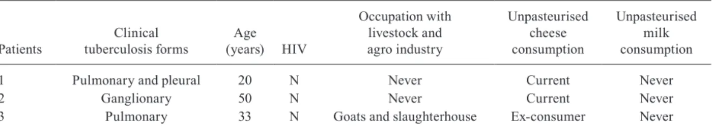
TABLE III
Comparative characteristics of three patients who were Mycobacterium bovis carriers, city of Juiz de Fora, state of Minas Gerais, Brazil, 2008-2010
Patients
Clinical tuberculosis forms
Age (years) HIV
Occupation with livestock and agro industry
Unpasteurised cheese consumption
Unpasteurised milk consumption
1 Pulmonary and pleural 20 N Never Current Never
2 Ganglionary 50 N Never Current Never
ACKNOWLEDGEMENTS
To the CITO, EBLEN, Cytopathology, Macro-regional and Ezequiel Dias Foundation, by kindly provide us the clini-cal specimens, and to Marcos Thadeu Rangel L’Hotellier, Mayumi Seito and Mércia Guadalupe Ramos, for assistance in the project.
REFERENCES
Barouni AS, Augusto CJ, Lopes MT, Zanini MS, Salas CE 2004. A
pncA polymorphism to differentiate between Mycobacterium bo-vis and Mycobacterium tuberculosis. Mol Cell Probes18: 167-170.
Cornejo BJ, Sahagun-Ruiz A, Suarez-Guemes F, Thornton CG, Ficht TA, Adams LG 1998. Comparison of C-18-carboxypropylbetaine and glass bead DNA extraction methods for detection of Myco-bacterium bovis in bovine milk samples and analysis of samples by PCR. Appl Environ Microbiol64: 3099-3101.
Corrêa CNM, Corrêa WM 1974. Tuberculose humana por bacilo bo-vino em São Paulo, Brasil. Arq Inst Biol41: 131-134.
Cosivi O, Grange JM, Daborn CJ, Raviglione MC, Fujikura T, Cous-ins D, RobCous-inson RA, Huchzermeyer HF, de Kantor I, Meslin FX 1998. Zoonotic tuberculosis due to Mycobacterium bovis in de-veloping countries. Emerg Infect Dis4: 59-70.
Dankner WM, Davis CE 2000. Mycobacterium bovis as a significant cause of tuberculosis in children residing along the United States-Mexico border in the Baja California region. Pediatrics105: e79.
Dankner WM, Waecker NJ, Essey MA, Moser K, Thompson M, Da-vis CE 1993. Mycobacterium bovis infections in San Diego: a clinicoepidemiologic study of 73 patients and a historical review of a forgotten pathogen. Medicine (Baltimore) 72: 11-37.
de Kantor IN, Ambroggi M, Poggi S, Morcillo N, da Silva Telles MA, Osório Ribeiro M, Garzón Torres MC, Llerena Polo C, Ribón W, García V, Kuffo D, Asencios L, Vásquez Campos LM, Rivas C, de Waard JH 2008. Human Mycobacterium bovis infection in ten Latin American countries. Tuberculosis(Edinb) 88: 358-365.
Dean AG, Dean JA, Coulombier D, Brendel KA, Smith DC, Burton AH, Dicker RC, Sullivan K, Fagan RF, Arner TG 1994. Epi Info Version 6.01. A word processing, database and statistics program for epidemiology on microcomputers, Centers for Disease Con-trol and Prevention, Atlanta, GA.
Drobniewski F, Strutt M, Smith G, Magee J, Flanagan P 2003. Audit of scope and culture techniques applied to samples for the diag-nosis of Mycobacterium bovis by hospital laboratories in England and Wales. Epidemiol Infect130: 235-237.
Evans JT, Smith EG, Banerjee A, Smith RMM, Dale J, Innes JA, Hunt D, Tweddell A, Wood A, Anderson C, Hewinson RG, Simth NH, Hawkey PM, Sonnenberg P 2007. Cluster of human tuberculosis caused by Mycobacterium bovis: evidence for person-to-person transmission in the UK. Lancet369: 1270-1276.
Gutiérrez MC, Galán JC, Blázquez J, Bouvet E, Vincent V 1999. Mo-lecular markers demonstrate that the first described multidrug-resistant Mycobacterium bovis outbreak was due to Mycobacte-rium tuberculosis. J Clin Microbiol37: 971-975.
Kent PT, Kubica GP 1985. Public health mycobacteriology: a guide for the level III laboratory, US Department of Health and Hu-man Services/Public Health Service/Centers for Disease Control, Atlanta, 207 pp.
Konno K, Feldman FM, McDermott W 1967. Pyrazinamide suscep-tibility and amidase activity of tubercle bacilli. Am Rev Respir Dis 95: 461-469.
LoBue PA, Betacourt W, Peter C, Moser KS 2003. Epidemiology of
Mycobacterium bovis disease in San Diego county, 1994-2000.
Int J Tuberc Lung Dis7: 180-185.
MAPA - Ministério da Agricultura, Pecuária e Abastecimento Brasil 2006. Programa Nacional de Controle e Erradicação da Bruce-lose e da TubercuBruce-lose Animal (PNCEBT), MAPA/SDA/DSA, Brasília, 188 pp.
MS - Ministério da Saúde Brasil 2005. Manual de bacteriologia da tuberculose, 3rd ed., Centro de Referência Professor Hélio Fra ga/ Secretaria de Vigilância em Saúde, Rio de Janeiro, 240 pp.
Ordoñez PT, Sauzo FM, Flores MAS, Casillas ICR 1999. Aislamiento e identificación de Mycobacterium bovis a partir de muestras de expectoración de pacientes humanos com problemas respiratóri-os crônicrespiratóri-os. Vet Mex 30: 227-229.
Pérez-Guerrero L, Milián-Suazo F, Arriga-Díaz C, Romero-Torres C, Escartín-Chávez M 2008. Epidemiología molecular de las tuber-culosis bovina y humana em uma zona endêmica de Querétaro, México. Salud Publica Mex50: 286-291.
Rocha A, Elias AR, Sobral LF, Soares DF, Santos AC, Marsico AG, Hacker MA, Caldas PC, Parente LC, Silva MR, Fonseca L, Suf-fys P, Boéchat N 2011. Genotyping did not evidence any contri-bution of Mycobacterium bovis to human tuberculosis in Brazil.
Tuberculosis (Edinb) 91: 14-21.
Rodwell TC, Kapasi AJ, Moore M, Milian-Suazo F, Harris B, Guer-rero LP, Moser K, Strathdee SA, Garfein RS 2010. Tracing the ori-gins of Mycobacterium bovistuberculosis in humans in the USA to cattle in Mexico using spoligotyping. Int J Infect Dis14: 29-35.
Samper S, Martín C, Pinedo A, Rivero A, Blázquez J, Baquero F, van Soolingen D, van Embden J 1997. Transmission between HIV-infected patients of multidrug-resistant tuberculosis caused by
Mycobacterium bovis. AIDS11: 1237-1242.
Scorpio A, Zhang Y 1996. Mutations in pncA, a gene encoding pyrazi-namidase/nicotinamidase, cause resistance to the antituberculous drug pyrazinamide in tubercle bacillus. Nat Med2: 635-636.
Silva MR, Guimarães MDC, de Oliveira VM, Moreira AS, da Costa RR, Abi-Zaid KCF, Rocha AS, Suffys PN 2011. Identification of Mycobacterium tuberculosis complex based on amplifica-tion and sequencing of the oxyR pseudogene from stored Ziehl-Neelsen-stained sputum smears in Brazil. Mem Inst Oswaldo Cruz106: 9-15.
Sreevatsan S, Escalante P, Pan X, Gillies DA 2nd, Siddiqui S, Khalaf CN, Kreiswirth BN, Bifani P, Adams LG, Ficht T, Perumaalla VS, Cave MD, van Embden JD, Musser JM 1996. Identification of a polymorphic nucleotide in oxyR specific for Mycobacterium bovis. J Clin Microbiol34: 2007-2010.
Taylor GM, Goyal M, Legge AJ, Shaw RJ, Young D 1999. Genotypic analysis of Mycobacterium tuberculosis from medieval human remains. Microbiology145: 899-904.
Van Embden JDA, Cave MD, Crawford JT, Dale JW, Eisenach KD, Gicquel B, Hermans P, Martin C, McAdam R, Shinnick TM 1993. Strain identification of Mycobacterium tuberculosis by DNA fin-gerprinting: recommendation for a standardized methodology.
J Clin Microbiol31: 406-409.
Wards BJ, Collins DM, de Lisle GW 1995. Detection of Mycobacte-rium bovis in tissues by polymerase chain reaction. Vet Microbiol 43: 227-240.
Zink AR, Nerlich AG 2004. Molecular strain identification of the
Mycobacterium tuberculosis complex in archival tissue samples.