Use o f fluo re sce nt pro be s to fo llo w
m e m brane traffic in ne rve te rm inals
Laboratório de Neurofarmacologia, Departamento de Farmacologia, Instituto de Ciências Biológicas, Universidade Federal de Minas Gerais, Belo Horizonte, MG, Brasil
C. Guatimosim, M.A. Romano-Silva, M.V. Gomez and M.A.M. Prado
Abstract
Optical tracers in conjunction with fluorescence microscopy have become widely used to follow the movement of synaptic vesicles in nerve terminals. The present review discusses the use of these optical methods to understand the regulation of exocytosis and endocytosis of synaptic vesicles. The maintenance of neurotransmission depends on the constant recycling of synaptic vesicles and important insights have been gained by visualization of vesicles with the vital dye FM1-43. A number of questions related to the control of recycling of synaptic vesicles by prolonged stimulation and the role of calcium to control membrane internalization are now being addressed. It is expected that optical monitoring of presynaptic activity coupled to appropriate genetic models will contribute to the understanding of membrane traffic in synaptic terminals.
Co rre spo nde nce
M.A.M. Prado
Laboratório de Neurofarmacologia Departamento de Farmacologia ICB, UFMG
Av. Antonio Carlos, 6627 31270-901 Belo Horizonte, MG Brasil
Fax: + 55-31-499-2695 E-mail: mprado@ icb.ufmg.br
Presented at the International Symposium: Biological Applications of Confocal Microscopy, Belo Horizonte, MG, Brasil, April 6-8, 1998.
Research supported by PADCT, PRPq-UFMG, FAPEMIG, CNPq, FINEP and PRO NEX.
Received May 14, 1998 Accepted June 3, 1998
Ke y wo rds
•Laser scanning confocal microscopy
•FM1-43
•Exocytosis
•Synaptic vesicles
•Endocytosis
Intro ductio n
In the nervous system, the most common type of communication between cells is pro-vided by local release of chemical mediators known as neurotransmitters. The activity of specific vesicular transporters accumulates these chemical messengers into synaptic vesicles from where they are released to the synaptic cleft. The release of neurotransmit-ters depends on the fusion (exocytosis) of the synaptic vesicle membrane with specialized regions of the plasma membrane. Further-more, in order to maintain constant the size of the membrane surface and especially to avoid depletion of the vesicle pool, synaptic vesicles are retrieved (endocytosis), recycled locally and refilled with neurotransmitter (1-3). Over the last six years, the introduction of
optical tracer methods has been responsible for improving our understanding about mem-brane trafficking in nerve terminals (4,5). Since nerve endings are generally too small for electrophysiological measurements, op-tical analysis of individual active zones is a promising technique to monitor membrane dynamics in intact terminals, and provides an attractive alternative to follow exo-en-docytosis linked to transmitter release (4).
In this review, we focus on recent ad-vances on how the use of optical tracers and fluorescence microscopy has helped to an-swer important questions related to the re-lease of neurotransmitters.
Pro te ins invo lve d in e xo cyto sis
vesicles store classical neurotransmitters for release (3). Under resting conditions, some of the vesicles can be found docked at spe-cialized plasmalemmal sites (active zones). The arrival of a nerve impulse in the terminal region is followed by calcium influx through different subtypes of voltage-activated cal-cium channels preferentially located in the active zone (6,7). A rise in intracellular cal-cium increases the probability of fusion of synaptic vesicles with the axolemma in the active zone, most likely due to a myriad of protein-protein interactions (8). This issue has been the focus of attention of several reviews in the past years and will not be covered in depth here; readers are directed to recent reviews of this topic (8,9).
Dodge Jr. and Rahamimoff (10) demon-strated a non-linear dependence of the exci-tatory post-synaptic potential on the external calcium concentration. They concluded that a cooperative action of four calcium ions is necessary to stimulate quantal release of neu-rotransmitters, suggesting the participation of a calcium receptor in the exocytotic ma-chinery. One of the proteins that fulfill many of the requirements for a calcium sensor is synaptotagmin I. This protein has two C2 domains that bind phospholipids and cal-cium (11,12). Indeed, genetic and biochemi-cal evidence has clearly shown the need of functional synaptotagmin I for fast exocyto-sis in a number of preparations, although its postulated role as a calcium sensor may be shared by other members of the min family (13). Unexpectedly, synaptotag-min has also been shown to have a role in endocytosis of synaptic vesicles (14,15).
It is likely that a number of protein-pro-tein interactions take place during the synap-tic vesicle cycle. Synaptobrevin, syntaxin and SNAP-25, proteins present in the synap-tic vesicle and plasma membrane, have been shown to form a stable complex in vitro (16). This may form the basis for binding of cyto-solic proteins, soluble NSF attachment
pro-teins (SNAPs) and NSF, to form a core com-plex which is of importance for exocytosis (16). The three former proteins are also the targets of clostridium toxins, proteases that impair synaptic transmission (17), which strongly implicates synaptobrevin, syntaxin and SNAP-25 in exocytosis.
There are controversies concerning the role played by each synaptic protein in mobi-lization, docking, priming, fusion, fission and recycling of synaptic vesicles. It is hoped that functional optical assays used concomi-tantly with appropriate genetic models could help understand some of the roles of synap-tic proteins in membrane traffic.
O ptical analysis o f e xo cyto sis
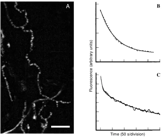
Several probes useful in following pre-synaptic membrane activity in live nerve terminals have been described (5). So far, the most popular way to visualize vesicle dynamics in synaptic terminals has been the use of the styryl dye FM1-43 (5). The evi-dence that supports the proposed mechanism for staining with FM1-43 can be summa-rized as follows: axonal terminals stain in an activity-dependent fashion with FM1-43, which generates fluorescent puncta (Figure 1A) located in close opposition to nicotinic receptors at the neuromuscular junction (NMJ) (4), and which colocalizes with synapsin I in hippocampal terminals (18).
Figure 1 - Staining and destaining of frog motor nerve terminals labeled w ith FM 1-43.
A, M otor nerve terminals from frog cutaneous pectoris muscle w ere labeled w ith FM 1-43 during electrical stimulation of the nerve (20 Hz, 5 min). After a w ashing period, FM 1-43-stained terminals w ere selected using a Zeiss fluorescence microscope (axiovert 100) w ith a 40-X w ater immersion objective (1.2 NA). The nerve terminals w ere imaged using an M RC 1024-BioRad argon UV laser scanning confocal microscope coupled to the Zeiss microscope. The preparation w as excited at 488 nm and light emitted above 515 nm w as collected for analysis. This figure show s a low magnification confocal image obtained after reconstruc-tion of a series of Z-secreconstruc-tions (0.5 µm apart, iris at 1.7). Note the presence of several fluorescent puncta, each of w hich corresponds to a cluster of synaptic vesicles labeled w ith FM 1-43. Bar, 30 µm.
B, FM 1-43-labeled motor nerve terminals such as those described in A w ere electrically stimulated through the nerve (20 Hz, 5 min) in dye-free medium. The fluorescent puncta destained progressively during stimulation and the loss of fluorescence w as quantified as mean fluorescence intensity in 256 levels of grey. The dashed curve represents the mean intensity of fluorescence destaining of 17 fluorescent puncta and the solid line corresponds to the fitting of the destaining curve by a single exponential equation (fluorescence (t) = Ae-t/τ + C). The destaining curve presented a τ = 58 ± 4 s (data from Ref. 32).
C, Rat cerebrocortical synaptosomes w ere labeled w ith FM 1-43 according to Ref. 27. After labeling, the synaptosomes w ere w ashed in dye-free medium and transferred to a fluorimeter cuvette. Fluorescence measurements w ere performed w ith a PTI spectrofluorimeter, at 488 nm (excitation) and 560 nm (emission). FM 1-43-labeled synaptosomes w ere destained under depolarization w ith 60 mM KCl. The destaining curve (dashed line) represents the mean of tw elve independent experiments and is described by a double exponential equation (fluorescence (t) = A1e-t/τ1 + A2e-t/τ2 + C), w ith time constants of 8 ± 1 s and 129 ± 9 s
for the first and second components, respectively. The solid line represents the fitting of the FM 1-43-destaining curve for synaptosomes.
Time (50 s/division)
F
lu
o
re
s
c
e
n
c
e
(
a
rb
it
ra
ry
u
n
it
s
)
A
B
C
A key characteristic of FM dyes is that they are several-fold more fluorescent when dissolved in the membrane than when in aqueous environment. Therefore, synaptic vesicle clusters labeled with FM1-43 are very bright (Figure 1A), but they dim due to dye release during exocytosis (4; see Figure 1B and C). Consequently, destaining fol-lows most of the characteristics of neurotrans-mitter release (21,22; see Figure 1B and C). Several model preparations are suitable for optical imaging of presynaptic activity with FM1-43. A number of studies have shown that FM1-43 staining and destaining are calcium dependent (4,18,23-25). In fact, in hippocampal and cerebellar granule cells in culture, as well as in rat brain synapto-somes, FM1-43 measurements have been used to investigate the nature of calcium channels coupled to exocytosis (23,26,27). Thus, in field stimulated cultured cells, N-type calcium channels blocked by ω -conotoxin GVIA have been shown to contri-bute distinctly to exocytosis in different popu-lations of nerve endings (23). Two classes of channels may be present in the same nerve terminal and act in concert to trigger and regulate exocytosis (28). In cerebellar gran-ule cells maintained in culture, identification of release sites and calcium imaging of neurites have also indicated a role for N- and P/Q type calcium channels in exocytosis (29). Interestingly, P/Q type calcium chan-nels seemed to be more effectively coupled to FM2-10 exocytosis than N-type channels (29), confirming previous observations that the former may be more effectively coupled to neurotransmitter release (30). In synapto-somes depolarized with KCl, only ω-agatoxin IVA-sensitive channels seemed to partici-pate in FM1-43 release, suggesting that P/Q channels (the targets of ω-agatoxin IVA) may be present in the majority of nerve terminals from rat cortex to control transmit-ter release (27).
Analysis of FM1-43 release from cul-tured neurons indicates that synaptic vesicle
pool depletion (100 to 200 vesicles) occurs with a τ of 20 s at 10-Hz stimulation (31). At the frog neuromuscular junction, pool deple-tion (500 to 1000 vesicles) occurs with a τ of 120 and 60 s at 10 Hz and 20 Hz of stimula-tion, respectively (22,32; see Figure 1B). FM1-43 release from frog NMJ can be fitted by a single exponential function at least dur-ing strong stimulation (22,32; see also Fig-ure 1B). In contrast, kinetic analysis of FM1-43 release from synaptosomes shows two components of destaining: a fast component with a τ of 8 ± 1 s, which probably reflects the time for the dye to be released from the membranes of rapidly exocytosed vesicles, and a second component which releases dye much slower (τ 129 ± 9 s; Figure 1C; 33). It is noteworthy that a recent abstract reports two rates of FM dye release for stimulated hippocampal neurons (34), similar to that observed in synaptosomes. Although these differences were unappreciated before, they may provide insight into the characteristics of exo-endocytosis of different preparations. For instance, dye release from synaptic vesicles probably slows down when unla-beled vesicles recycle back to the pool and start to compete for release sites in active zones. Thus, differences in exponential char-acteristics of dye release from the above preparations may underlie small differences in the rates of synaptic vesicle recycling and repriming.
in-crease exocytosis (37). Since one of the dif-ficulties in assigning a locus to long-term potentiation has always been the inability to assess presynaptic activity directly, the use of FM1-43 may provide new paradigms to study synaptic plasticity. Further experiments performed by the same group (38) concerned dye internalization by only a few synaptic vesicles. The authors found that the charac-teristics of dye internalization are compat-ible with the quantal nature of transmitter release, again an issue that has always been difficult to address in central synapses due to technical limitations imposed by post-syn-aptic detection of transmitter release. Thus, optical methods have provided unique in-sights into pre-synaptic mechanisms.
However, alternative approaches to fol-low synaptic vesicle and secretory granule dynamics in living cells have also contrib-uted to our knowledge of membrane traffic. One of these takes advantage of fluorophoro-conjugated antibodies against intraluminal domains of synaptotagmin I (39) to label synaptic vesicles, thus providing a probe to monitor long-term changes in synaptic activ-ity.
Recently, secretory granule movement has been assessed using the evanescent-wave setup of a laser beam that suffers total reflec-tion on the coverslip/cell interface (40). Com-bining the use of fluorescent probes such as green-fluorescent protein-tagged chromo-granin (40) and acridine orange spheres (41), in conjunction with evanescent-wave fluo-rescence microscopy, it was possible to fol-low the movement of the granules that are adjacent to the coverslip, beneath the plas-malemma (in a 300-nm thin layer of cytosol) in PC12 cells.
O ptical analysis o f synaptic ve sicle re cycling
Studies of release of neurotransmitters and exocytosis have gained by the use of optical tracers such as FM1-43, but it is on
the investigation of endocytosis and synap-tic vesicle recycling that opsynap-tical monitoring and confocal microscopy have had a greater impact. The fate of the membrane after exo-cytosis has been a difficult issue to investi-gate which was amenable only to electron microscopy in fixed cells. Optical methods allowed the investigation of the dynamics of synaptic vesicle endocytosis in living cells. A number of experiments performed with FM1-43 have led to the suggestion that after exocytosis recycled synaptic vesicles fully mix with the pool of pre-existing vesicles in a given vesicle cluster (21,31). However, synaptic vesicle clusters at the NMJ are quite stable, and it seems that little movement between vesicle clusters occurs, as shown by the lack of recovery of fluorescence after photobleaching experiments (42). It is of interest that a putative mechanism for the translocation of vesicles in the nerve termi-nal can be unmasked by the phosphatase inhibitor okadaic acid; however, how this mechanism works without pharmacological manipulation remains a mystery (43).
of neurotransmitters, the goldfish bipolar terminal, endocytosis measured by capaci-tance seems to occur faster, with a time constant of 2 s (45).
Pro te ins invo lve d in the e ndo cyto sis o f synaptic ve sicle s
There are a number of proteins that seem to be necessary for endocytosis of synaptic vesicles. Purification of vesicles from mam-malian brain indicated that most recycled vesicles contain clathrin (46). In fact, early electron microscopy evidence had led to the proposal that synaptic vesicles internalize after exocytosis through clathrin-coated pits (1). Moreover, suppression of the α-adaptin
gene in Drosophila generates mutants with impaired synaptic transmission due to a de-fect of vesicle recycling (47). This gene codes for a protein that is part of AP-2, a protein complex that seems to work as an adapter for the interaction of clathrin with the synaptic vesicle membrane. It is noteworthy that the AP-2 binding site in synaptic vesicles is the putative exocytotic calcium sensor synapto-tagmin (48). This result, together with evi-dence demonstrating lack of vesicle recy-cling during suppression of synaptotagmin activity (14,15), indicates a fundamental role for synaptotagmin in endocytosis.
Another protein, dynamin, a GTPase that is concentrated in nerve terminals (49), seems to play a key role in reformation of synaptic vesicles. Impairment of the GTPase activity of dynamin in the temperature-sensitive mu-tant shibire affects neurotransmission and immobilizes the fly in non-permissive tem-peratures by arresting endocytosis (50). Blockage of GTP hydrolysis by dynamin with GTPγS leads to the appearance of tubu-lar membrane invaginations coated with dynamin rings, thus pointing to a role of dynamin in the fission of vesicles from the plasma membrane or from an endosomal intermediary (51).
Many other key proteins that interact with
dynamin and/or clathrin are also required for the internalization of synaptic vesicle mem-brane. For example, amphiphysin is colocal-ized in nerve terminals with dynamin, inter-acting with the former by its SH3-domain. Recently, Shupliakov et al. (52) injected pep-tides that compete with amphiphysin for an SH3-binding site in dynamin and observed an impairment of synaptic vesicle endocyto-sis which resulted in depression of neu-rotransmitter release during prolonged stim-ulation. In addition, no dynamin rings were detected in the intermediate stage of endocy-tosis observed by blocking the interaction of amphiphysin with dynamin, suggesting that the former may act on the recruitment of dynamin during endocytosis (52).
It is thus possible that most of the en-docytosis occurring in nerve endings is clathrin dependent; however, it is not known whether this works similarly to other clathrin-mediated events or presents any specializa-tion at nerve endings. A particular point of interest is whether the availability of pro-teins involved in clathrin targeting or assem-bly may become rate limiting, when large amounts of synaptic vesicle membrane have to be retrieved from the terminal membrane. Nonetheless, perturbations in vesicle recy-cling seem to rapidly impair neurotransmis-sion, underlying the key role of recycling in maintaining the release of neurotransmit-ters.
Re gulatio n o f synaptic ve sicle re cycling
(44,54). However, at the NMJ, staurospo-rine has been reported to affect FM1-43 release in a manner that is consistent with a rapid opening and closure of a fusion pore, without complete flattening of the vesicle membrane (55).
Several factors may affect the rate of endocytosis during neuronal stimulation. For example, the duration of stimulation trains can delay the rate of endocytosis in frog NMJ (44). However, in cultured hippocam-pal cells, different periods (1 or 5 s at 20 Hz) of stimulation do not seem to change much the bulk endocytosis, although the initial rate of membrane internalization seems to be faster with longer periods of activity (5 s at 20 Hz) (54). Interestingly, in this prepara-tion, endocytosis seems to occur twice as fast at more physiological temperatures.
One of the factors that could have an important influence on different aspects of membrane cycling is the localized influx of calcium, which is necessary to elicit exocy-tosis. An early report using black widow spider venom shows that exocytosis can be evoked in the absence of calcium, but under these conditions membrane internalization is severely impaired in poisoned terminals. This ultrastructural study led to the sugges-tion that endocytosis involves a calcium-dependent step (56).
It is somewhat tricky to separate the ac-tions of calcium from exocytosis and en-docytosis, since calcium is a requirement for the former, and regulated endocytosis oc-curs only after synaptic vesicle incorpora-tion into the plasma membrane. Thus, there is some controversy whether calcium plays a role in endocytosis during physiological stim-ulation. The internalization of FM1-43 in hippocampal cells does not seem to be much altered by treatments aimed to change the intracellular calcium concentration (18,54). Consistent with that, the rate of dye internal-ization does not seem to correlate with the instantaneous level of intraterminal calcium at the frog NMJ (44). Although the above
observations suggest that the average level of calcium inside the terminal may not influ-ence much synaptic vesicle retrieval, there is a possibility that when calcium enters and evokes exocytosis, it could activate the pro-cess of membrane internalization.
In the shibire fly, where endocytosis is arrested in a temperatusensitive way, re-covery of synaptic vesicles at permissive temperatures in retinal cells occurs by two pathways, one located in the retinal dense bodies and dependent on the presence of extracellular calcium, and the other that is independent of calcium and seems to be located at ectopic sites (57). A drastic cal-cium effect has been observed in goldfish bipolar cells, where high internal calcium concentration seemed to arrest endocytosis (45). However, care must be taken since it is possible that a decrease in the rate of mem-brane retrieval observed by memmem-brane ca-pacitance may also occur due to increasing rates of exocytosis (58). Thus, independent measurements of endocytosis may have to be performed in order to confirm the sugges-tion of inhibisugges-tion of membrane retrieval by Ca2+ in goldfish bipolar cells. In addition, in
chromaffin cells and other secretory cells that secrete granules, fast endocytosis re-quires Ca2+ (59). Interestingly, Sr2+ or Ba2+
was not able to replace Ca2+, suggesting a
unique divalent cation requirement for the putative calcium sensor of secretory gran-ules (59).
The above observations prompted us to examine the recycling of synaptic vesicles by FM1-43 imaging using confocal micros-copy during substitution of Sr2+ by Ca2+ at
the frog NMJ (32). In these experiments, internalization of FM1-43 after trains of nerve stimulation (2400 or 4800 pulses) was im-paired in the presence of Sr2+ when
com-pared to Ca2+-bathed muscles. This
Sr2+ to 10 mM (1.8 mM was normally used)
during stimulation with 2400 action poten-tials. The exocytosis of vesicles was not altered by Sr2+ when compared to that seen
in Ca2+ medium, suggesting that the
inhibi-tion of FM1-43 internalizainhibi-tion by Sr2+ was
not due to a decrease of vesicle fusion (32). Taken together, the results suggested that Sr2+ is much less effective than Ca2+ in
sup-porting endocytosis, but upon reaching a certain threshold of intracellular concentra-tion it triggers endocytosis.
If indeed Ca2+ is required to control the
retrieval of synaptic vesicles, it becomes of interest to determine the identity of the puta-tive calcium sensor for endocytosis similar to the postulated role of calmodulin in chro-maffin granules (59). Synaptotagmin may be a candidate, in view of its ability to bind Ca2+
and to affect endocytosis. Dynamin also binds Ca2+ and its GTPase activity is controlled by
this divalent ion (60). Moreover, activation of protein kinases or phosphatases may also be a target through which Ca2+ could
influ-ence the internalization of synaptic vesicles. A number of important points therefore remain open, such as whether endocytosis presents distinct sensitivity to calcium com-pared to exocytosis, what is the identity of a putative calcium sensor for membrane inter-nalization, which factors may control the rates of endocytosis, and finally whether other steps related to the recycling of vesicles are influenced by the intracellular concen-tration of calcium. Thus, in the near future, using the optical methods described herein, we can expect novel insights into the dynam-ics of membrane trafficking.
Re fe re nce s
1. Reuser JE & Reese TS (1973). Evidence for recycling of synaptic vesicle mem-brane during transmitter release at the frog neuromuscular junction. Journal of
Cell Biology, 57: 315-344.
2. Ceccarelli B, Hurbult WP & M auro A (1973). Turnover of transmitter and syn-aptic vesicles at the frog neuromuscular junction. Journal of Cell Biology, 57: 499-524.
3. Kelly RB (1993). Storage and release of neurotransmitters. Cell,72 (Suppl): 43-53. 4. Betz WJ, M ao F & Bew ick GS (1992). Activity-dependent fluorescent staining and destaining of living vertebrate motor nerve terminals. Journal of Neuroscience, 12: 363-375.
5. Betz WJ, M ao F & Smith CB (1996). Imag-ing exocytosis and endocytosis. Current
Opinion in Neurobiology,6: 365-371.
6. Robitaille R, Adler EM & Charlton M P (1990). Strategic location of calcium chan-nels at transmitter release sites of frog neuromuscular junction. Neuron,5: 773-779.
7. M artin-M outot N, Charvin N, Leveque C, Sato K, Nishiki T, Kosaki S, Takahashi M & Seagar M (1996). Interaction of SNARE
complexes w ith P/Q-type calcium chan-nels in rat cerebellar synaptosomes.
Jour-nal of Biological Chemistry, 271:
6567-6570.
8. Augustine GJ, Burns M E, De Bello WM , Pettit DL & Schw eizer FE (1996). Exocy-tosis: proteins and perturbations. Annual
Review of Pharmacology and Toxicology,
36: 659-701.
9. Südhof TC (1995). The synaptic vesicle cycle: a cascade of protein-protein inter-actions. Nature, 375: 645-653.
10. Dodge Jr FA & Rahamimoff R (1967). Co-operative action of calcium ions in trans-mitter release at the neuromuscular junc-tion. Journal of Physiology, 193: 419-432. 11. Davletov BA & Sudhof TC (1993). A single C2 domain from synaptotagmin I is suffi-cient for high affinity Ca2+/phospholipid
binding. Journal of Biological Chemistry, 268: 26386-26390.
12. Chapman ER, An S, Edw ardson JM & Jahn R (1996). A novel function for the second C2 domain of synaptotagmin. Ca2+-triggered dimerization. Journal of
Biological Chemistry,271: 5844-5849.
13. Geppert M , Goda Y, Hammer RE, Li C, Rosahl TW, Stevens CF & Sudhof TC
(1994). Synaptotagmin I: a major Ca2+
sensor for transmitter release at a central synapse. Cell,79: 717-727.
14. Jorgensen EM , Hartw ieg E, Schuske K, Nonet M L, Jin Y & Horvitz HR (1995). Defective recycling of synaptic vesicles in synaptotagmin mutants of
Caenorhabdi-tis elegans. Nature,378: 196-199.
15. Fukuda M , M oreira JE, Lew is FM , Sugimori M , Niinobe M , M ikoshiba K & Llinas R (1995). Role of the C2B domain of synaptotagmin in vesicular release and recycling as determined by specific anti-body injection into the squid giant syn-apse preterminal. Proceedings of the
Na-tional Academy of Sciences, USA, 92:
10708-10712.
16. Sollner T, Whiteheart SW, Brunner M , Erdjument-Bromage H, Geromanos S, Tempst P & Rothman JE (1993). SNAP receptors implicated in vesicle targeting and fusion. Nature,362: 318-324. 17. M ontecucco C & Schiavo G (1993).
Teta-nus and botulinum neurotoxins: a new group of zinc proteases. Trends in
Bio-chemical Sciences,18: 324-327.
(1993). The kinetics of synaptic vesicle recycling measured at single presynaptic boutons. Neuron, 11: 713-724.
19. Henkel AW, Lubke J & Betz WJ (1996). FM 1-43 dye ultrastructural localization in and release from frog motor nerve termi-nals. Proceedings of the National
Acade-my of Sciences, USA,93: 1918-1923.
20. Angleson JK & Betz WJ (1997). M onitor-ing secretion in real time: capacitance, am perom et ry and f luorescence com -pared. Trends in Neurosciences,20: 281-287.
21. Betz WJ & Bew ick GS (1992). Optical a-nalysis of synaptic vesicle recycling at the frog neuromuscular junction. Science, 255: 200-203.
22. Betz WJ & Bew ick GS (1993). Optical monitoring of transmitter release and syn-aptic vesicle recycling at the frog neuro-muscular junction. Journal of Physiology, 460: 287-309.
23. Reuter H (1995). M easurements of exo-cytosis from single presynaptic nerve ter-minals reveal heterogeneous inhibition by Ca2+ channel blockers. Neuron,14:
773-779.
24. M effert M , Premack BA & Schulman H (1994). Nitric oxide stimulates Ca2+
-inde-pendent synaptic vesicle release. Neuron, 12: 1235-1244.
25. Smith CB & Betz WJ (1996). Simulta-neous independent measurement of en-docytosis and exocytosis. Nature, 380: 531-534.
26. Pocock JM , Cousin M A, Parkin J & Nicholls DG (1995). Glutamate exocytosis from cerebellar granule cells: the mech-anism of a transition to an L-type Ca2+
channel coupling. Neuroscience, 67: 595-607.
27. Guatimosim C, Romano-Silva M A, Cruz JS, Beirão PSL, Kalapothakis E, M oraes-Santos T, Cordeiro M N, Diniz CR, Gomez M V & Prado M AM (1997). A toxin from the spider Phoneutria nigriventer that blocks calcium channels coupled to exo-cytosis. British Journal of Pharmacology, 122: 591-597.
28. Reuter H (1996). Diversity and function of presynaptic calcium channels in the brain.
Current Opinion in Neurobiology, 6:
331-337.
29. Cousin M A, Hurst H & Nicholls DG (1997). Presynaptic calcium channels and field-evoked transmitter exocytosis from cul-tured cerebellar granule cells. Neurosci-ence,81: 151-161.
30. M intz IM , Sabatini BL & Regehr WG (1995). Calcium control of transmitter
re-lease at a cerebellar synapse. Neuron,15: 675-688.
31. Ryan TA & Smith SJ (1995). Vesicle pool mobilization during action potential firing at hippocampal synapses. Neuron, 14: 983-989.
32. Guatimosim C, Romano-Silva M A, Gomez M V & Prado M AM (1998). Recycling of synaptic vesicles at the frog neuromuscu-lar junction in the presence of strontium.
Journal of Neurochemistry, 70:
2477-2483.
33. Prado M AM , Romano-Silva M A, Beirão PSL, Collier B, Gomez M V & Guatimosim C (1996). Calcium control of exocytosis measured by FM 1-43 fluorescence in mammalian synaptosomes. Society for
Neuroscience Abstracts, 22: 506.
34. Klingauf J, Kavalali E & Tsien RW (1997). Evidence for fast endocytosis at single synapses of hippocampal neurons in cul-ture. Annals of the International Confer-ence “ La transmission synaptique et sa
modulation”, 92 (Abstract).
35. Liu G & Tsien RW (1995). Synaptic trans-mission at single visualized hippocampal boutons. Neuropharmacology, 34: 1407-1421.
36. Isaacson JS & Hille B (1997). GABA (B)-mediated presynaptic inhibition of excita-tory transmission and synaptic vesicle dy-namics in cultured hippocampal neurons.
Neuron,18: 143-152.
37. Ryan TA, Ziv NE & Smith SJ (1996). Po-tentiation of evoked vesicle turnover at individually resolved synaptic boutons.
Neuron,17: 125-134.
38. Ryan TA, Reuter H & Smith SJ (1997). Optical detection of a quantal presynaptic membrane turnover. Nature, 388: 478-482.
39. Kraszew ski K, M undigl O, Daniell L, Verderio C, M atteoli M & De Camilli P (1995). Synaptic vesicle dynamics in living cultured hippocampal neurons visualized w ith CY3-conjugated antibodies directed against the lumenal domain of synapto-tagm in. Journal of Neuroscience, 15: 4328-4342.
40. Lang T, Wacker I, Steyer J, Kaether C, Wunderlich I, Soldati T, Gerdes HH & Almers W (1997). Ca2+-triggered peptide
secretion in single cells imaged w ith green fluorescent protein and evanes-cent-w ave microscopy. Neuron,18: 857-863.
41. Steyer JA, Horstmann H & Almers W (1997). Transport, docking and exocytosis of single secretory granules in live chro-maffin cells. Nature,388: 474-478.
42. Henkel AW, Simpson LL, Ridge RM & Betz WJ (1996). Synaptic vesicle move-ments monitored by fluorescence recov-ery after photobleaching in nerve termi-nals stained w ith FM 1-43. Journal of
Neu-roscience,16: 3960-3967.
43. Betz WJ & Henkel AW (1994). Okadaic acid disrupts clusters of synaptic vesicles in frog motor nerve terminals. Journal of
Cell Biology,124: 843-854.
44. Wu LG & Betz WJ (1996). Nerve activity but not intracellular calcium determines the time course of endocytosis at the frog neuromuscular junction. Neuron,17: 769-779.
45. Von Gersdorff H & M atthew s G (1994). Inhibition of endocytosis by elevated cal-cium in a synaptic terminal. Nature,370: 652-655.
46. M aycox PR, Link E, Reetz A, M orris SA & Jahn R (1992). Clathrin-coated vesicles in nervous tissue are involved primarily in synaptic vesicle recycling. Journal of Cell
Biology,118: 1379-1388.
47. Gonzalez-Gaitan M & Jackle H (1997). Role of Drosophila alpha-adaptin in pre-synaptic vesicle recycling. Cell,88: 767-776.
48. Zhang JZ, Davletov BA, Sudhof TC & Anderson RG (1994). Synaptotagmin I is a high affinity receptor for clathrin AP-2: im-plication for membrane recycling. Cell,78: 751-760.
49. Pow ell KA & Robinson PJ (1995). Dephos-phin/dynamin is a neuronal phosphopro-tein concentrated in nerve terminals: evi-dence from rat cerebellum. Neuroscience, 64: 821-833.
50. Koenig JH, Saito K & Ikeda K (1983). Re-versible control of synaptic transmission in a single gene mutant of Drosophila
melanogaster. Journal of Cell Biology,96:
1517-1522.
51. Takei K, M cPherson PS, Schmid SL & De Camilli P (1995). Tubular membrane in-vaginations coated by dynamin rings are induced by GTP-γS in nerve terminals. Na-ture, 374: 186-190.
52. Shupliakov O, Low P, Grabs D, Gad H, Chen H, David C, Takei K, De Camilli P & Brodin L (1997). Synaptic vesicle endocy-tosis impaired by disruption of dynamin-SH3 domain interactions. Science, 276: 259-263.
53. M eldolesi J & Ceccarelli B (1981). Exocy-tosis and membrane cycling. Philosophi-cal Transactions of the Royal Society of
London, B296: 55-65.
endocyto-sis. Proceedings of the National Academy
of Sciences, USA,93: 5567-5571.
55. Henkel AW & Betz WJ (1995). Staurospo-rine blocks evoked release of FM 1-43 but not acetylcholine from frog motor nerve terminals. Journal of Neuroscience, 15: 8246-8258.
56. Ceccarelli B & Hurlbut W (1980). Ca2+
-dependent recycling of synaptic vesicles at the frog neuromuscular junction.
Jour-nal of Cell Biology, 87: 297-303.
57. Koenig JH & Ikeda K (1996). Synaptic vesicles have tw o distinct recycling path-w ays. Journal of Cell Biology,135: 767-808.
58. Lagnado L, Gomis A & Job C (1996). Con-tinuous vesicle cycling in the synaptic ter-minal of retinal bipolar cells. Neuron, 17: 957-967.
59. Artalejo CR, Elhamdani A & Palfrey HC
(1996). Calmodulin is the divalent cation receptor for rapid endocytosis, but not exocytosis, in adrenal chromaffin cells.
Neuron, 16: 195-205.
60. Liu JP, Zhang QX, Baldw in G & Robinson PJ (1996). Calcium binds dynamin and in-hibits its GTPase activity. Journal of