WORK PERFORMED AT:
Forest Biotechnology Laboratory
Instituto de Biologia Experimental e Tecnológica
Instituto de Tecnologia Química e Biológica
Universidade Nova de Lisboa
Av. da República
2780-157 Oeiras
Portugal
SUPERVISOR:
Aos meus pais
“Learn from yesterday, live for today, hope for tomorrow. The important thing is to not stop questioning”
I would like to thank Célia Miguel for the opportunity she offered me to be a member of her lab. For being not only my supervisor and advisor but also for her friendship throughout these years. I am truly grateful for all you have taught me, for your good advices, your encouragement and for always believing and making me believe that I would be able to fulfill my goals. Thanks for all the availability during the writing of the thesis, for all the suggestions, improvements, for always being optimistic and for all your dedication. In a few words, Thank You Very Much!
To Brian Jones for his availability and good advice. For being critical when necessary and for all the words of encouragement.
To Professor Pinto Ricardo and Professor Margarida Oliveira for having accepted to be part of my PhD thesis committee. Thank you for the availability, for the words of encouragement and for always caring about my work.
To all my colleagues who worked with me. To Liliana Marum for all the support, for her good mood and friendship. Thank you for being my partner in the discovery of the “Cork oak world”. No one like you to understand the difficulties of working with this so peculiar tree species. To Ana Milhinhos, for all the debates and scientific discussions in the “Universe of Populus”. Thanks for all the companionship and friendship and for your dedication. To José de Vega-Bartol for being my partner in the “transcriptomics”. Thank you for all the patience in the bioinformatics and for the availability.
company and for your friendship; to Andreia Rodrigues for your companionship and friendship. Thank you for always caring about my work. To Inês Chaves, Inês Modesto, Ilanit, Raissa, Mariagrazia, Ana Maria, Marta Simões, Marta Madeira. Thank you all for your companionship, good mood and for the team spirit.
To everyone at the GPS lab, for all your kindness and for always being helpful.
To Eugénia, Pilar, Sónia and all the staff at the washing rooms. Without your help the Populus subcultures would have taken so much longer.
To everyone at the ITQB and IBET that in some way helped me over the years and to Ana Maria Portocarrero and Fátima Madeira for all the help, especially in the last steps of thesis preparation and delivery.
Um especial obrigada aos meus pais por sempre me terem apoiado. Não há palavras que descrevam a importância do vosso apoio. Obrigada pela paciência e pelas palavras de conforto, por todo o incentivo e confiança. O vosso amor e os vossos ensinamentos foram, são e sempre serão fundamentais.
Ao Ricardo por ser o meu melhor amigo e companheiro. Obrigada por toda a compreensão, companheirismo e paciência. Obrigada por teres estado sempre presente, por todo o carinho e incentivo. Esta é mais uma das etapas ultrapassadas a dois!
momentos de alegria e de distração.
Aos pais do Ricardo, à irmã e avós, por todo o incentivo e palavras de conforto. Senti-me como uma filha, irmã e neta.
Aos meus amigos por me terem proporcionado momentos de descontração. Obrigada por terem estado presentes nesta fase.
Um muito obrigada a todos, sem vocês teria sido muito mais difícil!
ACT ACTIN
At Arabidopsis thaliana
BCIP 5-bromo-4-chloro-3’-indolyphosphate
bp Base pair
CAC CLATHRIN ADAPTOR COMPLEX
CaMV 35S Cauliflower mosaic virus 35S promoter cDNA Complementary DNA
CEI Cortex/endodermal initial
CK Cytokinin
CKX CYTOKININ OXIDASE
cm Centimetre
Cq Quantification cycle CODB Cork oak database
COG Clusters of Orthologous Groups CV Coefficient of variation
DAG Days after germination DNA Deoxyribonucleic acid DNase Deoxyribonuclease
DE Differentially expressed DEG Differentially expressed gene
E Efficiency
EN Internode
EST Expressed Sequence Tag
FAA Formaldehyde, Acetic acid, Ethanol FDR False Discovery Rate
g Gram
GO Gene ontology
HK Housekeeping
HD-ZIP III Class III homeodomain leucine zipper
kb Kilobase
KEGG Kyoto Encyclopedia of Genes and Genomes M Stability value
Mbp Mega base pair
min Minute
miRNA MicroRNA mm Millimeter mM Millimolar
mRNA Messenger RNA NBT Nitro blue tetrazolium
nM Nanomolar
NF Normalization factor
ORF Open Reading Frame
PCR Polymerase Chain Reaction
Pg Phellogen
Ph Phellem
QC Quiescent center Qi Quercus ilex
Qs Quercus suber
RAM Root apical meristem
RG Reference gene
RNA Ribonucleic acid RNA-Seq RNA sequencing
RR7 A-type response regulator; Cytokinin primary response gene RT-qPCR Reverse Transcription Quantitative Polymerase Chain Reaction
SCR SCARECROW scr scarecrow mutant
SHR SHORT-ROOT
shr short-root mutant TF Transcription factor
uidA Encodes the β-glucuronidase enzyme VC Vascular cambium
Vn/n+1 Pairwise variation
WT Wild-type
X-Gluc 5-bromo-4-chloro-3-indolyl β-D-glucuronide º C Celsius degrees
µg Microgram
µl Microliter
µm Micrometer
µM Micromolar
studies were performed in Arabidopsis. None of the cork oak gene sequences fully complemented the Arabidopsis shr2 mutant phenotype, thus indicating that different SHR-like genes may have evolved different functions (neo-functionalization) or the function of the original gene may have been distributed between the copies (sub-functionalization) after genome duplication following the divergence of Populus/Quercus and Arabidopsis lineages. Altogether, the results obtained in the experiments conducted in Populus and cork oak, as well as previously published data, point to the involvement of SHR genes in the regulation of secondary growth, where SHR1 appears to act in the regulation of vascular cambium and SHR2 in the regulation of the phellogen.
successful regeneration programs for the species.
privação de água, bem como com a regulação da transcrição e o metabolismo dos hidratos de carbono. Aproximadamente um quarto dos genes diferencialmente expressos codificam para possíveis fatores de transcrição associados a vários processos biológicos refletindo uma atividade transcricional muito ativa e fortemente regulada durante o desenvolvimento do fruto. Enquanto estes genes se encontram distribuídos quase igualmente ao longo de todos os estádios de desenvolvimento analisados, os transcritos envolvidos na resposta à água ou no metabolismo dos hidratos de carbono estavam sobre-representados em estádios específicos. Este conjunto de dados representa uma ferramenta muito útil para a compreensão de vários aspetos da biologia do sobreiro, nomeadamente ao nível da reprodução e desenvolvimento inicial, que se podem revelar cruciais para a implementação de programas de regeneração de sobreiro.
Acknowledgements ... v
List of abbreviations ... ix
Summary ... xiii
Sumário ... xvii
Chapter I
General Introduction ... 1 Chapter II
A SHORT-ROOT-like gene (PtSHR2B) is involved in Populus phellogen activity ... 63 Chapter III
Reference gene selection for quantitative real-time PCR normalization in Quercus suber ... 95 Chapter IV
SHORT-ROOT-like genes are differentially regulated during secondary growth in Quercus suber ... 131 Chapter V
Transcriptomic profiling during acorn development in cork oak: a contribute towards a reference transcriptome ... 175 Chapter VI
Chapter I
Plant meristems
Plants have an open body in the sense that they are able to form new organs and highly specialized tissues by post-embryonic development. This ability depends on the activity of specialized structures called meristems, in which pluripotent stem cells are localized. By exhibiting the capacity for self-renewal and for producing progeny that follow various differentiation pathways, stem cells are crucial for the development of multicellular organisms. While in animals stem cells are used mainly to compensate for cell loss during post-embryonic development, in plants they are used in a continuous way to generate new structures during the entire plant’s life-span. These new structures include organs like roots, leaves, flowers, fruits and stems, but also specialized tissues formed during secondary growth such as wood and cork.
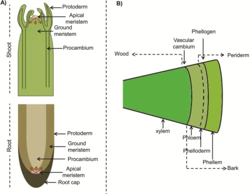
Based on their position in the plant body, meristems can be either apical or lateral (Esau, 1977). The apical meristems comprise the shoot and root apical meristems, termed as SAM and RAM, respectively, and are responsible for plant elongation through cell division that provides new cells for expansion, tissue differentiation or initiation of new organs (stems, leaves and roots). The cells produced in the apical meristems differentiate into primary tissues: protoderm, procambium and ground meristem (Fig. 1A). In the shoots the procambium will form the vascular tissues and the ground meristem will form the pith, whereas in the roots the procambium gives rise to the stele and the ground meristem to the cortex. The protoderm is responsible for the formation of the epidermis in both shoots and roots.
the formation of the periderm. A tree stem with secondary growth is characterized by the presence of a layer of wood derived from the vascular cambium, and a bark layer consisting of all the tissues outside the vascular cambium, comprehending the phloem and the periderm (Fig. 1B).
Fig. 1. Schematic representation of primary and secondary plant growth. (A) Primary growth in the shoot and root showing the apical meristems (SAM and RAM) and the three primary tissues: protoderm, procambium and ground meristem. The arrows show the cell proliferation direction. (B) Secondary growth in the stem showing the two lateral meristems: vascular cambium and cork cambium (phellogen). Periderm, bark and wood are also represented.
The vascular tissue system
vascular tissues allow the transport of water and nutrients along the plant making them less dependent on a very humid environment. Structural support is another essential function of the plant vasculature allowing growth in height as a way to compete for light. While the xylem transports water and minerals, the phloem is associated with the transport of organic nutrients. In addition, the vascular tissues provide a long-distance communication system, delivering information in the form of hormones and other molecules, for the coordination of developmental and physiological processes. During primary shoot growth the phloem is typically positioned parallel to the xylem and both conducting tissues are organized into vascular bundles.
Primary xylem and phloem develop from the activity of the procambium, a primary meristem formed during early embryogenesis, with narrow and cytoplasm dense cells organized in continuous strands (Scarpella and Meijer, 2004). The vascular cambium derives from the procambium within the vascular bundles (fascicular cambium) and from the parenchyma between the vascular bundles (interfascicular cambium), forming a continuous cylinder around the plant stem. Two types of cell divisions occur in the vascular cambium. The anticlinal divisions produce new cambial initials and the periclinal divisions of cambial initials and its derivatives produce secondary vascular tissues through differentiation into secondary xylem (wood) to the inside of the plant stem or root, and secondary phloem to the outside (Lachaud et al., 1999; Scarpella and Meijer, 2004), thus promoting growth in lateral directions (Esau, 1977). Cambial activity is seasonal and related to changes in temperature and humidity, and the length of the period of cambial activity depends on the species (Lachaud et al., 1999; Evert, 2006a).
Xylogenesis includes cell division and enlargement, cell wall thickening, lignification and programmed cell death (Fukuda, 1996; Myburg and Sederoff, 2001). Protoxylem cells are the first xylem cells to differentiate within the vascular bundles, developing in the innermost position in the shoot and next to the pericycle in the root, whereas the metaxylem cells differentiate later. Both xylem cell types can be distinguished based on their secondary cell wall. Protoxylem cells have ring-like (annular) or helical (spiral) cell wall thickenings, that can be stretched, making it possible to elongate in the growth direction, while metaxylem cells appear as net-like (reticulate) or porous (pitted) being more uniform and thicker than protoxylem and providing more support. During secondary cell wall thickening the deposition of substances like cellulose, lignin and hemicellulose occur.
Phloem is composed of sieve elements (sieve tubes and sieve cells) which are the conducting elements, and of non conducting cells such as fibers and sclereids, and parenchyma cells. Unlike the tracheary elements, the sieve elements are living cells and during differentiation sieve tubes lose most of their organelles, including nucleus (Raven et al., 2005; Schuetz et al., 2012). Fibers and sclereids are responsible for the mechanical support function and exhibit a rigid and very difficult to damage secondary cell wall.
Periderm development
the regulation of gas exchanges (Lendzian, 2006; Glover, 2010). An analogy between the cuticle-epidermis-stomata and the periderm has been made (Lendzian, 2006). During secondary growth the epidermis is replaced in the stems and roots by the periderm, resulting in an increased thickness of these organs (Evert, 2006b). It is composed by the phellogen or cork cambium, phellem, usually termed as cork, and phelloderm (Fig. 2). The function of stomata and cuticle are replaced by the lenticels and phellem cells, respectively (Lendzian, 2006).
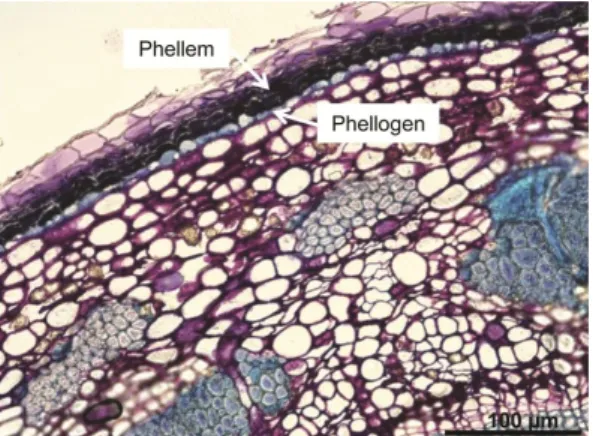
Fig. 2. Periderm development in a two year-old stem of hybrid aspen, Populus tremula L. × Populus tremuloides
Michx.; Clone T89. The phellogen and the phellem cells are highlighted by arrows.
Phellogen
The phellogen has a relatively simple structure consisting in only one cell type appearing rectangular in a transverse section, flattened radially and polygonal in tangential sections (Pereira, 2007). The first cell division as a precursor of phellogen formation is derived from a periclinal division that gives rise to two apparently similar cells. The inner daughter cell differentiates into phelloderm and no further divides while the outer cell undergoes a second periclinal division resulting again in the formation of two similar cells. The outer one differentiates into a phellem cell whereas the inner cell is the phellogen initial and continues to divide (Evert, 2006b; Pereira, 2007). However, more phellem cells are formed as a result of the division of the phellogen cells than phelloderm cells (Pereira, 2007). While most of the divisions are periclinal, some divisions have to occur in a radial anticlinal plane due to the increase in the circumference of the stems (Evert, 2006b). The number of phellem cell layers formed varies among species, and can be very large in the case of Quercus suber (Pereira, 2007).
seven months (Oliveira et al., 1994; Caritat et al., 1996; Fialho et al., 2001; Costa et al., 2002, 2003; Silva et al., 2005).
The annual rhythm of the two lateral meristems can overlap or can occur independently, also depending on the species (Waisel et al., 1967; Yáñes-Espinosa et al., 2010). The activity of the phellogen is considered relatively slow under normal conditions when compared to the activity of vascular cambium (Waisel et al., 1967; Liphschitz et al., 1984). Consequently, the stem radial growth has been mostly associated with the vascular cambium activity and to a lesser extent with the phellogen activity (Yáñes-Espinosa et al., 2010). The lifespan of the phellogen activity varies between species from less than one year to several years and the first phellogen remains functional for the entire tree life in only a few species. In most species other periderms form after the first one due to the development of a new phellogen (Pereira, 2007).
Phellem and phelloderm cells
confers resistance to pathogens (Lulai and Corsini, 1998). The number of phellem layers and their thickness also vary among species. In the genus Quercus, some species have an extensive production of phellem, as it is the case of Quercus suber, Quercus cerris and Quercus variabilis (Pereira et al., 1992; Graça and Pereira, 2004; Şen et al., 2011b; Miranda et al., 2012) whereas others, like Quercus faginea, produce a much thinner phellem layer (Quilhó et al., 2013). Quercus faginea and Quercus cerris usually produce 2-5 and 6-12 layers of phellem, respectively, but as a discontinuous layer around the trunk (Şen et al., 2011a,b; Quilhó et al., 2013). On the other hand, Quercus suber produces a continuous layer of 3 to 8 phellem layers surrounding the stems of one year-old branches and their numbers keep increasing reaching an annual production of 10-20 phellem layers (Graça and Pereira, 2004). In contrast, for example in Populus the thickness of the periderm seems to be constant in younger and older trees consisting of about 6 to 10 cell layers independently of the plant age (Kaufert, 1937).
Lenticels
Lenticels, also termed as lenticular channels, are special structures through which gas exchange can occur in the bark. They develop under the stomata (Evert, 2006b; Lendzian, 2006) and these regions are characterized by an intense phellogen activity (Graça and Pereira, 2004; Evert, 2006b) producing a tissue with numerous intercellular spaces as well as the lenticel phellogen. The lenticel phellogen is continuous with the other phellogen but bends inward and it forms filling tissue to the outside, that protrude above the surface through a fissure in the periderm, and phelloderm to the inside, which is usually thicker than usual (Evert, 2006b). The difference between phellem and the filling tissue varies among species but it is mainly due to the presence of intercellular spaces or to different cell shape (Evert, 2006b).
The presence of lenticels in the cork tissue contributes to its porosity and is a detrimental characteristic for industrial quality since large and abundant lenticular channels change cork properties and contributes to poor quality cork (Pereira et al., 1996; Anjos et al., 2008).
Regulatory mechanisms in plant meristems
Mechanisms regulating the apical meristems
Plant meristems are dynamic structures that are regulated to keep a tight balance between stem cell proliferation and differentiation. This balance ensures the maintenance of stem cell identity while keeping meristem size.
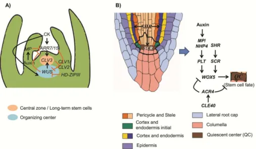
SHR/SCR in the establishment of the QC and stem cell position (Aida et al., 2004). PLT transcription is elevated by auxin accumulation and is dependent on AUXIN RESPONSE FACTORS (ARF) transcription factors, MP or the MP homolog NON-PHOTOTROPIC HYPOCOTYL4 (NPH4), that bind to the auxin-responsive elements (Aida et al., 2004) (Fig. 3B). Briefly, WUS and WOX5, as well as CLV3/CLV1 and CLE40/ACR4, seem to be functionally equivalent in stem cell control in the shoot and root, respectively.
Fig. 3. Schematic representation of equivalent regulatory mechanisms in the shoot (A) and root apical meristem (B). The thick black line in B) surrounds the quiescent center and the root stem cells. (A) Adapted from (Williams and Fletcher, 2005; Zhao et al., 2010) and (B) adapted from (Dinneny and Benfey, 2008; Stahl and Simon, 2009b).
Mechanisms regulating the vascular cambium
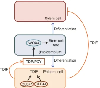
Fig. 4. Schematic representation of the TDIF-TDR-WOX4 signaling mechanism regulating the fate of procambial cells. Adapted from (Hirakawa et al., 2008, 2010; Miyashima et al., 2013).
In the Arabidopsis shoots the formation of the interfascicular cambium is initiated through cell divisions in parenchyma cells between primary bundles and in the starch sheath, the innermost cell layer of the cortex (Altamura et al., 2001; Sehr et al., 2010; Agusti et al., 2011). Identification of genes involved in the transition from primary to secondary growth in Arabidopsis demonstrated that stem weight triggers the transition from primary to secondary growth (Ko et al., 2004) but it is not sufficient to induce interfascicular cambium initiation, and instead it modulates the dynamics of secondary growth initiation and cambium activity (Ko et al., 2004; Sehr et al., 2010).
of the cambial activity. Some known regulator genes implicated in the development of the apical meristem were also found expressed in the Populus cambial zone (Schrader et al., 2004). One such example is the PopulusWUS -like genes and CLV1 that are also expressed during secondary growth (Schrader et al., 2004). This suggests an overlapping of molecular mechanisms and the presence of evolutionary conserved processes in the functioning of plant meristems (Schrader et al., 2004; Groover, 2005; Baucher et al., 2007; Spicer and Groover, 2010).
One of the first expressing transcriptional regulators in the preprocambial cells is the Class III HD-ZIP gene ATHB-8 (Donner et al., 2009). It is expressed in the procambium (Baima et al., 1995) induced by auxin, and acts as a positive regulator of proliferation and differentiation modulating the activity of procambial and cambial cells to differentiate (Baima et al., 2001). Other Class III HD-ZIP genes with a function in SAM and RAM (Otsuga et al., 2001; McConnell et al., 2001; Emery et al., 2003; Hawker and Bowman, 2004; Prigge and Otsuga, 2005; Barton, 2010) are also expressed in the cambial region of Populus (Schrader et al., 2004), regulating cambium initiation and secondary vascular tissues patterning, like Populus REVOLUTA (Ko et al., 2005; Robischon et al., 2011), while KANADY genes acts antagonistically to the Class III HD-ZIP (Emery et al., 2003; Hawker and Bowman, 2004; Schrader et al., 2004; Ilegems et al., 2010). SHR is also expressed in the cambial region of Populus (Schrader et al., 2004; Wang et al., 2011) and mediates, in a non-cell autonomous manner, Class III HD-ZIP expression and xylem patterning in the Arabidopsis root (Carlsbecker et al., 2010).
involved in the regulation of vascular cambium promoting meristem identity (Groover et al., 2006; Du et al., 2009). While ARK1 promotes meristematic cell fate and delays differentiation of daughter cells derived from the cambium, ARK2 is positively related with the timing of cambium formation, width of the cambial region and inhibition of cambial cells differentiation during secondary growth (Groover et al., 2006; Du et al., 2009) and both ARK1 and ARK2 regulate the expression of other genes involved in secondary growth. More recently, it was shown that the Populus LATERAL ORGAN BOUNDARY DOMAIN (LBD) regulates secondary phloem development in Populus stems suppressing meristem cell identity, activating phloem differentiation, promoting lateral stem growth and one of the main regulatory routes for its action is through modulation of ARK1 and ARK2 expression (Yordanov et al., 2010).
All these lines of evidence suggest that the apical meristems (RAM and SAM) and the lateral meristems may share common regulators. A relation between the regulation of secondary growth processes promoted by the two lateral meristems has also been briefly addressed (Soler, 2008). Soler (2008) reported that some genes involved in wood formation such as Class III HD-ZIP, KNOX and KANADY, are also expressed in cork tissues, indicating a possible role in cork formation.
Vanstraelen and Benková, 2012; Ursache et al., 2013; Milhinhos and Miguel, 2013). Auxin has been shown essential for (pro)cambium initiation and activity (Snow, 1935; Nilsson et al., 2008; Donner et al., 2009; Baba et al., 2011) and for fiber and xylem differentiation (Aloni, 1987; Tuominen et al., 1997; Zhong and Ye, 1999, 2001; Björklund et al., 2007; Bishopp et al., 2011). Cytokinins maintain stem cell pools by regulating cell differentiation, promoting cell proliferation in the vascular stem cells during secondary growth (Nieminen et al., 2008) and having a role in cambium formation and activity (Ye and Varner, 1994; Mähönen et al., 2000; Nieminen et al., 2008; Matsumoto-Kitano et al., 2008; Hejátko et al., 2009) and in xylem differentiation/specification (Mähönen et al., 2006; Bishopp et al., 2011) as well as fiber differentiation and programmed cell death (Aloni, 1982; Carimi et al., 2003, 2004; Zottini et al., 2006; Vescovi et al., 2012; Kunikowska et al., 2013a,b). Ethylene has a positive effect on cambial cell divisions and on xylem differentiation (Miller et al., 1984; Savidge, 1988; Love et al., 2009). Gibberellin has a role in xylem differentiation and cambial cell division (Ridoutt et al., 1996; Eriksson et al., 2000; Björklund et al., 2007). Brassinosteroids are involved in xylem development by inducing xylem differentiation in Zinnia cell cultures (Yamamoto et al., 1997, 2001; Ohashi-Ito et al., 2002, 2005; Ohashi-Ito and Fukuda, 2003) and in Arabidopsis (Caño-Delgado et al., 2004; Ibañes et al., 2009).
SHORT-ROOT
: a gene involved in the regulation of primary and
secondary growth
SHORT-ROOT controls radial patterning and stem cell maintenance in
the Arabidopsis root
irregular and the correct patterning is not restored when SCR is re-expressed in the QC region, suggesting that SHR has a role in QC function and stem cell maintenance, independent of the SCR transcription in this region (Sabatini et al., 2003). Overall, SHR and SCR are key regulators of root radial patterning (Laurenzio et al., 1996; Helariutta et al., 2000) and stem cell maintenance (Sabatini et al., 2003).
SHR protein movement and localization
cytoplasmic and nuclear localization, favoring a balance between nuclear import and export (Gallagher and Benfey, 2010).
A mechanism in which SCR tightly restricts SHR movement was proposed, describing that SCR sequesters SHR into the nucleus through protein complex formation preventing further SHR movement (Cui et al., 2007). Another gene belonging to a plant specific family of zinc finger proteins, JACKDAW (JKD), has a role in the control of radial pattern formation in the root meristem by restricting the range of SHR action, limiting its movement into the cortex and beyond the QC, by protein-protein interaction forming nuclear complexes in the cells where they are coexpressed and sequestering SHR in the nucleus (Welch et al., 2007).
A novel endosome-associated protein, SHORT-ROOT INTERACTING EMBRYONIC LETHAL (SIEL), is associated with intercellular trafficking and promotes movement of the SHR protein from the stele into the endodermis (Koizumi et al., 2011). This protein interacts with other non-cell-autonomous transcription factors and has a role in the movement of multiple different families of transcription factors. Whereas in the endodermis SHR up-regulates the expression of SCR and JKD, which in turn restrict SHR movement, it also promotes SIEL expression, suggesting that SHR may actively promote its own movement.
SHR regulates a large transcriptional network
Levesque et al. (2006) combined several microarray experiments to analyse global expression profiles after modulating SHR activity (Levesque et al., 2006). This study confirmed SCR as a direct target of SHR and revealed seven more putative SHR direct targets that are positively regulated: a SCARECROW-like 3 gene (SCL-3), two C2H2 zinc finger transcription factor genes, MAGPIE (MGP) and NUTCRACKER (NUC), a gene that encodes a receptor-like kinase (RLK), two genes that encode metabolic enzymes putatively involved in tropane alkaloid synthesis and in brassinosteroid biosynthesis (TRI and BR6ox2, respectively), and SNEEZY/SLEEPY2 (SNE) thought to play a role in gibberellin signaling. Many indirect target genes were also identified including transcription factors such as the Class III HD-ZIP genes PHABULOSA (PHB) and PHAVOLUTA (PHV) and genes involved in the response to hormones such as auxin, most of them regulated in a repressive manner. These results point to a major role of SHR in root development through regulation of a large transcription factor network and of hormonal and signaling pathways using receptor-like kinases.
CDKB2;2, together with CYCD6;1 seem to be involved in specific formative cell divisions downstream of the SHR/SCR network (Sozzani et al., 2010).
SHR function in the Arabidopsis root vascular system and in the
regulation of leaf growth
Although much has been known about the mechanisms by which SHR regulates ground tissue patterning, its role in the control of other aspects of root development has more recently started to be revealed. The localization of SHR transcript and protein in the stele is suggestive of a function in the development of the vascular tissues in the root. In fact, Levesque et al. (2006) observed that mutation of SHR results in a decrease in the number of stele initials, affecting their specification and the differentiation of the phloem. These data strongly supports a role for SHR in cell division and specification within the stele (Levesque et al., 2006), and thus in the development of the vascular tissues. Additional developmental defects in the root vascular system of shr mutants include aberrant specification and/or patterning in both protophloem and protoxylem elements (Yu et al., 2010). In the shr mutants the number of cells in the xylem strand is reduced and protoxylem is not even present, indicating that SHR specifically regulates the specification and/or patterning of protoxylem, but not metaxylem, affecting stele development in a non-cell-autonomous manner (Yu et al., 2010). Indeed, in shr and scr mutants metaxylem differentiates ectopically in place of protoxylem (Carlsbecker et al., 2010).
trigger the miR165/6, and it is through this activation mechanism that SHR/SCR function non-cell-autonomously in xylem patterning. Upon activation, miR165/6 moves into the stele to restrict HD-ZIPIII mRNA domains, thereby specifying protoxylem cell fate. While PHABULOSA (PHB) is the primary determinant for xylem patterning through its role on metaxylem specification, the other Class III HD-ZIP genes have a role as well in the determination of xylem type (Carlsbecker et al., 2010).
Gene regulation in the pericycle and vascular domains was also found directly related with SHR and novel SHR targets were found (Cui et al., 2011). Whereas in the root of shr the phloem and phloem-associated pericycle are enlarged, xylem and xylem-associated pericycle are reduced, indicating that SHR controls the balance between these vascular tissues and has a role in vascular patterning (Cui et al., 2011).
Inhibition of leaf growth is also observed in the shr and scr mutants (Benfey et al., 1993; Laurenzio et al., 1996; Dhondt et al., 2010), suggesting that SAM is partially inhibited (Dhondt et al., 2010). This effect was not due to the compromised root development but it is instead caused by a reduced cell division rate and early exit of the proliferation phase in the leaves (Dhondt et al., 2010). Both SHR and SCR are expressed in leaf proliferating cells and in the vascular system and, similarly to what happens in the root, SCR acts downstream of SHR in the shoot. In the leaf, SHR is activated simultaneously with the Class III HD-ZIP gene ATHB-8 and their expression domains coincide, suggesting that synchronous activation of their expression defines a reproducible cell state that announces vein appearance (Gardiner et al., 2011). Similar to the behavior observed in the Arabidopsis root, also in leaf development SHR moves to the contiguous cell layer, a region of non-vascular cells that surrounds leaf veins, suggesting activities in procambium-precursor cells beyond vein formation (Gardiner et al., 2011).
In addition to the previously described roles, both SHR and SCR regulate the radial organization of the shoot axial organs in Arabidopsis and are essential for normal shoot gravitropism, which in turn is dependent on a normal endodermis formation (Fukaki et al., 1998).
SHR acts in a dosage-dependent manner
Therefore, the pattern of cell division within the endodermis is sensitive to the SHR concentration and high levels of SHR prevent the formation of middle cortex whereas intermediate levels promote its formation (Koizumi et al., 2012).
Another example of the SHR mode of action through a dosage-dependent manner was reported in the aerial part of the Populus tree (Wang et al., 2011). The partial silencing of the ortholog of the Arabidopsis SHR (AtSHR) gene in Populus, PtSHR1, revealed an overall increase in plant size, with an enhancement of the primary (height) and secondary (girth) growth rates. Furthermore, by reducing the Arabidopsis SHR activity, rather than completely suppressing it, similar effects to those found in Populus were achieved.
Taken together, these results indicate that SHR acts in a concentration-dependent manner to regulate plant growth and development through the regulation of cell division and meristem activity, both in root and shoot.
SHR function in other plant species
hybridization analysis showed that it is also expressed in the root primordia and in the cambial region of hypocotyl cuttings. Its expression pattern suggests that PrSHR, like the AtSHR gene (Helariutta et al., 2000), has a function in root initiation and root development and a potential role in organs with active cell division (Solé et al., 2008). Furthermore, the authors suggest that SHR plays a role in the development of the vascular tissues possibly regulating asymmetric cell division in the hypocotyl cambial region or in the development and maturation of the vascular system (Solé et al., 2008). Whilst adventitious root formation in pine is dependent on the application of exogenous auxin, increased levels of SHR were observed in the cambial region and root competent cells of hypocotyl cuttings independently of the presence or absence of exogenous auxin (Solé et al., 2008).
In the model legume Medicago truncatula, SHR (MtSHR) was mainly expressed in the root tip and, like in P. radiata,its expression did not increase in response to exogenous auxin (Imin et al., 2007). In rice, SHR (OsSHR1) was found expressed during stomata development and its expression in the root was not restricted to vascular tissues but also occurred in the endodermis and some cortex cells. Therefore, the authors suggested that OsSHR1 may also be involved in root development but, due to the differential expression patterns, it may act in a somehow different way as compared to Arabidopsis (Kamiya et al., 2003). In a transcriptomic study in Solanum tuberosum to identify genes involved in periderm response to heat stress, SHR was found down-regulated in the skin (phellem) when compared to the phelloderm (Ginzberg et al., 2009).
Populus
as a model system for studies on secondary growth
complex system when compared to Arabidopsis. However, genes required for secondary growth are not exclusive of woody plants and are present in herbaceous species, such as Arabidopsis (Groover, 2005).
Arabidopsis has long been considered as a model species for molecular studies in dicotyledonous plants. This is not only due to the fact that this was the first plant species to have its genome sequenced, but also to other features like its small size, short life cycle, easy manipulation including genetic transformation, and the large collection of knock-out mutations (Pang and Meyerowitz, 1987; Goodman et al., 1995; Taylor, 2002). Under appropriate conditions Arabidopsis can exhibit secondary growth at some extent in the hypocotyls as a result of the activity of the vascular cambium and cork cambium, resembling secondary growth in trees (Chaffey et al., 2002). However, the use of Arabidopsis presents some limitations as it lacks some unique characteristics of the perennial growth in trees, becoming unsuitable for studies related to the cambial activity, which is seasonal and comprises periods of activity and dormancy (Bradshaw Jr et al., 2000; Chaffey et al., 2002). Moreover, when comparing Arabidopsis and Populus, ray parenchymatic cells are completely absent from the Arabidopsis xylem and cambial and wood cells are smaller (Chaffey et al., 2002).
The disadvantages of using Populus as a model include its size as it can achieve an appreciable dimension not very suitable for some studies and its dioecy, making impossible self and back-cross manipulations (Bradshaw Jr et al., 2000; Taylor, 2002). Nevertheless, it is still highly attractive when compared to other tree species where genetic and genomic tools are lacking (Groover, 2005).
Cork oak: a peculiar species with major ecological and economic
impact
The Fagaceae is a large plant family that comprises more than 900 species belonging to 8-10 genera spread throughout the Northern hemisphere, from the tropical to the boreal regions (Kremer et al., 2012). These species have a high socio-economic value, providing wood, biomass, food, timber, and are considered as keystone species in their native ecosystems. About half of these plants belong to the Quercus family.
Quercus suber L. or cork oak is an evergreen long-living species native from the Western Mediterranean region and North Africa (Bugalho et al., 2011). It is a monoecious wind-pollinated species with asynchronous development of its sexual organs in each tree, extending from April until the end of May, with the male flowers developing first followed by the female flowers, thus promoting a high degree of self-incompatibility (Boavida et al., 1999). Fertilization occurs in July and several ovules can be fertilized but only one continues development. Embryo development occurs during autumn and a monospermic seed matures giving rise to the acorn (Boavida et al., 1999), which is an elongated fruit with a short pedicel and covered by a half-spherical cupule.
structures evidencing the reproductive process and pollination phase (Varela and Valdiviesso, 1996). A peculiar characteristic in this species is the variability in its reproductive cycle, enabling the existence of two different seed cycles giving rise to annual and biennial acorns (Elena-Rosello et al., 1993). Annual biotypes require one season to complete its reproductive cycle while seed maturation in the biennial acorns occurs only one year after pollination (Elena-Rosello et al., 1993; Díaz-Fernández et al., 2004). This feature may represent an adaptive strategy to external conditions such as climate (Elena-Rosello et al., 1993; Díaz-Fernández et al., 2004; Pons and Pausas, 2012), and a single tree can exhibit both types of acorn maturation in the same year (Varela and Valdiviesso, 1996; Díaz-Fernández et al., 2004), demonstrating the plasticity of this species.
Cork oak woodlands, also termed “montados”, have a high social, ecological and economic value and are considered as reservoirs of biological diversity due to their richness in flora and fauna (Gil; Pereira et al., 2008). Its social and economic importance derives from the cork exploitation activity. Cork oak is the only plant species able of renewable production of cork with properties suitable for industrial applications (Şen et al., 2011b).
Cork is 100% recyclable and reusable and its uniqueness, given by its chemical, mechanical and physical properties makes it a material of choice for innumerous applications, of which the cork stoppers in wine and champagne industry have a high relevance. Additional applications include pavements, coverings and insulation, automobile industry, musical instruments, fashion accessories among others (Gil; APCOR, 2012).
Periderm development in cork oak
June and July when the phellogen is swollen and the thin newly formed phellem cells allow an easy rupture and separation from the underlying tissues (Fialho et al., 2001; Costa et al., 2003; Pereira, 2007). A dormancy period was reported for the remaining months (Natividade, 1950; Fialho et al., 2001; Costa et al., 2003; Silva et al., 2005; Pereira, 2007) or a low growth rate from early autumn to the next spring (Oliveira et al., 1994; Costa et al., 2003). The phellogen activity, and consequently cork growth, is dependent on external and internal factors, related to climatic conditions (Caritat et al., 1996, 2000; Costa et al., 2002; Pereira, 2007) and cork quality inherent to the tree and related to the cellular structure (Gonzalez-Adrados and Pereira, 1996; Pereira et al., 1996; Gonzalez-Adrados et al., 2000; Pereira, 2007).
One year-old cork oak stems already present a continuous ring of periderm with a uniform thickness, composed by three to six phellem cell layers to the outside of the phellogen and one phelloderm cell layer to its interior, with few and small lenticels. At this stage, the division of the phellogen only occurs after suberization of the previously formed phellem cell (Graça and Pereira, 2004). During the following year of growth the periderm increases in thickness and in four year-old branches a higher number of irregularities of the periderm is observed due to the increase of the lenticels. From the fifth to the seventh years of growth, phellem cells acquire the characteristics of “adult” cork cells showing empty lumens and thin suberized cell walls. At this point the activity of the phellogen increases producing 10-20 phellem cell layers each year with regular radial arrangement (Graça and Pereira, 2004).
cell wall characteristics and cellular structure, therefore affecting cork properties and quality. It has been reported that the chemical composition of cork depends on several factors, such as climate and soil as well as genetic origin, growth conditions and geographic location, tree dimensions and age (Silva et al., 2005). However, variation among trees seems to account for larger differences in cork properties than factors such as geographic location (Pereira, 2013).
The width of a cork ring is highly dependent on the phellogen age. The cork boards withdrawn from the cork oak tree have different thickness as result of differences in cork growth rates, and they are classified by caliber. Different cork boards thicknesses tend to have different properties like density and porosity (Natividade, 1950; Pereira et al., 1992).
Previous work on periderm and cork formation
The analysis of candidate genes for suberin biosynthesis and periderm formation, performed through a suppression subtractive hybridization library between the potato tuber skin (phellem) and tuber parenchyma flesh tissue (Soler et al., 2011), identified a set of genes that can be used in molecular studies focusing on suberin and periderm formation and regulation. More recently, genes involved in cell cycle and encoding structural cell wall proteins have been suggested to be involved in wound periderm formation (Neubauer et al., 2012).
A genomic approach to cork formation has been reported, however it mostly focused on suberin biosynthesis and investigation of gene expression patterns was performed solely in phellem tissues (Soler et al., 2007). Based on these results, some of the identified candidate genes representing metabolic pathways involved in cork biosynthesis and regulatory transcription factors were selected to analyse its seasonal variation in cork tissues. Structural genes of suberin pathways and the regulatory genes in analysis showed highest transcript accumulation in June, a crucial month for cork development (Soler et al., 2008). Proteomic analysis of cork formation was also performed in cork oak and several of the identified proteins had also been detected in studies performed in the potato tuber tissues (Ricardo et al., 2011).
Molecular and genomic tools in cork oak
need to obtain more information on cork oak have been decisive in the launching of a large scale transcriptomic project targeting the release of a reference transcriptome of cork oak. In this project (CorkOak EST Consortium) the sequencing of Expressed Sequence Tags (ESTs) in cork oak covered a wide range of tissues, developmental stages and stress conditions, allowing the assembly of the transcriptome (Pereira-Leal et al., 2014) and the creation of a database (CorkOakDB, http://corkoakdb.org/). A cork oak genome sequencing project is currently ongoing (GenoSuber project: http://www.genosuber.com/index.html) and it is expected to release a draft genome by the end of 2015. These novel tools will surely open new avenues for studying this peculiar tree allowing to further dissect the regulatory networks controlling cork formation, stress responses, reproductive biology among other processes.
Transcriptomic (Derory et al., 2006; Durand et al., 2010; Ueno et al., 2010, 2013; Santamaría et al., 2011) and genomic (Faivre Rampant et al., 2011; Lesur et al., 2011) data generated from other oak species have also become available in recent years and provide a helpful resource for ongoing molecular studies in cork oak. Quercus robur (pedunculate oak) and Quercus petraea (sessile oak) have been in the center of the studies in oaks. The development of an expressed gene sequence database resulted from the first large scale analysis of the oaks transcriptome and data were generated from mRNA extracted from several tissues of the two oak species, some of which subjected to abiotic stresses (Ueno et al., 2010). The analysis of orthologous relationships between oak and other species evidenced a higher number of orthologous gene pairs between oak and Vitis.
Gene expression analysis
In order to further characterize genes and gene networks identified in transcriptomic studies, accurate protocols for gene expression analysis need to be established.
recently, a work focusing on the expression of DNA methyltransferases in cork of different qualities described that the cork oak DNA methyltransferase DOMAIN REARRANGED METHYLTRANSFERASE 2, QsDRM2, was the most stable gene in the active phellogen and derived cells (Ramos et al., 2013).
Overall, the combination of molecular and genomic data retrieved from cork oak, or model plant species like Populus, will contribute to elucidate important aspects of its biology such as the regulation of secondary growth with an impact on cork and periderm formation, or molecular features of acorn development with a potential impact on the establishment of strategies to improve plant perpetuation and regeneration.
Research objectives and thesis layout
The peculiar and amazing biologic characteristics of cork oak, which are the drivers of its socio-economic relevance and recognition as tree of national interest, have also been the inspiration for this thesis. The general aim of the work here described was to provide novel insights into the regulation of specific post-embryonic developmental processes in cork oak, focusing on secondary growth and particularly the cork formation process, but also contributing new tools for the dissection of other processes with an emphasis on fruit development.
the mechanisms regulating secondary growth and search for a possible role in the regulation of the phellogen activity. The model tree Populus was used for this purpose, but additional studies have been pursued in Quercus suber (cork oak).
Another aim of this study was to perform a transcriptomic analysis of cork oak developing fruits as part of a larger effort to obtain a reference transcriptome of the cork oak, thus contributing to the understanding of important aspects of its reproductive biology which may be important for the implementation of successful regeneration programs.
The specific objectives of this work were:
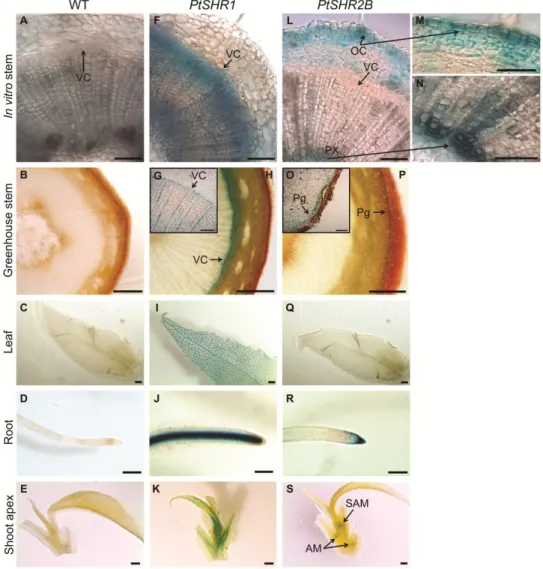
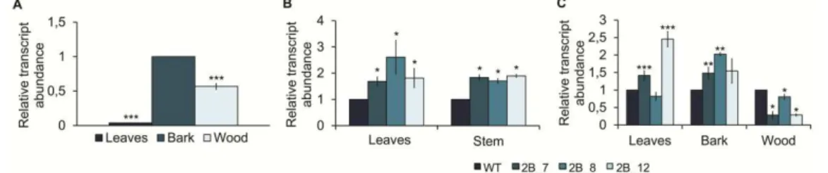
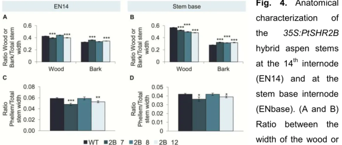
1. To understand the role of SHR-like genes during the secondary growth of Populus tremula L. x Populus tremuloides Michx.; Clone T89 (hybrid aspen), particularly PtSHR2B, by analyzing specific expression patterns and altering transcript levels to study the effects on plant growth and stem anatomy. (Chapter II)
2. To establish basic procedures for accurate transcript quantification in cork oak tissues through identification of adequate reference genes. (Chapter III)
3. To characterize the cork oak SHR homologs and their putative roles during secondary growth by analyzing their expression patterns along periderm development and phellogen dormancy/activity periods. Additional gene characterization approaches included comparative studies with a related species (holm oak) that does not produce a thick phellem layer, and functional studies in Arabidopsis. (Chapter IV)
oak reference transcriptome and allowing the identification of genes differentially regulated between different fruit developmental stages. Genes related to carbohydrate metabolism, response to water and transcriptional control were specifically targeted. (Chapter V)
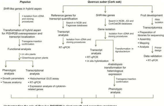
This thesis is in the form of articles, from Chapters II to V, and the work followed the steps described in Figure 5. Final conclusions and future perspectives are discussed in Chapter VI.
References
Agusti J, Lichtenberger R, Schwarz M, Nehlin L, Greb T. 2011. Characterization of transcriptome remodeling during cambium formation identifies MOL1 and RUL1 as opposing regulators of secondary growth. PLoS Genetics 7, e1001312.
Aida M, Beis D, Heidstra R, Willemsen V, Blilou I, Galinha C, Nussaume L, Noh Y, Amasino R, Scheres B, et al. 2004. The PLETHORA genes mediate patterning of the Arabidopsis root stem cell niche. Cell 119, 109–120.
Aloni R. 1982. Role of cytokinin in differentiation of secondary xylem fibers. Plant Physiology 70, 1631–1633.
Aloni R. 1987. Differentiation of vascular tissues. Plant Physiology 38, 179–204. Altamura MM, Possenti M, Matteucci A, Baima S, Ruberti I, Morelli G. 2001. Development of the vascular system in the inflorescence stem of Arabidopsis. New Phytologist 151, 381–389.
Andersen CL, Jensen JL, Ørntoft TF. 2004. Normalization of real-time quantitative reverse transcription-PCR data: a model-based variance estimation approach to identify genes suited for normalization, applied to bladder and colon cancer data sets normalization of real-time quantitative reverse. Cancer Research 64, 5245–5250. Anjos O, Pereira H, Rosa ME. 2008. Effect of quality, porosity and density on the compression properties of cork. Holz als Roh- und Werkstoff 66, 295–301.
APCOR. 2012. Cork 2012. , 1–76.
Artico S, Nardeli SM, Brilhante O, Grossi-de-Sa MF, Alves-Ferreira M. 2010. Identification and evaluation of new reference genes in Gossypium hirsutum for accurate normalization of real-time quantitative RT-PCR data. BMC Plant Biology 10, 49.
Baba K, Karlberg A, Schmidt J, Schrader J, Hvidsten TR, Bako L. 2011. Activity – dormancy transition in the cambial meristem involves stage-specific modulation of auxin response in hybrid aspen. Proceedings of the National Academy of Sciences of the United States of America 108, 3418–3423.
Baima S, Nobili F, Sessa G, Lucchetti S, Ruberti I, Morelli G. 1995. The expression of the Athb-8 homeobox gene is restricted to provascular cells in Arabidopsis thaliana.
Development 121, 4171–4182.
acts as a differentiation-promoting transcription factor of the vascular meristems. Plant Physiology 126, 643–655.
Barton MK. 2010. Twenty years on: the inner workings of the shoot apical meristem, a developmental dynamo. Developmental Biology 341, 95–113.
Basa B, Solti Á, Sárvári É, Támás L. 2009. Housekeeping gene selection in poplar plants under Cd-stress: comparative study for real-time PCR normalisation. Functional Plant Biology 36, 1–9.
Baucher M, El Jaziri M, Vandeputte O. 2007. From primary to secondary growth: origin and development of the vascular system. Journal of Experimental Botany 58, 3485–3501.
Benfey PN, Linstead PJ, Roberts K, Schiefelbein JW, Hauser MT, Aeschbacher RA. 1993. Root development in Arabidopsis: four mutants with dramatically altered root morphogenesis. Development 119, 57–70.
Bishopp A, Help H, El-Showk S, Weijers D, Scheres B, Friml J, Benková E, Mähönen AP, Helariutta Y. 2011. A mutually inhibitory interaction between auxin and cytokinin specifies vascular pattern in roots. Current Biology 21, 917–926.
Björklund S, Antti H, Uddestrand I, Moritz T, Sundberg B. 2007. Cross-talk between gibberellin and auxin in development of Populus wood: gibberellin stimulates
polar auxin transport and has a common transcriptome with auxin. The Plant Journal :
for cell and molecular biology 52, 499–511.
Boavida LC, Varela C, Feijó JA. 1999. Sexual reproduction in the cork oak (Quercus suber L.). I . The progamic phase. Sexual Plant Reproduction 11, 347–353.
Bolle C. 2004. The role of GRAS proteins in plant signal transduction and development. Planta 218, 683–692.
Bradshaw Jr HD, Ceulemans R, Davis J, Stettler R. 2000. Emerging Model Systems in Plant Biology: Poplar (Populus) as A Model Forest Tree. Journal of Plant Growth Regulation 19, 306–313.
Brand U, Fletcher J, Hobe M, Meyerowitz E, Simon R. 2000. Dependence of Stem Cell Fate in Arabidopsis on a Feedback Loop Regulated by CLV3 Activity. Science 289, 617–619.
Bugalho MN, Caldeira MC, Pereira JS, Aronson J, Pausas JG. 2011. Mediterranean cork oak savannas require human use to sustain biodiversity and ecosystem services. Frontiers in Ecology and the Environment 9, 278–286.
Caño-Delgado A, Yin Y, Yu C, Vafeados D, Mora-García S, Cheng J-C, Nam KH, Li J, Chory J. 2004. BRL1 and BRL3 are novel brassinosteroid receptors that function in vascular differentiation in Arabidopsis. Development 131, 5341–5351.
Caño-Delgado A, Lee J-Y, Demura T. 2010. Regulatory mechanisms for specification and patterning of plant vascular tissues. Annual review of Cell and Developmental Biology 26, 605–637.
Carimi F, Zottini M, Formentin E, Terzi M, Lo Schiavo F. 2003. Cytokinins: new apoptotic inducers in plants. Planta 216, 413–421.
Carimi F, Terzi M, De Michele R, Zottini M, Lo Schiavo F. 2004. High levels of the cytokinin BAP induce PCD by accelerating senescence. Plant Science 166, 963–969. Caritat A, Molinas M, Gutierrez E. 1996. Annual cork-ring width variability of
Quercus suber L. in relation to temperature and precipitation (Extremadura, southwestern Spain). Forest Ecology and Management 86, 113–120.
Caritat A, Gutiérrez E, Molinas M. 2000. Influence of weather on cork-ring width. Tree Physiology 20, 893–900.
Carlsbecker A, Lee J-Y, Roberts CJ, Dettmer J, Lehesranta S, Zhou J, Lindgren O, Moreno-Risueno MA, Vatén A, Thitamadee S, et al. 2010. Cell signalling by microRNA165/6 directs gene dose-dependent root cell fate. Nature 465, 316–321. Chaffey N, Cholewa E, Regan S, Sundberg B. 2002. Secondary xylem development in Arabidopsis: a model for wood formation. Physiologia Plantarum 114, 594–600. Chang E, Shi S, Liu J, Cheng T, Xue L, Yang X, Yang W, Lan Q, Jiang Z. 2012. Selection of reference genes for quantitative gene expression studies in Platycladus orientalis (Cupressaceae) using real-time PCR. PLoS One 7, e33278.
Clark SE, Jacobsen SE, Levin JZ, Meyerowitz EM. 1996. The CLAVATA and
SHOOT MERISTEMLESS loci competitively regulate meristem activity in Arabidopsis. Development 122, 1567–1575.
Costa A, Pereira H, Oliveira A. 2001. A dendroclimatological approach to diameter growth in adult cork-oak trees under production. Trees 15, 438–443.
Costa A, Pereira H, Oliveira A. 2003. Variability of radial growth in cork oak adult trees under cork production. Forest Ecology and Management 175, 239–246.
Cui H, Levesque MP, Vernoux T, Jung JW, Paquette AJ, Gallagher KL, Wang JY, Blilou I, Scheres B, Benfey PN. 2007. An evolutionarily conserved mechanism delimiting SHR movement defines a single layer of endodermis in plants. Science 316, 421–425.
Cui H, Hao Y, Kovtun M, Stolc V, Deng X-W, Sakakibara H, Kojima M. 2011. Genome-wide direct target analysis reveals a role for SHORT-ROOT in root vascular patterning through cytokinin homeostasis. Plant Physiology 157, 1221–1231.
Czechowski T, Stitt M, Altmann T, Udvardi MK. 2005. Genome-wide identification and testing of superior reference genes for transcript normalization. Plant Physiology 139, 5–17.
Demidenko N V, Logacheva MD, Penin AA. 2011. Selection and validation of reference genes for quantitative real-time PCR in buckwheat (Fagopyrum esculentum) based on transcriptome sequence data. PLoS ONE 6, e19434.
Derory J, Léger P, Garcia V, Schaeffer J, Hauser M-T, Salin F, Luschnig C, Plomion C, Glössl J, Kremer A. 2006. Transcriptome analysis of bud burst in sessile oak (Quercus petraea). The New Phytologist 170, 723–738.
Dettmer J, Elo A, Helariutta Y. 2009. Hormone interactions during vascular development. Plant Molecular Biology 69, 347–360.
Dhondt S, Coppens F, De Winter F, Swarup K, Merks RMH, Inzé D, Bennett MJ, Beemster GTS. 2010. SHORT-ROOT and SCARECROW regulate leaf growth in Arabidopsis by stimulating S-phase progression of the cell cycle. Plant Physiology 154, 1183–1195.
Díaz-Fernández PM, Climent J, Gil L. 2004. Biennial acorn maturation and its relationship with flowering phenology in Iberian populations of Quercus suber. Trees 18, 615–621.
Die J V, Román B, Nadal S, González-Verdejo CI. 2010. Evaluation of candidate reference genes for expression studies in Pisum sativum under different experimental conditions. Planta 232, 145–153.
Dinneny JR, Benfey PN. 2008. Plant stem cell niches: standing the test of time. Cell 132, 553–557.
Du J, Mansfield SD, Groover AT. 2009. The Populus homeobox gene
ARBORKNOX2 regulates cell differentiation during secondary growth. The Plant Journal 60, 1000–1014.
Du J, Groover A. 2010. Transcriptional regulation of secondary growth and wood formation. Journal of Integrative Plant Biology 52, 17–27.
Durand J, Bodénès C, Chancerel E, Frigerio J-M, Vendramin G, Sebastiani F, Buonamici A, Gailing O, Koelewijn H-P, Villani F, et al. 2010. A fast and cost-effective approach to develop and map EST-SSR markers: oak as a case study. BMC Genomics 11, 570.
Elena-Rosello J, Rio J, Valdecantos JG, Santamaria I. 1993. Ecological aspects of the floral phenology of the cork-oak (Q suber L.): why do annual and biennial biotypes
appear ? Annals of Forest Science 50, 114–121.
Elo A, Immanen J, Nieminen K, Helariutta Y. 2009. Stem cell function during plant vascular development. Seminars in Cell & Developmental Biology 20, 1097–1106. Emery JF, Floyd SK, Alvarez J, Eshed Y, Hawker NP, Izhaki A, Baum SF, Bowman JL. 2003. Radial patterning of Arabidopsis shoots by class III HD-ZIP and KANADI genes. Current Biology 13, 1768–1774.
Eriksson ME, Israelsson M, Olsson O, Moritz T. 2000. Increased gibberellin biosynthesis in transgenic trees promotes growth, biomass production and xylem fiber length. Nature Biotechnology 18, 784–788.
Esau K. 1977. Anatomy of seed plants. New York: John Wiley & Sons.
Etchells JP, Turner SR. 2010. The PXY-CLE41 receptor ligand pair defines a multifunctional pathway that controls the rate and orientation of vascular cell division. Development 137, 767–774.
Evert R. 2006a. Vascular cambium. Esau`s Plant Anatomy: Meristems, Cells, and Tissues of the Plant Body – Their Structure, Function and Development. New Jersey: John Wiley & Sons, Inc., 323-355.
Evert R. 2006b. Periderm. Esau’s Plant Anatomy: Meristems, Cells, and Tissues of
the Plant Body: Their Structure, Function, and Development. New Jersey: John Wiley & Sons, Inc., 427–445.