Genotypic analysis of secreted aspartyl proteinases in
vaginal
Candida albicans
isolates
Análise genotípica de aspartil proteases secretórias em isolados vaginais de
Candida albicans
Janaína S. Lima; Kaoane Regina G. S. Braga; Camila A. Vieira; Wérika Weryanne R. Souza; Juliana Helena Chávez-Pavoni; Claudinéia de Araújo; Letícia S. Goulart
Universidade Federal de Mato Grosso (UFMT), campus Rondonópolis, Mato Grosso, Brazil.
First submission on 09/18/17; last submission on 01/08/18; accepted for publication on 01/17/18; published on 02/20/18
ABSTRACT
Introduction:Candida albicans is the most common etiologic agent of fungal vaginitis. These yeasts produce secreted aspartyl proteinases encoded by a family of 10 genes (SAP1-10). Objective: The purpose of this study was to analyze the presence of genes SAP1-7 in vulvovaginal
C. albicans. Materials and method: The study included 26 C. albicans vaginal isolates. Detection of aspartyl proteases genes (SAP1-7)
was performed by polymerase chain reaction (PCR). Results: The most frequent gene in C. albicans isolated from colonization was SAP6
(93.33%), and from infection, SAP7 (100%). We observed a statistical difference (p = 0.049) in SAP1 gene frequency between isolates from vulvovaginal colonization and infection. Conclusion: High frequency of SAP genes was observed in vulvovaginal C. albicans. The results
suggest SAP1 participation in vulvovaginal candidiasis infection.
Key words: vulvovaginal candidiasis; Candida albicans; virulence factors.
INTRODUCTION
Vulvovaginal candidiasis (VVC) is an endogenous infection caused by yeasts of the genus Candida. The disease commonly develops when conditions exist favoring fungal growth, such as alterations in normal microbiota or lowered host immune response(1-5). The species of Candida are microorganisms
commonly found in the microbiota of the gastrointestinal and urogenital tracts without causing disease, but when the balance between fungus and host is disturbed, there is an increase in colonization, and the fungus invades tissues, initiating the infectious process(6, 7).
VVC is one of the most frequent diagnoses in clinical practice, with a rising incidence, becoming the second most common genital infection in the United States and in Brazil(1, 8, 9). Among
vulvovaginitides, VVC accounts for 39% of the cases, just behind bacterial vaginosis. Candida species can be found in up to 50% of healthy women without causing symptoms; however, around
70%-75% of women in childbearing age develop at least one episode of fungal vulvovaginitis during life. Among these, 50% will present two or more episodes, and approximately 6%-9% are likely to present recurrent VVC (RVVC), characterized by the presence of four or more symptomatic episodes in a year(10-14).
Candida albicans is the most prevalent etiologic agent in
VVC, accounting for 85%-90% of the cases. The virulence of this species is due to several mechanisms and abilities inherent in the fungus, as yeast-to-hypha transition, phenotypic switching, expression of adhesins and invasins on the cell surface, biofilm formation, and secretion of hydrolytic enzymes(3, 11, 15-17).
The most studied hydrolases related to Candida virulence are proteases, phospholipases, and lipases. Secreted aspartyl proteinases (Sap) form a family of 10 isoenzymes (Sap1-10) that participate in the infection process by degrading several host cell proteins, such as immunoglobulins, proteins of the complement system and extracellular matrix, contributing to tissue damage and the resulting invasion by the microorganism(18-21). These enzymes play different
roles depending on the environmental stimuli and are involved in
the host inflammatory response to fungus. The activation of these proteins is a well-regulated process at specific time points, what increases the infection potential of C. albicans(6, 16, 22, 23). Among the
SAP gene family, the most frequently studied in the literature and associated with pathogenicity of C. albicans are SAP1-7 genes. The
objective of this study was to evaluate the presence of these genes in
vulvovaginal C. albicans cell lines.
MATERIALS AND METHOD
Studied microorganisms
Twenty-six C. albicans cell lines were isolated from the vaginal mucosa of asymptomatic (n = 15) and symptomatic
(n = 11) women with VVC. The yeasts were obtained from vaginal
swabs as described by Goulart et al.(2016)(24). The clinical
samples were collected from women seen at basic health-care units in the municipality of Rondonópolis (MT), Brazil, regardless VVC symptoms. Participants were divided into two groups: 1) symptomatic patients, characterized by the presence of curd-like vaginal discharge, pruritus, edema and erythema of vulva and vagina; and 2) asymptomatic patients, which did not present the mentioned characteristics. Yeasts were stored at Sabouraud agar at 4°C. Previously, microorganisms were grown in Sabouraud broth at 37°C, under agitation [200 revolutions per minute (rpm)] for 24 h. Then the culture was centrifuged, the supernatant was discharged, and deoxyribonucleic acid (DNA) was extracted from the cell sediment with a kit (Nucleo Spin Tissue, Macherey-Nagel GmbH & Co. KG, Duren, Germany), following the instructions by the manufacturer. The yeast species was determined by the species-specific polymerase chain reaction (PCR) based on the protocol of
Liguori et al. (2010)(25), with modifications.
Identification of
SAP1-7
genes
Genes were identified by PCR based on the method proposed
by Bassyouni et al. (2015)(26), with some modifications.
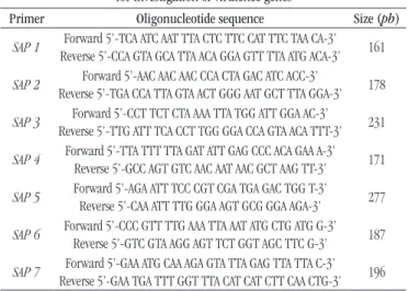
Reactions were performed at a final volume of 25 μl, containing approximately 20 ng of DNA, 12.5 μl of GoTaq Hot Start Green Master Mix (Promega, Madison, Wisconsin, USA) and 0.75 μl (20 pmol/μl) of each specific primer. Amplification conditions
for SAP1, 3, 4 and 7 genes were: initial denaturation at 94°C for
3 minutes, 30 denaturation cycles at 94°C for 30 seconds, annealing at 46°C for 30 seconds, extension at 72°C for 30 seconds and final extension at 72°C for 10 minutes. PCR for SAP2, 5 and 6 genes
was standardized from denaturation at 92°C for 3 minutes, 30 denaturation cycles at 94°C for 30 seconds, annealing at 52°C for
30 seconds, extension at 72°C for 30 seconds and final extension at 72°C for 10 minutes. Amplification products were analyzed on 1% agarose gel electrophoresis containing DNA stain (Promega, Madison, Wisconsin, USA) and visualized under ultraviolet light. The oligonucleotide sequences used in the study are described in
Table 1.
TABLE 1 − Oligonucleotide sequences used in PCR assays for investigation of virulence genes
Primer Oligonucleotide sequence Size (pb)
SAP 1 Reverse 5’-CCA GTA GCA TTA ACA GGA GTT TTA ATG ACA-3’Forward 5’-TCA ATC AAT TTA CTC TTC CAT TTC TAA CA-3’ 161
SAP 2 Reverse 5’-TGA CCA TTA GTA ACT GGG AAT GCT TTA GGA-3’Forward 5’-AAC AAC AAC CCA CTA GAC ATC ACC-3’ 178
SAP 3 Reverse 5’-TTG ATT TCA CCT TGG GGA CCA GTA ACA TTT-3’Forward 5’-CCT TCT CTA AAA TTA TGG ATT GGA AC-3’ 231
SAP 4 Forward 5’-TTA TTT TTA GAT ATT GAG CCC ACA GAA A-3’Reverse 5’-GCC AGT GTC AAC AAT AAC GCT AAG TT-3’ 171
SAP 5 Forward 5’-AGA ATT TCC CGT CGA TGA GAC TGG T-3’Reverse 5’-CAA ATT TTG GGA AGT GCG GGA AGA-3’ 277
SAP 6 Forward 5’-CCC GTT TTG AAA TTA AAT ATG CTG ATG G-3’Reverse 5’-GTC GTA AGG AGT TCT GGT AGC TTC G-3’ 187
SAP 7 Reverse 5’-GAA TGA TTT GGT TTA CAT CAT CTT CAA CTG-3’Forward 5’-GAA ATG CAA AGA GTA TTA GAG TTA TTA C-3’ 196
PCR: polymerase chain reaction.
Statistical analysis
Data were recorded in Excel 2016 spreadsheets and assessed in the statistical software Epi-info 7.2.0. Data analysis was carried out by descriptive statistics and non-parametric Fisher exact test, adopting a 5% significance level. We evaluated the correlation of the presence of SAP1-7 genes with the infection process and
vulvovaginal colonization.
RESULTS
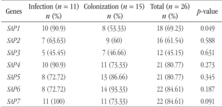
By means of PCR method, it was possible to detect the presence
of SAP1-7 genes in clinical isolates of C. albicans.After molecular
analysis of C. albicans isolated in women with symptoms of VVC, the presence of SAP1 gene was identified in 90.9% (10/11), SAP2
in 63.63% (7/11), SAP3 in 45.45% (5/11), SAP4 in 90.9% (10/11),
SAP5 in 72.72% (8/11), SAP6 in 72.72% (8/11), and SAP7 in
100% (11/11) of the samples. The frequency of virulence genes
in C. albicans from colonization was 53.33% (8/15) for SAP1, 60% (9/15) for SAP2,46.66% (7/15) for SAP3, 73.33% (11/15)
for SAP4, 86.66% (13/15) for SAP5,93.33% (14/15) for SAP6 and
When comparing the frequency of genes encoding aspartyl proteases (SAP1-7) among C. albicans isolated in asymptomatic and symptomatic women with VVC, we found a statistical difference
just for SAP1 gene (p = 0.049), with this being more prevalent in isolates associated with the infectious process. Although genes
SAP2-7 present distribution profiles distinct among the groups, this difference did not represent statistical difference. These pieces of data are shown in Table 2.
The results revealed genetic variability for the members of the SAP family in the studied cell lines, with 19 different genotypic patterns being found; just three isolates presented all the studied
genes (Table 3).
DISCUSSION
SAP genes greatly stand out in the pathogenesis of candidiasis, once they encode proteins able to degrade collagen, keratin, and peptides found in the surface of mucosas, ensuring, thus, an important and efficient proteolytic system to C. albicans, from the colonization process to active infection(20, 23, 27-30).
In this study, we observed that the most prevalent SAP genes in
C. albicans isolated from vulvovaginal infection and colonization
were SAP7 (100%) and SAP6 (93.33%), respectively. Monroy-Pérez
et al. (2013)(31) determined the frequency and the expression
of SAP1-10 genes isolates from women with VVC in Mexico and
identified that SAP4-6 genes were the most frequent (100%), besides observing that all SAP genes were expressed in a model of reconstituted human vaginal epithelium (RHVE), suggesting that aspartyl proteases play an important role in the pathogenesis of the infection. Bassyouni et al. (2015)(26) investigated the
presence of SAP1-8 genes in C. albicans isolated from vaginal
mucosa of diabetic and non-diabetic women and identified that SAP1 and SAP2 were the most frequently detected genes, followed by SAP5 in both groups; there was no difference in the
distribution of genes between diabetic and non-diabetic women. Kalkanci et al. (2005)(32) verified that SAP1-3 genes were the most
prevalent in vaginal isolates of C.albicans, with a 92.5% rate, followed by SAP6, with 12.5%, and SAP4-5, with 7.5%.
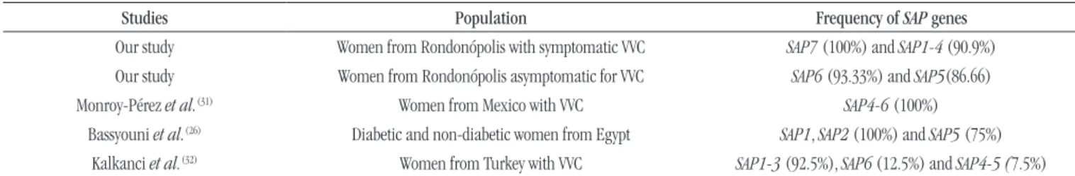
The most frequent SAP genes in the present research and in
other studies are available in Table 4.
Predominance of the SAP6 gene in the studied colonization
isolates and SAP7 in the studied infection isolates suggests the necessity of further studies to evaluate the expression of these
genes in vivo. The SAP6 gene participates with the SAP4-6
subfamily in the development of hyphae, an essential process for the fungus(33, 34) capacity of invasion. Besides, Sap6 protein was
associated with integrity maintenance of cell surface and capacity of producing inflammatory response in the host(33, 35, 36). The SAP7
gene encodes the most divergent protein within the Sap family and can be preferably associated with infections of human mucosas(36).
The expression of this gene was associated with the initial adaptation of C. albicans to human cells of the intestinal tract, while transcripts of SAP6-7 were associated with tissue damage in the early phase of infection, after 24 h, at a RHVE(37, 38) model.
This study revealed high (90.9%) presence of SAP4 gene in
vaginal cell lines of C. albicans involved in infection. The proteins
encoded by these genes are associated with adherence of the fungus to human cells and alterations in the morphogenesis of the yeast, playing an important role in biofilm formation. The SAP4 gene
TABLE 2 − Frequency of SAP1-7 genes in C. albicans isolated from vulvovaginal infection and colonization
Genes Infection (n = 11) n (%)
Colonization (n = 15)
n (%)
Total (n = 26)
n (%) p-value
SAP1 10 (90.9) 8 (53.33) 18 (69.23) 0.049
SAP2 7 (63.63) 9 (60) 16 (61.54) 0.588
SAP3 5 (45.45) 7 (46.66) 12 (45.15) 0.631
SAP4 10 (90.9) 11 (73.33) 21 (80.77) 0.273
SAP5 8 (72.72) 13 (86.66) 21 (80.77) 0.345
SAP6 8 (72.72) 14 (93.33) 22 (84.61) 0.187 SAP7 11 (100) 11 (73.33) 22 (84.61) 0.091
TABLE 3 − Genotypic patterns identified in vaginal isolates of C. albicans
Pattern Genotype n (%)
1 SAP1-7 3 (16)
2 SAP1/SAP2/SAP4/SAP5/SAP6/SAP7 3 (16)
3 SAP1/SAP3/SAP4/SAP5/SAP6/SAP7 2 (10)
4 SAP2/SAP4/SAP5/SAP6 2 (10)
5 SAP2/SAP5/SAP6/SAP7 2 (10)
6 SAP1/SAP2/SAP3/SAP4/SAP5/SAP7 1 (5)
7 SAP1/SAP2/SAP3/SAP5/SAP6/SAP7 1 (5)
8 SAP1/SAP2/SAP3/SAP4/SAP6/SAP7 1 (5)
9 SAP3/SAP4/SAP5/SAP6/SAP7 1 (5)
10 SAP1/SAP3/SAP4/SAP6/SAP7 1 (5)
11 SAP1/SAP2/SAP4/SAP6/SAP7 1 (5)
12 SAP1/SAP2/SAP3/SAP4/SAP7 1 (5)
13 SAP1/SAP4/SAP5/SAP6/SAP7 1 (5)
14 SAP2/SAP3/SAP4/SAP5/SAP6 1 (5)
15 SAP4/SAP5/SAP6/SAP7 1 (5)
16 SAP1/SAP5/SAP6/SAP7 1 (5)
17 SAP1/SAP4/SAP5/SAP7 1 (5)
18 SAP1/SAP4/SAP7 1 (5)
has also been implicated in evasion of phagocytosis(30, 39, 40). The
expression of SAP4 genes was demonstrated in vivo in the vaginal
mucosa of women who were pregnant (40%), post-menopausal (50%), and in childbearing age (33%)(41).
The statistical analysis showed significant difference (p = 0.049) for the SAP1 gene frequency among the C. albicans cell lines isolated from infection (90.9%) and colonization (53.33%). This suggests a probable participation of SAP1 in the infectious process
of VVC and points to a possible aim of further studies to distinguish colonization from active infection. This result can also contribute to researches aimed at developing new diagnostic methods for VVC. Sap1 protein is known to be linked to the capacity to cause lesions in the mucosa and to the development of systemic infections(30).
At a previous work, the expression of SAP1 in C. albicans from
patients with VVC was observed in 80% of the studied isolates(42).
The SAP1-7 genes can be expressed by C. albicans in the vaginal mucosa, both in colonization processes and in infections, but a differential expression of these genes is observed when comparing the transcript levels in isolates of carriers and of active VVC, besides a predominance of certain members of the SAP family during
vaginal infection(43).
A study of genic expression of C. albicans revealed that SAP1,
SAP3, and SAP6-8 genes are related with active VVC. Moreover, SAP1 and SAP3 are preferably expressed in the vaginal mucosa,
when compared with the oral infection, indicating that the differential expression of genes encoding hydrolytic enzymes varies according to the phase of the disease and the anatomical location(18). Lian and Liu (2007)(43) evaluated the expression of
SAP1-7 genes in C. albicans isolated from asymptomatic women,
TABLE 5 − Expression of SAP genes in different studies
Study Population Expression of SAP genes
Nas et al.(41)
Women from Turkey with VVC Expression of SAP4: pregnant (40%), post-menopausal (50%) women, and women in childbearing age (33%)
Lian and Liu(43) Symptomatic, asymptomatic women,
and women with RVVC from China SAP1 and SAP3 were present only in VVC and RVVC isolates
Lin et al.(44) Women from China with acute VVC SAP2 and SAP5 were most commonly expressed in the vaginal mucosa
VVC: vulvovaginal candidiasis; RVVC: recurrent vulvovaginal candidiasis.
TABLE 4 − Frequency of SAP genes in different studies
Studies Population Frequency of SAP genes
Our study Women from Rondonópolis with symptomatic VVC SAP7 (100%) and SAP1-4 (90.9%)
Our study Women from Rondonópolis asymptomatic for VVC SAP6 (93.33%) and SAP5(86.66) Monroy-Pérez et al.(31) Women from Mexico with VVC SAP4-6 (100%)
Bassyouni et al.(26) Diabetic and non-diabetic women from Egypt SAP1, SAP2 (100%) and SAP5 (75%) Kalkanci et al.(32) Women from Turkey with VVC SAP1-3 (92.5%), SAP6 (12.5%) and SAP4-5 (7.5%)
VVC: vulvovaginal candidiasis.
and women with symptomatic and recurrent VVC. The authors observed that the transcripts for SAP1 and SAP3 genes were present
only in isolates of VVC and RVVC. Lin et al. (2007)(44) determined
by real-time PCR (RT-PCR) that SAP2 and SAP5 were the most
commonly expressed genes in the vaginal mucosa of women with acute VVC. The expression of SAP genes in the different studies is shown in Table 5.
Medeiros et al. (2017)(45) did not find differences in the
expression of virulence factors between C. albicans isolated from
patients with sporadic VVC and those obtained from patients with RVVC, suggesting that the ability to express virulence factors is important in the pathogenesis of VVC, but it seems not to be crucial for the transition from colonization to infection. We did not identify statistical difference for the presence of SAP2-7 genes
in isolates from infection and vulvovaginal colonization. This suggests that other factors that control the genic fungal expression and characteristics inherent in the host are likely to influence in the infection/colonization process. Also, further studies comprising a larger number of samples must be carried out.
CONCLUSION
C. albicans isolated from symptomatic women with VVC and
asymptomatic women presented different patterns of distribution
for SAP1-7 genes, with predominance of the SAP6 gene in
colonization and SAP7 gene in infection. The SAP1 gene was
REFERENCES
1. Álvares CA, Svidzinski TIE, Consolaro MEL. Candidíase vulvovaginal: fatores predisponentes do hospedeiro e virulência das leveduras. J Bras Patol Med Lab. 2007; 43(5): 319-27.
2. Achkar JM, Fries BC. Candida infections of the genitourinary tract. Rev Clin Microbiol. 2010; 23(2): 253-73.
3. Sobel JD. Genital candidiasis. Med. 2010; 38: 6.
4. Rossi T, Lozovoy MAB, Silva RV, et al. Interações entre Candida albicans e hospedeiro. Semina Cienc Biol Saúde. 2011; 32(1): 15-28.
5. Cassone A. Vulvovaginal Candida albicans infections: pathogenesis, immunity and vaccine prospects. BJOG. 2015; 122(6): 785-94.
6. Costa KRCD. Aspectos fenotípicos e moleculares da adesão e atividade enzimática de Candida sp. isolados de pacientes com sinais clínicos de candidíase oral [thesis]. Programa de Pós-Graduação em Biociências Aplicadas a Farmácia, Universidade de São Paulo; 2009.
7. Barbedo LS, Sgarb DBG. Candidíase. DST-J Bras Doenças Sex Transm. 2010; 22(1): 22-38.
8. Kim J, Sudbery P. Candida albicans, a major human fungal pathogen. J Microbiol. 2011; 49(2): 171-7.
9. Medeiros MAPD. Aspectos de patogenicidades e relacionamento genético de isolados clínicos vaginais e anais de Candida albicans oriundos de pacientes com candidíase vaginal [dissertation]. Programa de Pós-Graduação em Ciências Farmacêuticas, Universidade Federal do Rio Grande do Norte; 2013.
10. Feuerschuette OHM, Silveira SK, Feuerschuette I, Corrêa T, Grando L, Trepani A. Candidíase vaginal recorrente: manejo clínico. Femina. 2010; 38(2).
11. Mayer FL, Duncan W, Hube B. Candida albicans pathogenicity mechanisms. Virulence. 2013; 4(2): 119-28.
12. Ministério da Saúde. Secretária de Ciências, Tecnologia e Insumos Estratégicos. Protocolo clínico e diretrizes terapêuticas infecções sexualmente transmissíveis: relatório de recomendações. Brasília (DF); 2015.
13. Sobel JD. Recurrent vulvovaginal candidiasis. AJOG. 2016; 214(1): 15-21.
RESUMO
Introdução:Candida albicans é o principal agente etiológico das vaginites fúngicas. Essas leveduras produzem aspartil proteases
secretórias que são codificadas por uma família de 10 genes (SAP1-10). Objetivo: O objetivo deste estudo foi avaliar a presença dos genes SAP1-7 em linhagens vulvovaginais de C. albicans. Materiais e método: O estudo incluiu 26 isolados vaginais de
C. albicans. Os genes de aspartil proteases (SAP1-7) foram detectados por reação em cadeia da polimerase (PCR). Resultados: O gene mais frequente em C. albicans isolado de colonização foi SAP6 (93,33%), e de infecção, SAP7 (100%). Foi observada diferença estatística (p = 0,049) na frequência do gene SAP1 entre isolados oriundos de colonização e infecção vulvovaginal. Conclusão:
Constatou-se alta frequência dos genes SAP em linhagens vaginais de C. albicans. Os resultados sugerem uma participação de
SAP1 no processo infeccioso da candidíase vulvovaginal.
Unitermos: candidíase vulvovaginal; Candida albicans; fatores de virulência.
14. Brandolt TM, Klafke GB, Gonçalves CV, et al. Prevalence of Candida spp. in cervical-vaginal samples and the in vitro susceptibility of isolates. Braz J Microbiolv. 2017; 48(1): 145-50.
15. Kumamoto CA, Vinces MD. Contributions of hyphae and hypha-co-regulated genes to Candida albicans virulence. Cell Microbiol. 2005; 7(11): 1546-54.
16. Geraldino TH, Costa TMPS, Brunnquell CR, et al. Dimorfismo, produção de enzimas funcionais e adesinas de Candida albicans: minirrevisão. Biosaúde. 2012; 14(1): 26-41.
17. Boatto HF, Moraes MS, Machado AP, Girão MJBC, Fischman O. Correlação entre os resultados laboratoriais e os sinais e sintomas clínicos das pacientes com candidíase vulvovaginal e relevância dos parceiros sexuais na manutenção da infecção em São Paulo, Brasil. Rev Bras Ginecol Obstet. 2007; 29(2): 80-4.
18. Naglik JR, Rodgers CA, Shirlaw PJ, et al. Differential expression of Candida albicans secreted aspartyl proteinase and phospholipase B genes in humans correlates with active oral and vaginal infections. JID. 2003; 188(3): 469-79.
19. Calle-Rodríguez NL, Santa-Vélez C, Cardona-Castro N. Factores de virulencia para la infección de tejidos queratinizados por Candida albicans y hongos dermatofitos. Rev CES Med. 2012; 26(1): 43-55. 20. Santana DP, Ribeiro EL, Menezes ACS, Naves PLF. Novas abordagens sobre os fatores de virulência de Candida albicans. Rev Ciênc Méd Biol. 2013; 12(2): 229-33.
21. Taylor BN, Staib P, Binder A, et al. Profile of Candida albicans-secreted aspartic proteinase elicited during vaginal infection. Infect Immun. 2005; 1828-35.
22. Giolo MP, Svdizinski TIE. Fisiopatogenia, epidemiologia e diagnostico laboratorial da candidemia. J Bras Patol Med Lab. 2010; 46(3): 225-34. 23. Naglik JR, Moyes DL, Wächtler B, Hube B. Candida albicans interactions with epithelial cells and mucosal immunity. Microbes Infec. 2011; 13: 963-76.
24. Goulart LS, Santiago EF, Ramon JL, et al. Species distribution and antifungal susceptibility to vulvovaginal Candida spp. in Southern Mato Grosso State, Brazil. J Bras Patol Med Lab. 2016; 52(4): 233-7.
26. Bassyouni RH, Wegdan AA, Abdelmoneim A, et al. Phospholipase and aspartyl proteinase activities of Candida species causing vulvovaginal candidiasis in patients with type 2 diabetes mellitus. J Microbiol Biotechnol. 2015; 25(10): 1734-41.
27. Hube B, Naglik J. Candida albicans proteinases: resolving the mystery of a gene family. Microbiology. 2001; 147: 1997-2005.
28. Lermann U, Morschhäuser J. Secreted aspartic proteases are not required for invasion of reconstituted human epithelia by Candida albicans. Microbiology. 2008; 154: 3281-95.
29. Naglik J, Albrecht A, Bader O, Hube B. Candida albicans proteinases and host/pathogen interactions. Cell Microbiol. 2004; 6(10): 915-26. 30. Costa CR. Fatores de virulência de isolados de Candida de pacientes imunocomprometidos. Caracterização molecular de Candida albicans suscetíveis e resistentes ao fluconazol [thesis]. Programa de Pós-Graduação em Medicina Tropical e Saúde Pública, Universidade Federal de Goiás; 2009.
31. Monroy-Pérez E, Paniagua-Contreras G, Vaca-Paniagua F, Negrete-Abascal E, Vaca S. SAP expression in Candida albicans strains isolated from Mexican patients with vaginal candidosis. Int J Clin Med. 2013; 4: 25-3.
32. Kalkanci A, Bozdayi G, Birib A, Kustimur S. Distribution of secreted aspartyl proteinases using a polymerase chain reaction assay with SAP specific primers in Candida albicans isolates. Folia Microbiol. 2005; 50(5): 409-13.
33. Naglik JR, Challacombe SJ, Hube B. Candida albicans secreted aspartyl proteinases in virulence and pathogenesis. Microbiol Mol Biol Rev. 2003; 67(3): 400-28.
34. Buu LM, Chen YC. Impact of glucose levels on expression of hypha-associated secreted aspartyl proteinases in Candida albicans. J Biomed Sci. 2014; 21(1): 21-2.
35. Silva NC. Análise de aspartato protease (Sap) como fator associado à virulência de linhagens de Candida albicans e Candida não-albicans [dissertation]. Universidade Federal de Alfenas; 2013.
36. Gabrielli E, Sabbatini S, Rosellett E, et al. In vivo induction of neutrophil chemotaxis by secretory aspartyl proteinases of Candida albicans. Virulence. 2016; 7(7): 819-25.
37. Staniszewska M, Bondaryk M, Zukowski K, Chudy M. Role of SAP7-10 and morphological regulators (EFG1, CPH1) in Candida albicans’ hypha formation and adhesion to colorectal carcinoma Caco-2. Pol J Microbiol. 2015; 64(3): 203-10.
38. Schaller M, Bein M, Korting HC. The secreted aspartyl proteinases Sap1 and Sap2 cause tissue damage in an in vitro model of vaginal candidiasis based on reconstituted human vaginal epithelium. Infect Immun. 2003; 3227-34.
39. Dantas CV. Produção de enzimas relacionadas à virulência de Candida albicans [monography]. Secretaria de Estado da Saúde, Coordenadoria de Controle de Doenças, Instituto Adolfo Lutz; 2015.
40. Winter MB, Salcedo EC, Lohse MB, et al. Global identification of biofilm-specific proteolysis in Candida albicans. MBio. 2016; 7(5): e01514-16.
41. Nas T, Kalkanci A, Ozkan S. Expression of ALS1 and SAP4 genes in Candida albicans strains isolated from women with vaginitis. Folia Microbiol. 2008; 53(2): 179-83.
42. Dabiri S, Shams-Ghahfarokhi M, Razzaghi-Abyaneh M. SAP (1-3) gene expression in high proteinase producer Candida species strains isolated from Iranian patients with different candidosis. J Pure Appl Microbiol. 2016; 10(3): 1891-6.
43. Lian CH, Liu WD. Differential expression of Candida albicans secreted aspartyl proteinase in human vulvovaginal candidiasis. Mycoses. 2007; 50: 383-90.
44. Lin N, Feng J, Tu Y, Feng A. Expression of Candida albicans secreted aspartyl proteinase in acute vaginal candidiasis. J Huazhong Univ Sci Technol Med Sci. 2007; 27(3): 333-5.
45. Medeiros MAP, Melo APV, Sousa AMM, et al. Characterization of virulence factors of vaginal and anal isolates of Candida albicans sequentially obtained from patients with vulvovaginal candidiasis in north-east Brazil. J Mycol Med. 2017; 27(4): 567-72.
CORRESPONDING AUTHOR
Letícia Silveira Goulart
Avenida dos Estudantes, 5005; Cidade Universitária; CEP: 78735-901; Rondonópolis-MT, Brasil; e-mail: [email protected].