Pedro Miguel Veríssimo Mateus
Dissertation presented to obtain the Ph.D degree in Chemistry
Instituto de Tecnologia Química e Biológica | Universidade Nova de Lisboa
Oeiras,
August, 2011
Pedro Miguel Veríssimo Mateus
Dissertation presented to obtain the Ph.D degree in Chemistry
Instituto de Tecnologia Química e Biológica | Universidade Nova de Lisboa
Oeiras, August, 2011
”Un savant dans son laboratoire n'est pas un technicien; c'est aussi un enfant
placé en face des phénomènes naturels qui l'impressionnent comme un conte de
fées.”
Marie Curie
iii
Table of Contents
Foreword xiii
Acknowledgments xv
List of publications and communications xvii
Keywords xix
Summary xxi
Resumo xxv
Index of Figures xxix
Index of Schemes xxxix
Index of Tables xliii
List of compounds and complexes discussed in this work xlvii
List of compounds synthesized and studied in this work xlix
List of abbreviations and symbols li
Chapter 1 - Introduction 1
1.1 Supramolecular Chemistry 3
1.2. Molecular recognition 4
1.2.1 Molecular interactions in molecular recognition 5
1.2.2 Complementarity and preorganization 5
1.2.3 The solvent 7
1.2.4 Binding constant 8
1.2.5 Selectivity 9
1.2.6 Receptor molecules 11
1.2.6.1 The macrobicyclic architecture 13
1.2.6.2 Strategies of synthesis of macrobicycles 14
1.3 Supramolecular chemistry of anions 16
1.3.1 Anions as targets for molecular recognition 16
1.3.2 The development of supramolecular chemistry of anions 17
1.3.4 Natural anion receptors 18
1.3.4.1 Chloride channels 18
1.3.4.2 Sulfate and phosphate binding proteins 19
1.3.4.3 Nitrate and bicarbonate binding proteins 20
1.3.4.4 Periplasmic sensor domain of Sinorhizobium meliloti 21
1.3.4.5 Menaquinol:fumarate oxidoreductase 22
1.3.5 Synthetic anion receptors 23
1.3.5.1 Neutral receptors 24
1.3.5.2 Charged receptors 25
1.3.5.2.1 Amine based receptors 26
1.3.5.2.2 Polyamine macrobicycles 28
1.3.5.2.2.1 Synthesis of polyamine macrobicycles 28
1.3.5.2.2.2 Polyamine macrobicycles as anion receptors 30
1.4 Work aims and contribution to the field of anion recognition 49
1.5 References 51
Chapter 2 - Selective recognition of tetrahedral dianions by a hexaaza
cryptand receptor 57
2.1 Summary 59
2.2 Introduction 59
2.3 Results and discussion 61
2.3.1 Synthesis of the cryptand 61
2.3.2 Potentiometric studies 61
2.3.2.1 Acid-base behaviour 61
2.3.2.2 Binding studies 63
2.3.3 NMR studies 67
2.3.4 Electrospray mass spectrometry studies on sulfate cryptate 70
2.3.5 Crystallographic studies 71
2.3.6 Molecular mechanics and molecular dynamics calculations 74
v
2.5.1 General considerations 81
2.5.2 Synthesis 81
2.5.2.1 Schiff’s base of xyl 81
2.5.2.2 xyl 82
2.5.2.3 Crystals of [(H6xyl)(SO4)(H2O)6](SO4)2·9.5H2O 82
2.5.3 Potentiometric measurements 83
2.5.3.1 Reagents and solutions 83
2.5.3.2 Equipment and working conditions 83
2.5.3.3 Measurements 84
2.5.3.4 Calculation of equilibrium constants 84
2.5.4 NMR studies 85
2.5.4.1 1H-NMR of the cryptates 85
2.5.4.2 Job’s plots 85
2.5.5 Mass spectrometry studies 86
2.5.6 Crystallography 86
2.5.7 Molecular modelling simulations 87
2.6 Acknowledgements 88
2.7 References 89
Chapter 3 - Polyaza cryptand receptor selective for dihydrogen
phosphate 91
3.1 Summary 93
3.2 Introduction 93
3.3 Results and discussion 95
3.3.1 Synthesis of pyr 95
3.3.2 Potentiometric studies 95
3.3.2.1 Acid-base behaviour 95
3.3.2.2 Binding studies 97
3.3.3 31P-NMR studies 102
3.3.4 Crystallographic studies 104
3.5 Experimental 111
3.5.1 General considerations 111
3.5.2 Synthesis 111
3.5.2.1 Schiff’s base of pyr 111
3.5.2.2 pyr 112
3.5.2.3 Crystals of [(H6pyr)(NO3)3(H2O)3](NO3)3·4H2O 112
3.5.2.4 Crystals of [(H6pyr)(SO4)2(H2O)4](HSO4)2·6H2O 113
3.5.2.5 Crystals of [(H6pyr)(HPO4)2(H2PO4)(H2O)2](H2PO4)·16H2O 113
3.5.3 Potentiometric measurements 113
3.5.3.1 Reagents and solutions 113
3.5.3.2 Equipment and working conditions 113
3.5.3.3 Measurements 114
3.5.3.4 Calculation of equilibrium constants 114
3.5.4 31P-NMR studies 115
3.5.5 Crystallography 115
3.6 Acknowledgements 116
3.7 References 116
Chapter 4 - A trinuclear copper(II) cryptate and its μ3-CO3 cascade
complex: thermodynamics, structural and magnetic properties 119
4.1 Summary 121
4.2 Introduction 121
4.3 Results and discussion 123
4.3.1 Potentiometric studies 123
4.3.1.1 Acid-base behaviour 123
4.3.1.2 Complexation studies 123
4.3.2. Crystallographic studies 126
4.3.3 CO2 fixation 128
4.3.4 IR and ESI mass spectra 131
vii
4.4 Conclusions 139
4.5 Experimental 140
4.5.1 General considerations 140
4.5.2 Synthesis of crystals 141
4.5.2.1 [Cu3pyr(μ3-CO3)](NO3)4·9H2O 141
4.5.2.2 [Cu3pyr(μ3-CO3)](ClO4)4·H2O 141
4.5.3 Potentiometric measurements 141
4.5.3.1 Reagents and solutions 141
4.5.3.2 Equipment and working conditions 142
4.5.3.3 Measurements 142
4.5.3.4 Calculation of equilibrium constants 143
4.5.4 Crystallography 143
4.5.5 ESI MS 144
4.5.6 Magnetic measurements 144
4.5.7 Computational details 145
4.6 Acknowledgements 146
4.7 References 146
Chapter 5 - Recognition of oxalate by a new copper(II) polyaza
macrobicyclic complex 151
5.1 Summary 153
5.2 Introduction 153
5.3 Results and discussion 156
5.3.1 Synthesis of the cryptand 156
5.3.2 Acid-base behaviour of the synthesized compounds 157
5.3.3 Crystal structure 158
5.3.4 Binding affinity of the protonated forms of btpN7 towards
dicarboxylates 160
5.3.5 Copper(II) coordination studies 161
5.3.6 Cascade species formed by the copper(II) complexes of btpN7
5.3.7 Study of selective uptake of oxa2- anion from protonated forms
of the cryptate over other dicarboxylate anions 167
5.3.8 Spectroscopic studies 168
5.3.9 Indicator-displacement assay 172
5.4 Conclusions 173
5.5 Experimental 174
5.5.1 General considerations 174
5.5.2 Synthesis 175
5.5.2.1 Tripodal trialdehyde 175
5.5.2.2 Schiff’s base of btpN7 176
5.5.2.3 btpN7 176
5.5.2.4 Crystals of [H7btpN7(H2O)(MeOH)Cl2]5+ 177
5.5.3 Potentiometric measurements 177
5.5.3.1 Reagents and solutions 177
5.5.3.2 Equipment and working conditions 178
5.5.3.3 Measurements 178
5.5.3.4 Calculation of equilibrium constants 179
5.5.4 Absorption and X-band EPR spectra 180
5.5.5 Indicator-displacement assay 180
5.5.6 Crystallography 180
5.6 Acknowledgements 181
5.7 References 182
Chapter 6 - Thermodynamics and structural aspects of dicarboxylate
binding by two polyaza macrobicyclic receptors 185
6.1 Summary 187
6.2 Introduction 187
6.3 Results and discussion 189
ix
6.3.1.2 Binding affinity of the protonated forms of btpN7 and t2pN8
towards dicarboxylates 191
6.3.2 NMR studies 196
6.4 Conclusions 200
6.5 Experimental 202
6.5.1 General considerations 202
6.5.2 Potentiometric measurements 202
6.5.2.1 Reagents and solutions 202
6.5.2.2 Equipment and working conditions 203
6.5.2.3 Measurements 203
6.5.2.4 Calculation of equilibrium constants 204
6.5.3 NMR studies 204
6.5.3.1 Spectra of the supermolecules 204
6.5.3.2 Job’s plots 205
6.5.3.3 H/D exchange experiments 205
6.6 Acknowledgements 206
6.7 References 206
Chapter 7 - Two new polyoxapolyaza macrobicyclic receptors for
recognition of zwitterionic amino acids 209
7.1 Summary 211
7.2 Introduction 211
7.3 Results and discussion 213
7.3.1 Synthesis 213
7.3.2 Potentiometric studies 214
7.3.2.1 Acid-base behaviour of the cryptands 214
7.3.2.2 Binding affinity of the protonated forms of btpN7 and t2pN8
towards amino acids 218
7.4 Conclusions 220
7.5.1 General considerations 221
7.5.2 Synthesis 221
7.5.2.1 4-(bromomethyl)benzaldehyde 221
7.5.2.2 2-(4-bromomethyl-phenyl)-5,5-dimethyl-[1,3]dioxane 222
7.5.2.3 Tripodal trialdehyde (3) 222
7.5.2.4 btpN4O3 223
7.5.2.5 Tripodal trialdehyde (6) 224
7.5.2.6 t2pN5O3 225
7.5.2.7 Crystals of btpN4O3•MeCN 226
7.5.2.8 Crystals of [H3t2pN5O3(H2O)]Cl3•7(H2O) 226
7.5.3 Potentiometric measurements 226
7.5.3.1 Reagents and solutions 226
7.5.3.2 Equipment and working conditions 227
7.5.3.3 Measurements 227
7.5.3.4 Calculation of equilibrium constants 228
7.6 Acknowledgements 228
7.7 References 228
Chapter 8 – Final Conclusions 231
8.1 Final conclusions 233
8.2 References 241
Appendix
A1 Determination of protonation, association and stability constants by
the potentiometric method A3
A1.1 The potentiometric method A3
A1.2 Determination of protonation, association and stability
constants. A6
A1.2.1 Protonation constants A6
xi
A1.2.4 Stability constants of cascade complexes A9
A1.3 References A10
A2 Supplementary Information of Chapter 2 A11
A3 Supplementary Information of Chapter 3 A17
A4 Supplementary Information of Chapter 4 A25
A5 Supplementary Information of Chapter 5 A27
A6 Supplementary Information of Chapter 6 A37
xiii
Foreword
This thesis represents the culmination of four years of work in the
Coordination and Supramolecular Chemistry group at Instituto de Tecnologia
Química e Biológica – UNL, under the supervision of Professor Rita Delgado.
All the work contained in this thesis was directly planned and carried out by
the candidate, with the following exceptions:
- Elemental analysis and ESI-MS data were carried out by M. Conceição Almeida
from the Elemental Analysis and Mass Spectrometry Service at the ITQB.
- The X-ray diffraction of all crystals obtained in this work was performed by Dr.
Paula Brandão from the Universidade de Aveiro, Portugal, as well as the
preliminary solving of the crystal structures presented in Chapter 7.
- Structure solving of the crystal structures present in Chapters 2-5 was done by
Prof. Vítor Félix, from the Universidade de Aveiro, Portugal, as well as the
description and discussion of the crystal structures in Chapters 2-5.
- The molecular mechanics and molecular dynamics calculations and respective
discussion in Chapter 2 were performed by Dr. Sílvia Carvalho and Prof. Vítor
Félix, from the Universidade de Aveiro, Portugal.
- The magnetic properties, the DFT calculations and the magneto-structural
correlations of the trinuclear copper(II) cryptate and respective discussion in
Chapter 4 were performed by Dr. Joan Cano and Prof. Francesc Lloret, from the
xv
Acknowledgements
It is my pleasure to take this opportunity to acknowledge the people that have
contributed directly or indirectly to the successful outcome of this work.
First and foremost I owe my deepest gratitude to my supervisor, Prof. Rita
Delgado, for receiving me in her group and for providing invaluable support and
guidance, whilst granting me freedom in the planning and conduction of the work.
I’m truly thankful not only for all the knowledge and important values of honesty
and rigor I’ve acquired from Prof. Rita Delgado but also for the friendship and
advice, which meant a great deal to me.
Very special thanks to my lab colleagues Luís Lima, Feng Li, Catarina
Esteves and Nicolas Bernier for all the help and friendship. A warm thanks also to
Prof. Judite Costa for introducing me to the acquisition of potentiometric data and
her friendship.
A big thanks to all my friends, too numerous to mention here but they know
who they are.
I wish to acknowledge Dr. Paula Brandão and Prof. Vítor Félix for the X-ray
crystallographic data acquisition and analysis and also Dr. Sílvia Carvalho for the
molecular mechanics and molecular dynamics calculations.
I am grateful to Dr. Joan Cano and Prof. Francesc Lloret from the Universitat
de València for the study of the magnetic properties, DFT calculations and
magneto-structural correlations of the trinuclear copper(II) cryptate.
A note of appreciation to Dr. Helena Matias and Dr. Pedro Lamosa from
CERMAX for support with the NMR, to Dr. Manuela Pereira for support with the
EPR machine, to Dr. Filipe de Oliveira for help with the EPR spectra simulations,
to Conceição Almeida from the Elemental Analysis and Mass Spectrometry
Service at the ITQB for providing elemental analysis and ESI-MS data and to Prof.
Christopher Maycock for taking the time to correct the English of the Title and
Summary of this thesis.
I would like to thank ITQB and its staff for the great research conditions
offered, namely the NMR spectrometers which are part of the National NMR
Scientific Re-equipment, contract REDE/1517/RMN/2005, with funds from POCI
2010 (FEDER) and Fundação para a Ciência e a Tecnologia (FCT). FCT is also
acknowledged for the B.I. grant under project POCI/QUI/56569/2004 and for the
PhD grant, SFRH/BD/36159/2007.
Last but not least, I am overwhelmingly grateful to my family for their endless
support and encouragement, especially to my dearest mother to whom I owe
more than I can ever repay. Very special thanks to my wife and son for their love,
xvii
List of publications and communications
Thesis publications:
1) "Selective recognition of tetrahedral dianions by a hexaaza cryptand
receptor", P. Mateus, R. Delgado, P. Brandão, S. Carvalho, V. Félix, Org.
Biomol. Chem.2009, 7, 4661–4637.
2) "Polyaza cryptand receptor selective for dihydrogen phosphate", P. Mateus,
R. Delgado, P. Brandão, V. Félix, J. Org. Chem.2009, 74, 8638–8646. 3) "Recognition of oxalate by a copper(II) polyaza macrobicyclic complex", P.
Mateus, R. Delgado, P. Brandão, V. Félix, Chem. Eur. J. 2011, 17, 7020– 7031.
4) "A trinuclear copper(II) cryptate and its μ3-CO3 cascade complex:
thermodynamics, structural and magnetic properties", P. Mateus, R.
Delgado, F. Lloret, J. Cano, P. Brandão, V. Félix, Chem. Eur. J. 2011, 17, 11193–11203.
5) "Thermodynamics and structural aspects of dicarboxylate binding by two
polyaza macrobicyclic receptors", P. Mateus, R. Delgado, P. Brandão, V.
Félix, preliminary manuscript.
6) "Two new polyoxapolyaza macrobicyclic receptors for recognition of
zwitterionic amino acids", P. Mateus, R. Delgado, P. Brandão, V. Félix,
preliminary manuscript.
Other publications:
1) "Recognition of anions by polyammonium macrocyclic and cryptand
receptors. Influence of the dimensionality on the binding behavior", P.
Mateus, N. Bernier, R. Delgado, Coord. Chem. Rev., 2010, 254, 1726–1747. 2) "Properties of metal complexes of a new dioxadiaza macrocycle containing
a dibenzofuran unit and acetate pendant arms", P. Mateus, F. Li, R.
Communications at international scientific meetings:
Oral
1) "New hexaaza macrobicyclic cyclophane for recognition of tetrahedral
dianions" (FC14), 1st Portuguese Young Chemists Meeting (PYCheM),
15-17 de October 2008, Lisbon, Portugal. Awarded with “1st Prize for Best Flash Communication”.
2) "Recognition of oxalate by a new copper(II) polyaza macrobicyclic complex"
(OC20), International Symposium on Metal Complexes, ISMEC2010, 7-11
June 2010, Bilbao, Spain.
Poster
1) "Properties of metal complexes of a new dioxadiaza macrocycle containing
a dibenzofuran unit and acetate pendant arms" (PSB 7), II International
Symposium on Macrocyclic & Supramolecular Chemistry, 24-28 June 2007,
Salice Terme, Italy.
2) "A polyaza cryptand host for the recognition of tetrahedral dianions" (P161),
IV International Symposium on Macrocyclic & Supramolecular Chemistry,
21-25 June 2009, Maastricht, The Netherlands.
3) "Trinuclear copper(II) complex of a macrobicyclic ligand" (P76), 8th
Inorganic Chemistry Conference, 16 and 17 October 2009, Curia, Portugal.
4) "A new copper(II) polyaza macrobicyclic complex as a receptor for the
recognition of carboxylates" (P103), 2nd Portuguese Young Chemists
Meeting, 21-24 April 2010, Aveiro, Portugal.
5) "Anion recognition by polyamine macrobicyclic receptors. Selectivity tuning
through small structural changes" (IIIa.039), 3rd EuCheMS Chemistry
xix
Keywords
Supramolecular Chemistry
Molecular recognition
Cryptand receptors
Anionic substrates
Cascade complexes
Triangular copper(II) complexes
Indicator displacement assays
xxi
Summary
Anions are ubiquitous and very important species in biological, medicinal,
industrial and environmental processes. In biology, anions are essential for
normal metabolic functions, where their specific recognition, transport and
detection play a very important role. On the other hand, the uncontrolled release
of anions into the environment poses a significant threat.
The development of synthetic receptors capable of sequestering anions
should, therefore, provide solutions to a number of problems of current interest.
To be suitable for real-life applications, as for instance the detection and
quantification of biologically active anions in clinical laboratories or the
environmental monitoring and/or removal of pollutants, synthetic receptors need
to be able to function in aqueous solution. However, due to the high dielectric
constant and good hydrogen bond donor and acceptor capabilities, water is the
most challenging medium for anion recognition.
Polyamine macrobicycles are one of the most successful groups of
compounds used in the recognition of anions in aqueous solutions, due to the
binding properties of the ammonium group and the encapsulating abilities of the
macrobicyclic architecture. However, although polyamine macrobicycles have
proved to be extremely versatile compounds and possess very interesting anion
binding properties, the reported cases of true selectivity, where one analyte is
bound without any interference from all possible competitors, are scarce. This
fact is possibly related, at least in part, to the inability of existing receptors to
have the necessary rigidity and complementarity for the recognition of the partner.
Thus there is still much to be done for the improvement of the design of
polyamine macrobicycles.
The objective of this work was to take advantage of the ease of modification
of the macrobicyclic architecture and, by small structural changes, try to increase
its preorganization and to add building blocks that would allow selectivity for a
Bearing this in mind, the initial work aimed at obtaining a more rigid
macrobicyclic architecture by using the 2,4,6-triethylbenzene scaffold instead of
the tren [tris(2-aminoethyl)amine] subunit commonly used as building block in the
design of polyammonium cryptands. Therefore, a receptor with m-xylyl groups as
spacers (xyl) was synthezised and its ability to encapsulate anions of different
shape, size and charge was evaluated. These studies, described in detail in
Chapter 2, revealed a remarkable selectivity for dianionic tetrahedral species by
the protonated receptor. Association constants determined by potentiometric
measurements fell within the range 5.03–5.30 log units for the dianionic species
and 1.49–2.97 log units for monoanionic ones. Single crystal X-ray determination
of [(H6xyl)(SO4)(H2O)6](SO4)2.9.5H2O showed that one sulfate anion was
encapsulated into the receptor cage.
The work described in Chapter 3 shows how the selectivity pattern can be
modified by slight structural changes in the receptor framework. Using the same
macrobicyclic scaffold as xyl and changing from m-xylyl spacers to pyridyl ones,
a cavity containing hydrogen bond acceptors was produced (pyr). This had a very
significant impact in the selectivity pattern. Apparently, as in naturally occurring
phosphate-binding protein (PBP), the presence of hydrogen bond acceptors in
the Hnpyrn+ receptor enhanced the affinity for hydrogen phosphate. Therefore, at
low pH dihydrogen phosphate had an affinity of the order of sulfate, in spite of the
higher charge of the latter anion and it was capable of effectively competing with
sulfate at the receptor at higher pH. At a pH of about 7.0 the Hnpyrn+ receptor was
selective for hydrogen phosphate.
The structural motifs of the pyr compound, the C3 symmetry and the three
pyridyl spacers, suggested that it would be interesting to study its copper(II)
complexes in solution and in the solid state (Chapter 4). Unexpectedly, crystals of
the trinuclear copper complex grown at pH ≈ 6, revealed the presence of
carbonate (formed by spontaneous CO2 uptake from the air) bridging the three
copper centres. The CO2 fixation was likely derived from the nucleofilic attack of
xxiii
played a crucial role in lowering the pKa of coordinated water molecules. This
allowed hydroxo complexes to be formed in slightly acidic media that in turn
permitted CO2 fixation to occur without the need for a high pH. This resembled
the reactivity of the carbonic anhydrase enzyme. The production of [Cu3pyr(μ3
-CO3)]4+ in the presence of large amounts of NO3-, indicated that carbonate
bridging was preferred to nitrate in spite of the same geometry of both anions and
the higher concentration of nitrate in solution. It was also found that the
architecture of pyr was responsible for the interesting magnetic properties of
[Cu3pyr(μ3-CO3)]4+ observed.
Chapter 5 describes the synthesis of a new heteroditopic polyamine
macrobicyclic compound (btpN7), in which one of the head units was appropriate
for the coordination of copper(II) while the other head was available for additional
hydrogen bonding and electrostatic interactions with substrates. These studies
revealed a clear preference for oxalate by the receptor [CuHhbtpN7H2O](2+h)+ over
other dicarboxylate substrates of varied chain length, arising from cooperativity
between metal-anion coordination, electrostatic and hydrogen bonding
interactions. Indeed, this is in accordance with the ideal size of this dicarboxylate,
which allowed it to take full advantage of the potential binding sites of the
receptor. A qualitative indicator-displacement study, which was in agreement with
the potentiometric studies, demonstrated that the copper(II) cryptate receptor
could be used as a selective visual sensor for oxalate.
The dicarboxylate binding abilities of btpN7 were further investigated, this
time using the compound only in its protonated forms (Chapter 6). Its selectivity
pattern was compared with that of its bis-tren analogue, in order to shed light on
the influence of the introduction of the 2,4,6-triethylbenzene head unit on the
binding properties and selectivity pattern. The results revealed that both
compounds were able to form stable associations with the dianionic substrates in
competitive aqueous solution and that although the selectivity pattern was
unaffected by the introduction of 2,4,6-triethylbenzene head unit, the affinity
Finally, in Chapter 7 two new mixed polyoxapolyaza heteroditopic
macrobicyclic compounds are described which were designed to have cavities
containing both cationic (ether groups) and anionic binding sites (ammonium
groups) with the intention of using them for the recognition of zwitterionic amino
acids in aqueous solution. The results showed that HnbtpN4O3n+ could bind the
amino acids in mixed methanol/water solution, although the determined
association constants showed only very moderate affinity by the receptor for the
zwitterionic substrates.
Throughout this research different types of binding sites have been
introduced into the macrobicyclic architecture in conjunction with slight structural
changes. The change of building blocks allowed selectivity to be shifted from
sulfate to phosphate and yielded a receptor appropriate for dicarboxylate
recognition and a receptor suitable for zwitterionic amino acids. Thus a total of
five different previously unreported polyamine macrobicyclic compounds were
obtained with varied and interesting anion binding behaviour and selectivities in
xxv
Resumo
Os aniões são espécies muito importantes em áreas como a biologia, a
medicina, a indústria e o ambiente. Na biologia os aniões são essenciais para o
normal funcionamento do metabolismo onde o seu reconhecimento, transporte e
detecção desempenham um papel muito importante. Por outro lado, a libertação
descontrolada de aniões no meio ambiente representa uma ameaça significativa.
O desenvolvimento de receptores sintéticos capazes de encapsular aniões
pode, portanto, fornecer soluções para alguns dos problemas actuais. Em
aplicações concretas, como por exemplo a detecção e doseamento de aniões
biologicamente activos em laboratórios clínicos ou a monitorização e/ou remoção
de poluentes do meio ambiente, os receptores sintéticos têm de ser capazes de
funcionar em solução aquosa. No entanto, a água é o meio mais competitivo no
reconhecimento de aniões, devido à sua elevada constante dieléctrica e às suas
capacidades de doação e aceitação de ligações de hidrogénio.
As poliaminas macrobicíclicas estão entre os compostos com maior sucesso
no reconhecimento de aniões em solução aquosa, devido às propriedades de
ligação do grupo amónio e às capacidades de encapsulamento da arquitectura
macrobicíclica. No entanto, apesar de as poliaminas macrobicíclicas se terem
mostrado extremamente versáteis e possuidoras de propriedades muito
interessantes no que diz respeito ao reconhecimento de aniões, os casos de
verdadeira selectividade descritos na literatura, em que um analito é reconhecido
sem interferência de todos os possíveis competidores, são escassos. Este facto
está possivelmente relacionado, pelo menos em parte, com a incapacidade dos
receptores existentes possuírem a rigidez e a complementaridade necessárias
para o reconhecimento de um único substrato. Há assim ainda muito por fazer
no sentido de melhorar a concepção de poliaminas macrobicíclicas.
Neste trabalho pretendeu-se tirar partido da facilidade de modificação da
arquitectura macrobicíclica e com pequenas alterações estruturais tentar
estruturais que permitam aumentar a selectividade para um anião em particular
na presença dos seus competidores.
Com este objectivo, o trabalho inicial visou a obtenção de uma estrutura
macrobicíclica mais rígida por utilização de 2,4,6-trietilbenzeno como plataforma
estrutural em vez da tren [tris(2-aminoetil)amina], esta última muito utilizada na
construção de criptandos do tipo poliamónio. Deste modo, foi sintetizado um
receptor com espaçadores do tipo m-xililo (xyl) e avaliada a sua capacidade de
agarrar aniões de diferentes formas, tamanhos e cargas. Estes estudos,
descritos em detalhe no Capítulo 2, revelaram que o receptor na sua forma
protonada possui uma selectividade notável para espécies dianiónicas
tetraédricas. As constantes de associação, determinadas por método
potenciométrico, são da ordem de 5,02–5,30 unidades logarítmicas para as
espécies dianiónicas e de 1,49–2,97 unidades logarítmicas para as
monoaniónicas. Uma estrutura determinada por difracção de raios-X de cristal
único mostrou um anião sulfato encapsulado pelo receptor.
O trabalho descrito no Capítulo 3 mostra como a selectividade pode ser
modificada através de pequenas alterações no receptor. Utilizando a mesma
estrutura macrobicíclica que no caso do composto xyl mas substituindo os
espaçadores m-xililo por piridilo, construiu-se uma cavidade contendo
aceitadores de ligações de hidrogénio (pyr). Este procedimento teve um impacto
muito significativo na selectividade. A presença de aceitadores de ligações de
hidrogénio no receptor Hnpyrn+ faz aumentar a afinidade para o ião
hidrogenofosfato, tal como acontece na proteína específica para fosfato (PBP).
Deste modo, o anião dihidrogenofosfato apresenta uma afinidade para o receptor
da mesma ordem de grandeza da do ião sulfato a baixo pH, apesar da maior
carga deste último ião, e é capaz de competir com o ião sulfato para valores de
pH mais elevados. A pH aproximadamente 7,0 o receptor Hnpyrn+ é selectivo
para o ião hidrogenofosfato.
Os motivos estruturais do composto pyr, a simetria C3 e os três espaçadores
piridilo, sugeriram o estudo dos seus complexos de cobre(II) em solução aquosa
xxvii
revelaram a presença de carbonato (produzido por absorção espontânea de CO2
do ar) coordenado simultaneamente pelos três iões cobre(II). A fixação de CO2
advém possivelmente de um ataque nucleofílico do grupo hidróxido do complexo
[Cu3pyrOH]5+ ao carbono electrofílico do CO2. A capacidade do composto pyr de
coordenar os três iões metálicos e de os colocar a curta distância uns dos outros
parece desempenhar um papel crucial na diminuição do pKa de uma molécula de
água coordenada ao metal. Isto permite que a fixação de CO2 ocorra a um pH
relativamente baixo, o que se assemelha ao funcionamento do enzima anidrase
carbónica. A formação de [Cu3pyr(μ3-CO3)]4+ na presença de grande quantidade
de NO3-, indicou que a coordenação de ião carbonato é preferida à do nitrato,
apesar de ambos os aniões possuírem a mesma geometria e de o ião nitrato se
encontrar em concentração mais elevada em solução. Também se verificou que
a arquitectura do composto pyr é responsável pelas propriedades magnéticas
interessantes do ião complexo [Cu3pyr(μ3-CO3)]4+.
No Capítulo 5 descreve-se a síntese de um novo composto macrobicíclico
heteroditópico do tipo poliamina (btpN7), no qual uma das unidades estruturais é
apropriada para a coordenação de cobre(II) enquanto a outra pode servir para
estabelecimento de ligações de hidrogénio e interacções electrostáticas
adicionais. Estes estudos revelaram uma preferência do receptor
[CuHhbtpN7H2O](2+h)+ para o oxalato em relação a outros substratos do tipo
dicarboxilato com cadeias alifáticas de comprimento variado, resultante da
cooperatividade entre a coordenação ao metal, interacções electrostáticas e
ligações de hidrogénio. A preferência do anião oxalato para o
[CuHhbtpN7H2O](2+h)+ advém do tamanho ideal do substrato que tira partido de
todos os potenciais locais de ligação do receptor. Um estudo baseado no método
de deslocamento de indicador, concordante com os estudos potenciométricos,
demostrou que o criptato de cobre(II) pode ser utilizado como sensor visual
selectivo para iões oxalato.
Investigou-se, também, a capacidade do receptor HnbtpN7n+ para captar iões
dicarboxilato (Capítulo 6). A sua selectividade foi comparada com a do seu
2,4,6-trietilbenzeno nas capacidades de associação do composto. Os resultados
revelaram que ambos os compostos são capazes de formar associações
estáveis com os substratos dianiónicos em meio aquoso muito competitivo e que,
apesar da selectividade não ser afectada pela introdução de 2,4,6-trietilbenzeno,
a afinidade para com os dicarboxilatos é significativamente menor.
Finalmente, no Capítulo 7 são descritos dois novos compostos
macrobicíclicos heteroditópicos do tipo polioxapoliaza que foram concebidos
para possuírem cavidades contendo sítios de ligação para catiões (grupos éter)
e também para aniões (grupos amónio), com a intenção de os utilizar como
receptores para aminoácidos zwiteriónicos em solução aquosa. Os resultados
mostraram que o receptor HnbtpN4O3n+ pode associar-se a aminoácidos no
solvente metanol/água, apesar das constantes de associação determinadas
corresponderem apenas a uma afinidade moderada do receptor para com os
substratos zwiteriónicos.
Ao longo deste trabalho foram introduzidos numa arquitectura macrobicíclica
diferentes tipos de sítios de ligação assim como pequenas alterações estruturais.
A alteração de elementos estruturais permitiu que a selectividade se deslocasse
do ião sulfato para o ião fosfato, originou um receptor apropriado para o
reconhecimento de dicarboxilatos e um receptor adequado para aminoácidos
zwiteriónicos. Assim, sintetizaram-se cinco compostos macrobicíclicos diferentes
do tipo poliamina inteiramente novos, com comportamento variado e interessante
no que diz respeito à sua interacção com aniões e à sua selectividade, em meio
xxix
Index of Figures
Page
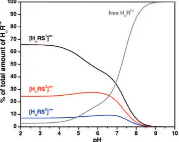
Figure 1.1 Distribution diagram of the overall amount of supramolecular species formed between a receptor of general formula HnRn+ and substrates 1 (S1), 2 (S2)
and 3 (S3), with HnRn+, S1, S2 and S3 in equimolar
amounts at a given solvent, ionic strength and temperature.
11
Figure 1.2 Binding site of the chloride channel from S. Typhimurium.
19
Figure 1.3 Binding site of SBP from S. typhimurium (a) and PBP from E. coli (b).
19
Figure 1.4 Binding site of NrtA (a) and CmpA (b) from
Synechocystis 6803.
20
Figure 1.5 Binding site of DctB from Sinorhizobium meliloti
occupied by succinate (a) and malonate (b).
21
Figure 1.6 Binding site of QFR from E. coli occupied by fumarate (a), oxaloacetate (b) and glutarate (c).
23
Figure 1.7 Crystal structures of the supermolecules formed by F -(a) and Cl- (b) anions with H616+ receptor, in (c) of
H616+ in presence of Br- with the Br- anions outside the
cavity, and in (d) the structure of the included H2O into
the cavity of H414+ receptor with several iodide anions
outside the cavity.
32
Figure 1.8 Crystal structure of the supermolecule formed by Cl -anion with H626+ receptor.
33
Figure 1.9 X-ray structures of te association of H646+ with F- (a),
Cl- (b), Br- (c) and N3- (d).
34
Figure 1.10 Distribution diagrams of the overall amount of supramolecular species formed between Hn5n+ and
the mixture of halide anions, Cl-, Br- and I- in the 1:1:1:1 ratio (a) and a mixture of the halides and sulfate (b). Ccryp = CA- = 2×10⁻³ mol dm-3.
Figure 1.11 Crystal structure of the supermolecules formed by association of F- (a) and Cl- (b) anions with receptor H686+. Crystal structure of and [H62(Cl)]5+ (c) is also
presented for comparison.
36
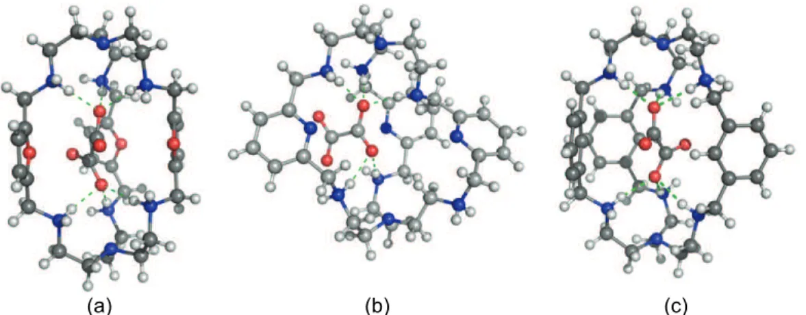
Figure 1.12 Crystal structures of supermolecules formed between H6106+ (a), H6116+ (b) and H612n+ (c) and the oxalate
anion.
38
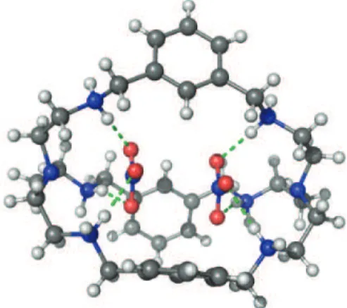
Figure 1.13 Crystal structure of the supermolecule formed by H6126+ with nitrate.
39
Figure 1.14 X-ray structures of the association of H8138+ with Cl
-(a) and NO3- (b).
40
Figure 1.15 Crystal structures of [H614(F)2(H2O)]4+ (a),
[H614(Cl)(H2O)]5+ (b), [H614(Br)(H2O)]5+ (c) and
[H414(H2O)4][I]4 (d).
41
Figure 1.16 Crystal structure of the cryptate formed by association of H6156+ with the tph2- dianion (a) and snapshot of
[H617(tph)]4+ taken at 10.5 ns of MD simulation (b).
42
Figure 1.17 Crystal structures of the supermolecules formed by F -and H4184+ (a); by Cl- with the H3183+ receptor (b); by
H3183+ in presence of Br- (c) and I- (d) with the anions
outside the cavity.
43
Figure 1.18 Crystal structures of the supermolecules formed by: F -(a) and Cl- (b) with H4194+ receptor; by Br- (c) with
H3193+ receptor; in (d) the structure of the included
H2O into the cavity of H3193+ receptor with several I
-anions outside the cavity and in (d) encapsulation of NO3- by H4194+.
44
Figure 1.19 Crystal structure of the cascade complex of 22 with CO32- anion.
46
Figure 1.20 Crystal structures of the cascade complexes of 25
with CN- (a) and N3- (b).
47
xxxi
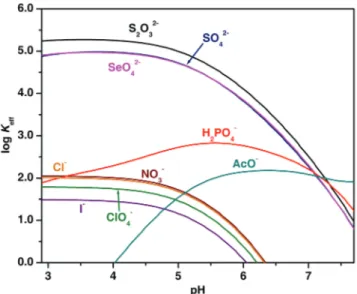
Figure 2.2 Plots of the effective association constant Keff (in log
units) versus pH for the supramolecular species formed between protonated xyl and the anions studied.
65
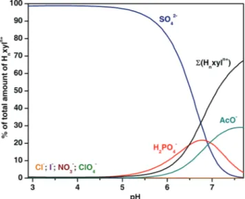
Figure 2.3 Distribution diagram of the overall amounts of supramolecular species formed between the receptor, Hnxyln+, and each anion.
67
Figure 2.4 1H NMR spectra of KTsO (A) and of H6xyl(TsO)6 (B) in
D2O at pD = 3.80 and 298.2 K.
67
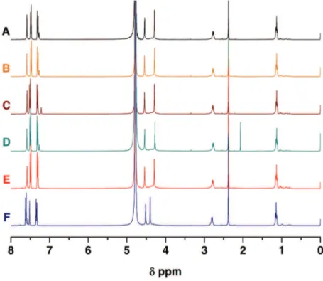
Figure 2.5 1H NMR spectra of solutions of the H6xyl(TsO)6
receptor (A) and of the receptor with each anionic substrate in equimolar amounts, Cl– (B), NO3– (C),
AcO– (D), H2PO4– (E) and SO42– (F), respectively, in
D2O at pD = 3.80 and 298.2 K.
68
Figure 2.6 Job’s plot between H6xyl6+ (R) and sulfate in D2O at
pD = 3.80 and 298.2 K.
69
Figure 2.7 ESI mass spectra of a solution of 1:1 receptor to sulfate stoichiometry in H2O/MeOH (50:50 v/v) at pH =
3.80.
70
Figure 2.8 ESI mass spectra of a solution of 1:1:1
receptor/sulfate/nitrate stoichiometry in H2O/MeOH
(50:50 v/v) at pH = 3.80.
71
Figure 2.9 Perspective views illustrating different structural features in the associated entity formed between SO42– and H6xyl6+ receptor.
72
Figure 2.10 Lowest energy conformations of H6xyl6+ (left),
[(H6xyl)(SO4)]4+ (centre) and [(H6xyl)(Cl)]5+ (right)
found in the conformational analyses
74
Figure 2.11 Evolution of the sulfate (left) and chloride (right) distances to the binding pocket centre in H2O:MeOH
solution.
75
Figure 2.12 Snapshots of [(H6xyl)(SO4)]4+ taken at 6 ns (left) and
13.7 ns (right) of MD simulation showing the anion located at the centre (left) and at the entrance (right) of the receptor pocket.
Figure 2.13 Rdfs for O–H···O=S and O–H···CN distances between
the water (blue) and methanol (red) molecules and the centre of mass of the receptor (left) and the anion (right).
78
Figure 2.14 Snapshots of the association between H6xyl6+ and
chloride taken at 3.0 ns (left) and 1.6 ns (right) of MD simulation showing one and two chloride anions accommodated into the cryptand cage.
79
Figure 3.1 Species distribution diagram for the protonation of pyr. 97
Figure 3.2 Plots of the effective association constant Keff (in log
units) versus pH for the supramolecular associations between Hnpyrn+ (a), Hnxyln+ (b) and the studied
anions.
99
Figure 3.3 Distribution diagram of the overall amounts of supramolecular species formed between the receptor, Hnpyrn+, and each anion.
100
Figure 3.4 Plot of phosphate/sulfate selectivity profile as a function of pH for Hnpyrn+ and other polyamine
compounds.
101
Figure 3.5 31P-NMR spectra of 1:1 Hnpyrn+:hydrogen phosphate
solutions in H2O/MeOH at pH = 3.8 (b) and 7.2 (e),
and for free hydrogen phosphate (a) and (d) at 298.2 K.
102
Figure 3.6 31P-NMR spectra of 1:1:1 Hnpyrn+:hydrogen
phosphate:sulfate solutions in H2O/MeOH at pH = 3.8
(c) and 7.2 (f) at 298.2 K.
103
Figure 3.7 Perspective views illustrating different structural features of the association between nitrate and the H6pyr6+ receptor:
105
Figure 3.8 Three views illustrating different structural features of the association of sulfate with H6pyr6+ receptor.
106
Figure 3.9 Two views illustrating different structural features of the association between phosphate and H6pyr6+
xxxiii
Figure 4.1 Species distribution diagram calculated for
copper(II):pyr solutions.
125
Figure 4.2 Molecular structure of [Cu3pyr(μ3-CO3)]4+ in complex
salt 1.
127
Figure 4.3 Molecular structure of [Cu3pyr(μ3-CO3)]4+ in complex
salt 2.
128
Figure 4.4 Temperature dependence of the product of the magnetic susceptibility with temperature for [Cu3pyr(μ3-CO3)]4+.
132
Figure 4.5 Magnetization curves for [Cu3pyr(μ3-CO3)]4+ at
different temperatures.
133
Figure 4.6 Variation of the spin state energies (E/Jversus the x =
j/J ratio) for an isosceles triangle with local spins S1 =
S2 = S3 = 1/2, using the common notation |S,S*>,
where S is the total spin (S = S1 + S2 + S3) and S* = S1
+ S3.
134
Figure 4.7 Contour map showing the dependence of the
magnetic coupling constant J (in cm–1) on the CuOO angles (in degrees).
137
Figure 4.8 Contour map showing the dependence of the
magnetic coupling constant J (in cm–1) on the CuOO angles (in degrees).
138
Figure 5.1 Species distribution diagrams of the protonation of btpN7 (a) and tbN4 (b). CbtpN7 = CtbN4 = 1.0×10⁻³ mol
dm–3.
157
Figure 5.2 X-ray crystal structure in two different views depicting [H7btpN7(H2O)(MeOH)Cl2]Cl6·(H3O)·3(H2O)·3MeOH:
159
Figure 5.3 Species distribution diagram calculated for the complexes of Cu2+ with of btpN7 (a) and tbN4 (b).
162
Figure 5.4 Species distribution diagram calculated for the system [CuHhbtpN7(oxa)]h+.
Figure 5.5 Distribution diagram of the overall amounts of supramolecular species formed between [CuHhbtpN7](2+h)+ and each dicarboxylate.
167
Figure 5.6 Absorption and X-band EPR spectra of
[CuHhbtpN7](2+h)+ at several pH values.
168
Figure 5.7 Absorption and X-band EPR spectra of
[CuHhbtpN7(oxa)]h+ at several pH values.
171
Figure 5.8 Vis absorbance spectra of aqueous solutions of PV. 173
Figure 6.1 Species distribution diagram of the protonation of btpN7 (a) and t2pN8 (b).
190
Figure 6.2 Plots of the log Keff versus pH for the associations
formed between the indicated dicarboxylate anions and HnbtpN7n+ (a) or Hnt2pN8n+ (b).
193
Figure 6.3 Distribution diagrams of the overall amounts of supramolecular species formed between the dicarboxylate anions and HnbtpN7n+ (a) or Hnt2pN8n+ (b)
in equimolar ratio.
194
Figure 6.4 Distribution diagrams of the overall amounts of supramolecular species formed between fumarate and maleate anions and HnbtpN7n+ (a) or Hnt2pN8n+ (b) in
equimolar ratio.
195
Figure 6.5 1H NMR spectra of solutions of the H6t2pN8(TsO)6 free
receptor (1), free substrates, mal2- (3a), suc2- (4a), glu2- (5a), male2- (6a) and fum2- (7a) and of the association of the receptor with each dicarboxylate substrate in equimolar amounts, oxa2- (2), mal2- (3b), suc2- (4b), glu2- (5b), male2- (6b) and fum2- (7b) , in D2O at pD = 4.5 and 298.2 K.
197
Figure 6.6 1H NMR spectra of solutions of the H6btpN7(TsO)6 free
receptor (1), free substrates, mal2- (3a), suc2- (4a), glu2- (5a), male2- (6a) and fum2- (7a) and of the association of the receptor with each dicarboxylate substrate in equimolar amounts, oxa2- (2), mal2- (3b), suc2- (4b), glu2- (5b), male2- (6b) and fum2- (7b) , in D2O at pD = 4.5 and 298.2 K.
xxxv
Figure 6.7 1H NMR signals of malonate recorded in the course of the H/D exchange in the absence of catalyst (A) and in the presence of H6btpN7(TsO)6 (B) and
H6t2pN8(TsO)6 (C).
200
Figure 7.1 X-ray crystal structure of btpN4O3 showing an
encapsulated MeCN.
215
Figure 7.2 Species distribution diagram of the protonation of btpN4O3 (a) and t2pN5O3 (b).
216
Figure 7.3 Perspective views illustrating different structural features in the associated entity formed between H3t2pN5O33+ and a water molecule:
218
Figure 7.4 Plot of the Keff versus pH for the associations formed
between the indicated amino acids and HnbtpN4O3n+.
220
Figure A2.1 Evolution of the six S···N distances over 15 ns long simulation
A11
Figure A2.2 Rdfs for O–H···CN and O–H···Cl distances between the
water (blue) and methanol (in red) molecules and the centre of mass of the receptor (left) and the anion (right).
A11
Figure A2.3 1H and 13C NMR spectra of the hexaimine in CDCl3. A12
Figure A2.4 HMQC spectra of the hexaimine in CDCl3. A12
Figure A2.5 1H and 13C NMR spectra of xyl in CDCl3. A13
Figure A2.6 COSY spectra of xyl in CDCl3. A13
Figure A2.7 HMQC spectra of xyl in CDCl3. A14
Figure A2.8 NOESY spectra of xyl in CDCl3. A14
Figure A3.1 1H and 13C NMR spectra of the hexaimine in CDCl3. A17
Figure A3.2 1H and 13C NMR spectra of pyr in CDCl3. A17
Figure A3.3 COSY spectrum of pyr in CDCl3. A18
Figure A3.4 HMQC spectrum of pyr in CDCl3. A18
Figure A3.5 NOESY spectrum of pyr in CDCl3. A19
Figure A3.6 ESI mass spectrum of pyr in MeOH. A19
Figure A4.1 FTIR spectrum of the free ligand pyr (KBr pellet). A25
Figure A4.2 FTIR spectrum of [Cu3pyr(μ3-CO3)]·(ClO4)4·H2O (KBr
pellet).
A25
Figure A4.3 ESI mass spectrum of [Cu3pyr(μ3-CO3)]·(ClO4)4,
H2O/MeOH (50:50 v/v), pH = 6.0.
A25
Figure A5.1 Experimental and simulated X-band EPR spectrum of [CuHhbtpN7](2+h)+ at pH = 6.3.
A28
Figure A5.2 Experimental and simulated X-band EPR spectrum of [CuHhbtpN7](2+h)+ at pH = 11.4.
A29
Figure A5.3 Experimental and simulated X-band EPR spectrum of [CuHhbtpN7(oxa)]h+ at pH = 7.0.
A29
Figure A5.4 Distribution diagram of the overall amounts of supramolecular species formed between [CuHhbtpN7](2+h)+ and each dicarboxylate.
A30
Figure A5.5 1H NMR spectrum of the tripodal trialdehyde in CDCl3. A30
Figure A5.6 13C NMR spectrum of the tripodal trialdehyde in CDCl3.
A31
Figure A5.7 1H NMR spectrum of the triimine in CDCl3. A31
xxxvii
Figure A5.10 13C NMR spectrum of btpN7 in CDCl3. A33
Figure A5.11 COSY spectrum of btpN7 in CDCl3. A33
Figure A5.12 HMQC spectrum of btpN7 in CDCl3. A34
Figure A5.13 NOESY spectrum of btpN7 in CDCl3. A34
Figure A5.14 ESI mass spectrum of btpN7 in MeOH. A35
Figure A6.1 Job’s plot between HnbtpN7n+ (R) and oxalate in D2O
at pD = 4.50 and 298.2 K.
A39
Figure A6.2 1H NMR spectrum of btpN7 in CDCl3. A39
Figure A6.3 1H NMR spectrum of t2pN8 in CDCl3. A40
Figure A7.1 1H NMR spectrum of 4-(bromomethyl)benzaldehyde in CDCl3.
A42
Figure A7.2 13C NMR spectrum of 4-(bromomethyl)benzaldehyde in CDCl3.
A42
Figure A7.3 1H NMR spectrum of 2-(4-bromomethyl-phenyl)-5,5-dimethyl-[1,3]dioxane in CDCl3.
A43
Figure A7.4 13C NMR spectrum of 2-(4-bromomethyl-phenyl)-5,5-dimethyl-[1,3]dioxane in CDCl3.
A43
Figure A7.5 1H NMR spectrum of the tripodal trialdehyde (3) in CDCl3.
A44
Figure A7.6 13C NMR spectrum of the tripodal trialdehyde (3) in CDCl3.
A44
Figure A7.7 1H NMR spectrum btpN4O3 in CDCl3. A45
Figure A7.8 13C NMR spectrum of btpN4O3 in CDCl3. A45
Figure A7.9 HMQC spectrum of btpN4O3 in CDCl3. A46
Figure A7.11 NOESY spectrum of btpN4O3 in CDCl3. A47
Figure A7.12 ESI mass spectrum of btpN4O3 in MeOH. A47
Figure A7.13 1H NMR spectrum the tripodal trialdehyde (6) in CDCl3.
A48
Figure A7.14 13C NMR spectrum of the tripodal trialdehyde (6) in CDCl3.
A48
Figure A7.15 1H NMR spectrum t2pN5O3 in CDCl3. A49
Figure A7.16 13C NMR spectrum t2pN5O3 in CDCl3. A49
Figure A7.17 HMQC spectrum of t2pN5O3 in CDCl3. A50
Figure A7.18 COSY spectrum of t2pN5O3 in CDCl3. A50
Figure A7.19 NOESY spectrum of t2pN5O3 in CDCl3. A51
xxxix
Index of Schemes
Page
Scheme 1.1 Preorganisation and complementarity effects in the coordination of K+ by polyether receptors.
6
Scheme 1.2 The association process in solution. 7
Scheme 1.3 Different types of receptors. Upper row, from left to right: podand, crown ether, cryptand, spherand and calixarene. Lower row from left to right: cavitand, carcerand and cryptophane.
12
Scheme 1.4 Homoditopic and heteroditopic compounds. 13
Scheme 1.5 General structure of macrobicyclic compounds. 14
Scheme 1.6 Synthetic strategies (a-b, c-d and e) for the construction of the macrobicyclic architecture.
15
Scheme 1.7 Examples of the most common anion binding sites. 23
Scheme 1.8 Examples of neutral anion receptors: a) amide based macrobicycle; b) urea based tripodal compound; c) thiourea based macrocycle; d) pyrrole based receptor;
e) urea/amide based macrocycle; f)
amide/urea/pyrrole based podand.
24
Scheme 1.9 Examples of charged anion receptors: a) macrocyclic polyammonium compound; b) polyguanidinium tripodal compound; c) dinuclear copper macrobicyclic complex; d) tripodal copper complex combined with ammonium binding sites; e) podand copper complex combined with guanidinium binding sites.
25
Scheme 1.10 First reported anion receptors, “katapinand”, “soccer ball cryptand and O-bistren.
26
Scheme 1.11 Examples of polyamine compounds of different topologies used as anion receptors in their protonated forms: a) tripodal; b) bis-macrocyclic; c) macrocyclic; d) macrobicyclic and e) macrotricyclic.
27
Scheme 1.13 Synthesis of O-bistren by a modified Richman-Atkins procedure.
29
Scheme 1.14 Synthesis of a macrobicyclic polyamine by Schiff base condensation followed by reduction.
30
Scheme 1.15 Polyamine cryptands with aliphatic spacers used as anion receptors in their protonated forms.
31
Scheme 1.16 Representation of bifluoride binding by H636+. 33
Scheme 1.17 Polyamine cryptands with aromatic spacers used as anion receptors in their protonated forms.
37
Scheme 1.18 Dinuclear copper(II) complexes of polyamine cryptands used as receptors for anions.
45
Scheme 1.19 Polyaza cryptands synthesized and studied in this work.
50
Scheme 1.20 Mixed polyazapolyaza cryptands synthesized and studied in this work.
51
Scheme 2.1 Synthetic procedure of xyl. 61
Scheme 3.1 Polyaza cryptands discussed in this work. 95
Scheme 4.1 pyr 122
Scheme 4.2 Proposed mechanism for CO2 fixation by [Cu3pyr]6+. 130
Scheme 4.3 (a) Definition of the CuOO angles. Schematic views of the overlapping between magnetic orbitals in the (b)
syn-syn, (c) syn-anti and (d) anti-anti conformations.
135
Scheme 5.1 The new compound btpN7, and the two heteroditopic
heptaamine cryptands already reported.
xli
Scheme 5.2 Copper(II) cryptate of btpN7 and its cascade species
formed with the studied dicarboxylates.
155
Scheme 5.3 Tripodal compound used for comparison purposes. 155
Scheme 5.4 Synthetic procedure of btpN7. 156
Scheme 5.5 Representation of possible conformational
rearrangement of H6btpN76+ needed for dicarboxylate
binding.
161
Scheme 5.6 Proposed change of geometry of the copper site upon oxa2- binding.
171
Scheme 5.7 Indicator-displacement assay. 172
Scheme 6.1 Target dicarboxylate substrates. 189
Scheme 6.2 Macrobicyclic compounds studied in this work. 189
Scheme 6.3 Representation of possible conformational
rearrangement of H6btpN76+ and H6t2pN86+ upon
dicarboxylate binding.
196
Scheme 7.1 Target amino acid substrates 212
Scheme 7.2 Macrobicyclic compounds studied in this work. 213
xliii
Index of Tables
Page
Table 2.1 Overall (βiH) and stepwise protonation (KiH) constants
of xyl in H2O/MeOH (50:50 v/v).
62
Table 2.2 Overall (log βHhLlAa) and stepwise (log KHhLlAa) association constants for the indicated equilibria in H2O/MeOH (50:50 v/v).
64
Table 2.3 Variations in N···X distances (Å) of [(H6xyl)(A)]n+ (A =
SO42–, n = 4; A = Cl–, n =5 ) over the MD simulations
in H2O:MeOH (50:50 v/v) solution at r.t.
77
Table 2.4 Variations in the number of water and methanol enclosing the receptor and anion estimated with cut-off radius of 4.2 Å and 3.5 Å, respectively.
78
Table 3.1 Overall (βiH) and stepwise protonation (KiH) constants
of pyr in H2O/MeOH (50:50 v/v).
96
Table 3.2 Stepwise association constants (log KHhLlAa) for the indicated equilibria in H2O/MeOH (50:50 v/v).
98
Table 4.1 Overall (βiH) and stepwise (KiH) protonation constants
of pyr in H2O/MeOH (50:50 v/v).
123
Table 4.2 Overall (log βMmHhLl) and stepwise (log KMmHhLl) stability constants of the copper(II) complexes of pyr in H2O/MeOH (50:50 v/v).
124
Table 4.3 Selected bond distances (Å) and angles (º) for [Cu3pyr(μ3-CO3)]4+ in salt complexes 1 and 2.
129
Table 4.4 Magneto-structural data for the compounds where a carbonate group acts as bridge between three copper(II) ions through only the OCO pathway.
Table 5.1 Overall (βiH) and stepwise protonation (KiH) constants
of btpN7 and tbN4 in H2O.
157
Table 5.2 Overall (βHhLlAa) and stepwise (KHhLlAa) association constants for the indicated equilibria in H2O.
160
Table 5.3 Overall (βMmHhLl) and stepwise (KMmHhLl) stability constants of the copper(II) complexes of btpN7 and
tbN4 in aqueous solution.
163
Table 5.4 Overall (βMmHhLlAa) and stepwise (KMmHhLlAa) association constants for the indicated equilibria in H2O.
165
Table 5.5 X-band EPR[a] and absorption spectra[b] data for the Cu2+ complexes of btpN7 and respective associations
with oxalate.
169
Table 6.1 Overall (βiH) and stepwise protonation (KiH) constants
of btpN7 and t2pN8 in H2O.
190
Table 6.2 Stepwise association constants (log KHhLlAa) of HnbtpN7n+ and Hnt2pN8n+ receptors with oxa2-, mal2-,
suc2-, glu2-, male2- and fum2- anions in H2O.[
192
Table 6.3 Chemical shifts (Δδ) and respective effective association constants (Keff), for the binding of
dicarboxylate substrates by HnbtpN7n+ and Hnt2pN8n+.
199
Table 7.1 Overall (βiH) and stepwise protonation (KiH) constants
of btpN4O3 and t2pN5O3 in MeOH/H2O.
216
Table 7.2 Stepwise association constants (KHhLlAa) for the indicated equilibria involving HnbtpN4O3n+ and Hgly,
Hbala, Htau, Hgaba, H2amp and H2aep in MeOH/H2O.
219
Table A2.1 Stepwise protonation (log KiH) constants of the anions
in H2O/MeOH (50:50 v/v).[
xlv
Table A3.1 Overall (log βHhLlAa) association constants for the indicated equilibria in H2O/MeOH (50:50 v/v)
A20
Table A3.2 N–H···O hydrogen bond dimensions of
[(H6pyr)(NO3)3(H2O)3](NO3)3·4H2O
A21
Table A3.3 Hydrogen bond dimensions of
[(H6pyr)(SO4)2(H2O)4](HSO4)2·6H2O
A22
Table A3.4 N–H···O hydrogen dimensions of
[(H6pyr)(HPO4)2(H2PO4)(H2O)2](H2PO4)·16H2O
A23
Table A3.5 Crystal data and selected refinement parameters for anion binding complexes
A24
Table A4.1 Crystal data and refinement details of [Cu3pyr(μ3
-CO3)](ClO4)4·(MeOH)·2H2O 1 and [Cu3pyr(μ3
-CO3)](NO3)4·9H2O 2.
A26
Table A5.1 Hydrogen bond dimensions of
[H7btpN7(H2O)(MeOH)Cl2]Cl6·(H3O) 3(H2O) 3MeOH
A27
Table A5.2 Overall (βiH) and stepwise protonation (KiH) constants
of the studied dicarboxylates in aqueous solution.
A27
Table A5.3 Overall (log βMmHhAa) and stepwise (log KMmHhAa)
stability constants of the copper(II) complexes of the studied dicarboxylates in aqueous solution.
A28
Table A6.1 Stepwise protonation (KiH) constants of btpN7 in H2O. A37
Table A6.2 Overall (βiH) and stepwise protonation (KiH) constants
of the studied dicarboxylates in aqueous solution.
A37
Table A6.3 Overall association constants (log βHhLlAa) for the indicated equilibria in H2O.
Table A7.1 Overall (βiH) and stepwise protonation (KiH) constants
of the studied amino acids in aqueous solution.
A41
Table A7.2 Overall association constants (log βHhLlAa) for the indicated equilibria between the HnbtpN4O3n+ receptor
and Hgly, Hbala, Htau, Hgaba, H2amp and H2aep
substrates in MeOH/H2O.
xlvii
xlix
li
List of abbreviations and symbols
1D one dimension
2D two dimensions
3J
(H,H) vicinal proton-proton coupling constant
AcOH acetic acid
adi2- Adipate
ADP adenosine diphosphate
AMP adenosine monophosphate
ATP adenosine triphosphate
BM Bohr magneton
btc3- benzenetricarboxylate
ch3- cyclohexanetricarboxylate
ci concentration of species i
ci⊖ concentration of species i in the standard state
CmpA bicarbonate binding protein
COSY correlation spectroscopy
D zero-field splitting parameter
Dct C4-dicarboxylate transport system
DctB periplasmic sensor domain
DFT density functional theory
DHP 3,4-dihydro-2H-pyran
DMF dimethylformamide
DMSO dimethylsulfoxide
DNA deoxyribonucleic acid
E electromotive force
Ej liquid junction potential
Eº´ formal electrode potential
EPR electron paramagnetic resonance
ESI electrospray ionization
EtOH ethanol
FAD flavin adenine dinucleotide
fum2- fumarate
g g factor
glu2- glutarate
h Planck constant
H magnetic field
H2aep 2-aminoethylphosphonate
H2amp aminomethylphosphonate
Hgly glycine
Hbala β-alanine
Hgaba γ-aminobutyrate
Htau taurine
HMQC heteronuclear multiple quantum coherence
I ionic strength
iph2- isophthalate
IR infrared
J coupling constant
k Boltzmann constant
Kass association (or binding) constant
Keff effective association (or binding) constant
KHhLlAa stepwise protonation constant
KiH stepwise protonation constant
KMmHhLl stepwise stability constants
KMmHhLlAa stepwise stability constants of cascade associations
Kw ionic product of water
M magnetization
mal2- malonate
male2- maleate
liii
MeOH methanol
MM molecular mechanics
MsCl mesylenesulphonyl chloride
NMR nuclear magnetic resonance
NOESY nuclear Overhauser effect spectroscopy
NrtA nitrate binding protein
oxa2- oxalate
PBP phosphate binding protein
pD –log [D+]
ph2- phthalate
PhOH phenol
pim2- pimelate
QFR menaquinol:fumarate oxidoreductase
R receptor
r.t. room temperature
RNA ribonucleic acid
S substrate
SBP sulfate binding protein
suc2- succinate
T temperature
THF tetrahydrofuran
THP tetrahydro-2H-pyran-2-yl
TMS tetramethylsilane
tph2- terephthalate
tren tris-(aminoethyl)amine
trpn tris-(aminopropyl)amine
Ts toluenesulphonyl
TsCl toluenesulphonyl chloride
TsOH toluenesulphonic acid
UV ultraviolet
vis visible
X molar fraction
βHhLlAa overall association constant
βiH overall protonation constant
βMmHhLl overall stability constants
βMmHhLlAa overall stability constants of cascade associations
δ chemical shift
ΔΔG difference in binding free energies
θ Weiss parameter
ν frequency
lv
Colour code for the crystal structure diagrams
The colour labelling scheme adopted in the molecular diagrams representing
the crystal structures, except otherwise stated in the respective figure caption, is
the following:
Hydrogen White
Carbon Grey
Nitrogen Blue
Oxygen Red
Sulfur Yellow
Copper Golden yellow
Fluorine Pink
Chlorine Green
Bromine Brown
Iodine Purple
Phosphorus Orange
Hydrogen bonds Green dashed lines
Molecular diagrams depicted in Chapter 1 were drawn with PyMOL (W. L.
DeLano, The PyMOL Molecular Graphics System DeLano Scientific, San Carlos,
Chapter 1
Introduction
3
1.1 Supramolecular Chemistry
Throughout the years chemists have strived to attain control over the
formation and breaking of the covalent bond and by doing so, numerous methods
for constructing new molecules of increasing complexity were developed.[1] About 40 years ago a new paradigm emerged that sought to achieve increasing
complexity by taking advantage of the non-covalent intermolecular forces
(electrostatic interactions, hydrogen bonding, van der Waals interactions, etc),
giving rise to a new field called Supramolecular Chemistry. The term was
introduced by one of its pioneers, Jean-Marie Lehn, Nobel prize laureate in 1987,
whom defined it as the "...chemistry of molecular assemblies and of the
intermolecular bond" or "the chemistry beyond the molecule",[1] as it deals with the complex entities formed by the association of two or more chemical species
held together by non-covalent intermolecular forces.
Many of supramolecular chemistry’s concepts, terminology and definitions
were already known for quite some time: in 1894 Fischer introduced the notion of
complementarity with the famous lock and key principle, twelve years later
Ehrlich created the concept of receptor by observing that molecules only act
when bound, and the term supermolecule was introduced in 1937 by Wolf.
However, it was only with the work of Nobel Prize winners, Pedersen, Lehn and,
Cram in the late 1960´s and early 1970´s that Supramolecular Chemistry was
conceptualized as new field of chemistry.[2]
In the beginning, the field dealt mainly with the so called host-guest chemistry,
but the area rapidly evolved to include self-assembly, self-replication, catalysis by
artificial enzymes and enzyme models, molecular devices and other processes
that involve non-covalent interactions. Nowadays Supramolecular Chemistry is a
highly interdisciplinary field interfacing chemistry, biology, and physics and
Chapter 1
1.2 Molecular recognition
Molecular recognition is defined by the selection of a substrate (or substrates)
by a given receptor (or host) molecule through the establishment of non-covalent
interactions between them, and it is at the basis of every aspect of
supramolecular chemistry.[1]
The receptor is the molecular entity possessing convergent binding sites
located in strategic points of its architecture of a well defined size and shape. The
binding sites are characterized by their electronic properties such as charge,
polarity, polarisability, size, shape, number and location in the receptor
framework. The substrate (or guest) in turn can be viewed as a species (cationic,
anionic or neutral) with diverging binding sites, complementary to those of the
receptor. Together they form a third entity called supermolecule, with its own
thermodynamic and structural properties.[1]
It should be stressed, however, that binding alone cannot be called
recognition. Recognition requires a high degree of geometrical and interactional
complementarity between the receptor with respect to a given substrate, which
allows the latter to be selected in the presence of any other competitor, including
the solvent. In order to achieve selective binding several aspects must be taken
into account:
a) steric complementarity – receptor and substrate must present matching size
and shape;
b) interactional complementarity – presence of complementary binding sites in
the correct disposition on the receptor and on the substrate, such as
positive/negative charge, charge/dipole, dipole/dipole, hydrogen bond
donor/acceptor, etc;
c) multiple interaction sites – the weak nature of non-covalent interactions
requires that the binding sites are numerous, well located and acting

![Figure 1.15 Crystal structures of [H 6 14(F) 2 (H 2 O)] 4+ (a), [H 6 14(Cl)(H 2 O)] 5+ (b), [H 6 14(Br)(H 2 O)] 5+ (c) and [H 4 14(H 2 O) 4 ][I] 4 (d)](https://thumb-eu.123doks.com/thumbv2/123dok_br/15766170.640522/103.765.96.681.360.628/figure-crystal-structures-h-f-h-cl-br.webp)