113 113 113 113 113 Mem Inst Oswaldo Cruz, Rio de Janeiro, Vol. 100(Suppl. I): 113-120, 2005
M echanisms of leukocyte lipid body formation and function
in inflammation
Patrícia T Bozza+, Christianne Bandeira-M elo
Laboratório de Imunofarmacologia, Departamento de Fisiologia e Farmacodinâmica, Instituto Oswaldo Cruz-Fiocruz, Av. Brasil 4365, 21040-900 Rio de Janeiro, RJ, Brasil
An area of increasingly interest for the understanding of cell signaling are the spatio-temporal aspects of the different enzymes involved in lipid mediator generation (eicosanoid-forming enzymes, phospholipases and their regulatory kinases and phosphatases) and pools of lipid precursors. The compartmentalization of signaling compo-nents within discrete and dynamic sites in the cell is critical for specificity and efficiency of enzymatic reactions of phosphorilation, enzyme activation and function. We hypothesized that lipid bodies – inducible non-membrane bound cytoplasmic lipid domains – function as specialized intracellular sites of compartmentalization of signaling with major roles in lipid mediator formation within leukocytes engaged in inflammatory process. Over the past years substantial progresses have been made demonstrating that all enzymes involved in eicosanoid synthesis localize at lipid bodies and lipid bodies are distinct sites for eicosanoid generation. Here we will review our current knowl-edge on the mechanisms of formation and functions of lipid bodies pertinent to inflammation.
Key words: inflammation - lipid bodies - eicosanoids
The cells of both innate and adaptive immune response express a great variety of receptors that transduce and integrate an enormous amount of information enabling the cells to mount effective immune responses. Over the past several years major advances have been made to elucidate the biochemical and molecular details of cell activation and depicting signaling cascades that leads to inflammatory mediator production. However the temporal and spatial organization of signaling and its impact on cell function is less clear. Recent evidence indicates that immune receptors and components of their signaling cas-cade are spatially organized within the cell and this spa-tial organization plays a key role in the regulation and amplification of cell signaling.
Many studies have delineated roles for lipid domains including plasma membrane-associated domains – lipid rafts and caveolae – and intracellular lipid-rich domains – named lipid bodies –, in cellular signaling. The functions of rafts and caveolae in cell signaling have been recently reviewed elsewhere (Anderson & Jacobson 2002, Dykstra et al. 2003, Pike 2003, van Deurs et al. 2003, Helms & Zurzolo 2004). Cytoplasmic lipid bodies are osmiophilic organelles present in virtually all mammalian cells, which are surrounded by a monolayer of phospholipids with a unique fatty acid composition, have a neutral lipid-rich core, and present variable protein composition (Murphy 2001, van Meer 2001, Tauchi-Sato et al. 2002). Members of the PAT family of proteins [perilipin,
adipose-differentia-Financial support: Howard Hughes Medical Institute (to PTB), Fogarty International Center-NIH, Pronex-MCT, CNPq, Profix, Faperj
+Corresponding author. E-mail: [email protected]
Received 8 November 2004 Accepted 30 December 2004
tion related protein (ADRP) and TIP 47], in particular ADRP, have been ubiquitously associated to lipid bodies in different cell types, including leukocytes, and although their function are not fully understood, they seem to act as structural proteins surrounding lipid bodies serving as nucleation center for lipid assembling and/or acting as an anchor for subcellular locations within the cell (Heid et al. 1998, Nakamura & Fujimoto 2003, Brasaemle et al. 2004). It is hypothesized that lipid body lipidic and proteic compo-sition varies according to the cell type and stimulatory conditions, with impacts to their function in the cell. Here we review the evidence that leukocyte lipid body forma-tion and compartmentalizaforma-tion of enzymes within lipid bodies is a highly regulated cellular event and that lipid bodies play a key role in the heightened capacity of leu-kocytes to generate eicosanoids in inflammatory condi-tions.
Mechanisms of leukocyte lipid body formation
syn-114 114 114 114
114 Leukocyte lipid bodies function in inflammation • Patrícia Bozza, Christianne Bandeira-M elo
drome (HES) and in tissue eosinophils in biopsies from Crohn’s disease (Solley et al. 1976, Beil et al. 1995, Bozza et al. 1998), in synovial leukocytes from patients with ar-thritis (Weinstein 1980, Schlesinger et al. 1982, Reginato et al. 1985, Bozza et al. 1996b) and lung and blood leuko-cytes from septic and/or ARDS patients (Triggiani et al. 1995, Pacheco et al. 2002).
Genesis of new lipid bodies is not a manifestation of cellular injury or simply attributable to excess substrate
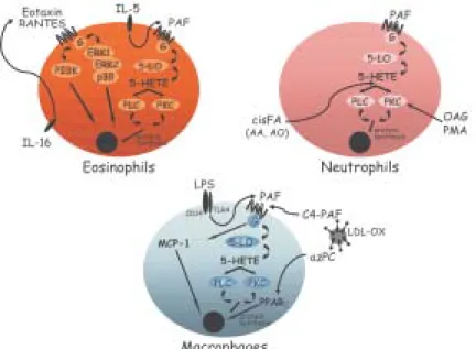
fatty acids, on the contrary, it is a rapid, highly regulated phenomenon that is stimulus and cell specific (Fig. 2). For instance, lipid bodies can be elicited in vivo and in vitro after stimulation of cells with either lipid agonists such as, cis-unsaturated fatty acids, PAF, LPS or PKC activa-tors (Weller et al. 1989, 1991 Bozza et al. 1996a, b, Pacheco et al. 2002) or protein stimuli such as chemokines and interleukines (Bozza et al. 1998, Bartemes et al. 1999, Bandeira-Melo et al. 2001a, 2002a).
Neutrophil lipid bodies, identical to lipid bodies in leu-kocytes in vivo as evidenced by light and electron mi-croscopy (EM), can also be rapidly (within 15-60 min) elic-ited in vitro after stimulation with cis-unsaturated fatty acids, PAF, or PKC activators (Weller et al. 1989, 1991, Bozza et al. 1996a, b). Although incubation of leukocytes with exogenous phospholipids and fatty acids provides a source of lipids for incorporation into newly formed lipid bodies, mechanisms other than simple availability of lipid precursors seem to be involved in lipid body formation. First, specific unsaturated fatty acids, including non-esterifiable analogs, in a stereochemically-restricted man-ner, can elicit de novo formation of lipid bodies in human leukocytes, whereas fully saturated fatty acids are not active (Weller et al. 1989, 1991, Bozza & Weller 2001).
Moreover, PAF but not its precursor and metabolite, lyso-PAF, that shares the lipid structure of PAF but has no receptor agonistic activity (Prescott et al. 2000), have the capacity to induce lipid body formation (Bozza et al. 1996a, 1997, de Assis et al. 2003). In addition, it has been demonstrated that PAF-induced lipid body formation in leukocytes involves the regulated activation of different signaling pathways (Fig. 1). PAF stimulated lipid body is a receptor-dependent phenomenon, inhibitable by PAF receptor antagonist and pertussis toxin (Bozza et al. 1996a, 1997, 1998). PAF, acting via its G-protein-linked receptor induces lipid body formation via a downstream signaling involving, sequentially, LO activation to generate 5-HETE that per se activates its G-protein-linked receptor, Fig. 1:in vivo lipid body formation within infiltrating leukocytes
during experimental endotoxin-induced pleurisy in mice. LPS ad-ministration induces formation of new lipid bodies within resident macrophages and infiltrating neutrophils (C, D). Such phenom-enon is easily observed in cell preparations stained with osmium tetroxide (C), which show several cytoplasmic punctate osmio-philic inclusions within macrophages (MΦ) and neutrophils (NΦ). Pleural leukocytes from LPS-stimulated mice stained with May-Grünwald-Giemsa do not display cytoplasmic lipid bodies (D), which were dissolved during such alcoholic fixation/staining procedure, only exhibiting a vacuolated cytoplasm (arrow heads). Leukocytes from PBS. Stimulatted animals are shown in A, B.
Fig. 2: lipid body formation is a cell and stimulus specific, highly regulated phenomenon. Different stimuli, including proteic and lipidic agonists, can trigger lipid body formation within eosinophils, neutrophils, and macrophages.
A B
115 115 115 115 115 Mem Inst Oswaldo Cruz, Rio de Janeiro, Vol. 100(Suppl. I), 2005
and protein kinase C activation (Bozza et al. 1996a). Inter-estingly other neutrophil agonists IL8, C5a, and LTB4 act-ing through G protein coupled receptors were not capable of inducing neutrophil lipid body formation (Bozza et al. 1996a).
Of special relevance to allergic inflammation, key me-diators of the allergic response in addition to PAF are capable of eliciting newly formed lipid bodies within hu-man eosinophils. IL-5 (a stimuli proteic in nature) alone or combined to GM-CSF in the absence of exogenous lipids as well as immobilized IgG lead to significant increase in lipid body numbers (Bozza et al. 1998, Bartemes et al. 1999), suggesting that receptor mediated stimuli results in intra-cellular lipid remodeling and lipid body formation. More-over, chemokines acting via CCR3 receptors, including RANTES, eotaxin, eotaxin-2, and eotaxin-3, can initiate intracellular signaling in eosinophils, but not neutrophils, that also culminate in de novo formation of lipid bodies (Bandeira-Melo et al. 2001a, b). Differently from the sig-naling pathways described by PAF, eotaxin and RANTES activating CCR3 receptor signals through phospho-inositide 3-kinase (PI3K) and the ERK1/2 and p38 MAP kinases (Bandeira-Melo et al. 2001a).
Increased lipid body formation in neutrophils and macrophages could also be observed in experimentally induced inflammation triggered either by LPS or OxLDL (oxidized low density lipoprotein) but not by native LDL (Pacheco et al. 2002, Silva et al. 2002). LPS administration into mice induced a dose- and time-dependent increase in lipid body numbers. Moreover, LPS failed to form lipid bodies in C3H/HeJ (TLR4 mutated) mice or in cells treated with neutralizing anti-CD14, demonstrating a requisite role for LPS receptors in lipid body formation (Pacheco et al. 2002). Infection by T. cruzi also leads to increase in lipid body numbers in macrophages (Melo et al. 2003), whether this reaction would also involve pattern recognition re-ceptors are now under investigation.
PAF and PAF-like lipids are believed to play an impor-tant role in lipid body formation induced by LPS or by oxLDL in vivo since the pretreatment with three structur-ally unrelated PAF receptor antagonists completely in-hibited this phenomenon (Pacheco et al. 2002, Silva et al. 2002). Although PAF may act at intracellular binding sites to induce cell activation (Bazan et al. 1994), results are suggestive that PAF is acting in a paracrine/autocrine way to induce lipid body formation, since the PAF-receptor antagonist used in those studies would act preferentially at membrane receptors and confirmed by findings that LPS or oxLDL induced lipid body formation were inhib-itable by treatment with extracellular PAH-acetylhidrolase (Silva et al. 2002).
It has been demonstrated that dietary fatty acids may have impacts on cellular lipid domains (Yaqoob 2003). Consumption of the long chain n-3 PUFA has been dem-onstrated to modify composition of lipid rafts and caveolae (Stulnig et al. 2001, Fan et al. 2003, 2004, Ma et al. 2004). We have recently demonstrated that fatty acid dietary intake may also modulate leukocyte lipid body formation. Indeed, dietary intake of extra virgin olive oil lead to inhi-bition of LPS-induced lipid body formation and decreased generation of MCP-1 and eico-sanoids (Leite et al. 2005).
Interestingly, the pretreatment of leukocytes with pro-tein synthesis inhibitors acting on transcription (actino-mycin D) and translation (cycloheximide) significantly, although partially, inhibited lipid body formation induced by PAF and fatty acids in vitro but not by eotaxin and RANTES, thus indicating that according to the stimulated conditions induction of lipid bodies depends on new pro-tein synthesis and its likely that specific and trans-criptionaly regulated early response genes are activated during the process of lipid body formation (Bozza et al. 1996a, b, 1997, Bandeira-Melo et al. 2001a, Pacheco et al. 2002). A role for peroxisome proliferator-activated recep-tors (PPARs), members of the nuclear receptor supergene family that function in ligand-activated transcription, in leukocyte lipid body formation has recently been sug-gested. Indeed, we observed that treatment of mouse mac-rophages with BRL 49653, a preferential PPARγ ligand, failed to induce lipid body formation. However, BRL 49653 treatment significantly potentiated lipid body formation induced by ox-LDL or PAF-like agonists, suggesting that PPAR have a role in regulating leukocyte lipid body for-mation (de Assis et al. 2003). However, we anticipate that differences in PPAR expression and role in lipid body for-mation may vary according to the cell type and stimuli studied and further studies are needed to characterize the roles of PPAR in lipid body formation and the involve-ment of PPAR targeted genes in this phenomenon.
The formation of structurally distinct lipid bodies re-flects mechanisms whereby proteins and lipids coalesce in a deliberate, highly regulated manner. Lipid body as-sembly can result from mobilization and reorganization of endogenously derived lipids as well as incorporating ex-ogenous lipid. Thus, although lipid bodies are not con-ventional membrane-bound organelles, they appear to represent specialized intracellular domains whose induced formation is centrally related to activating mechanisms within the cells. Moreover, lipid body size and numbers are characteristically increased in leukocytes following activation in vitro as well as in in vivo inflammatory disor-ders, and can be used as a marker of leukocyte activation.
Leukocyte lipid body functions in inflammation
Eicosanoidforming enzyme compartmentalization
116 116 116 116
116 Leukocyte lipid bodies function in inflammation • Patrícia Bozza, Christianne Bandeira-M elo
ERK2, p85, and p38, were localized at lipid bodies. In addi-tion, isolated lipid body fractions contained cPLA2 enzy-matic activity and released AA from sn-2-position of phos-phatidylcholine (Yu et al. 1998).
The two major enzymes, 5-LO and COX, involved in the enzymatic conversion of AA into eicosanoids were also shown to localize within lipid bodies. By using immu-nocytochemistry at the light level and ultrastructural postembedding immunogold, cyclooxygenase has been reported to localize at eosinophil lipid bodies, both in natu-rally formed lipid bodies in eosinophils from HES patients and in PAF-induced lipid bodies (Dvorak et al. 1992, 1993, Bozza et al. 1997, 1998). Previous studies on the intracel-lular localization of 5-LO have shown that 5-LO localiza-tion is cell type-specific and also varies according to the activation state of the cell. 5-LO was shown to localize within the nuclear environment (nuclear membrane and euchromatin) of alveolar macrophages and basophilic leu-kemia cells, whereas 5-LO was found to be predominantly cytosolic in human neutrophils and resting peritoneal macrophages (Peters-Golden & Brock 2000, Bandeira-Melo & Weller 2003). In addition to the nuclear environment, we have demonstrated the compartmentalization of the key enzyme for leukotriene production, 5-LO, within hu-man eosinophil and basophil lipid bodies (Bozza et al. 1997, 1998, Bandeira-Melo et al. 2001a). In addition, im-muno-reactivity for 5-LO was also detected in lipid bod-ies from enucleated human eosinophils (Bozza et al. 1997). Human eosinophils upon stimulation preferentially produce LTC4 as their 5-LO product. LTC4-synthase is the terminal LT-forming enzyme and is present selectively in eosinophils, basophils, and mast cells (Bandeira-Melo et al. 2002a). Similarly to that observed for 5-LO, LTC4 -synthase was shown to co-localize within eosinophil lipid bodies (Bozza et al. 1997).
Sub-cellular localization of eicosanoid forming en-zymes in leukocytes in vivo is starting to be unveiled. Li-pid bodies, in both ox-LDL- and LPS-induced models of inflammation, compartmentalize 5-LO and COX-2 enzymes within recruited leukocytes (Pacheco et al. 2002, de Assis et al. 2003). Similarly, eosinophils attracted to the site of allergic reaction showed 5-LO and COX-2 immuno-reac-tivity within newly formed lipid bodies. In addition, lipid bodies within leukocytes obtained from clinical inflamma-tory conditions including sepsis and arthritis were also demonstrated to be domains for intracellular compartmen-talization of 5-LO and COX-2 (Pacheco et al. 2002, Roimicher, unpub. data).
Together these findings support a role for lipid bodies to function as specific sites for eicosanoid formation. The compartmentalization of arachidonate substrate, cPLA2, and eicosanoid-forming enzymes provides in one locale an efficient mean to regulate arachidonate release and di-rectly couple it with the enzymes to form eicosanoids (Table).
Involvement of lipid bodies in enhanced generation of eicosanoids by inflammatory cells - Because leuko-cyte lipid bodies are sites of intracellular localization of eicosanoid-forming enzymes and also stores of the eicosanoid precursor arachidonic acid, it has been hy-pothesized that increases in lipid body numbers in leuko-cytes would result in enhanced capacity of eicosanoid production by leukocytes. Indeed, stimuli known to prime leukocytes to induce eicosanoid generation, including PKC activators, arachidonate and PAF, are also active in stimulating lipid body formation. Accordingly, others and we observed a significant correlation between lipid body formation and enhanced generation of both LO- and
COX-TABLE
Proteins found within leukocyte lipid bodies
Proteins Cell type References
Structural proteins (PATs)
. ADRP Murine RAW macrophages, murine inflammatory Chen et al. 2002
leukocytes, human U937, and 2H3 monocytes
Eicosanoid-forming enzymes
. PLA2 Human monocytic U937 cells Yu et al. 1998
. 5-LO, 15-LO, COX, LTC4 synthase Guinea pig peritoneal macrophages, line 10 Dvorak et al. 1993 hepatocarcinoma cells, human circulating Bozza et al. 1997, 1998 eosinophils and basophils, and murine Bandeira-Melo et al. 2001 macrophages from endotoxemic reaction Pacheco et al. 2002
Kinases
. MAPs: ERK1, ERK2, p38 Human monocytic U937 cells Yu et al. 1998
. PI3K subunits: p55, p85α, p85β Human U937 monocytes and murine RAW Yu et al. 2000 macrophages
. PKC Murine RAW macrophages Chen et al. 2002
Cytokines/Chemokines
. TNF-α Cells of Crohn’s disease biopsies, and Beil et al. 1995
neutrophilsof septic patients or murine Pacheco et al. 2002 endotoxemic inflammation
. RANTES / IL-16 Human eosinophils Lim et al. 1996
117 117 117 117 117 Mem Inst Oswaldo Cruz, Rio de Janeiro, Vol. 100(Suppl. I), 2005
derived eicosanoids in vitro (Bozza et al. 1996a, b, 1997, 1998, Bartmenes et al. 1999) as well as in vivo (Pacheco et al. 2002, Silva et al. 2002, de Assis et al. 2003, Melo et al. 2003).
Analogously, agents that inhibited lipid body forma-tion also resulted in inhibited priming for eicosanoid pro-duction. Indeed, lipid body formation induced by unsat-urated fatty acids is inhibited by non-steroidal anti-in-flammatory drugs (NSAIDs) including aspirin, indometha-cin and, the non-cyclooxygenase inhibitor, sodium sali-cylate (Bozza et al. 1996b). Moreover, these NSAIDs block the priming response for increased prostanoid and leukotriene formation through a mechanism independent of their effects on cyclooxygenase but correlated with their capacity to inhibit lipid body formation (Bozza et al. 1996b, 2002). In addition, pretreatment of granulocytes with the protein synthesis inhibitors actinomycin D or cycloheximide inhibited not only PAF-induced lipid body formation, but also priming for LTC4 and PGE2 release by leukocytes, under conditions where they failed to inhib-ited calcium ionophore-induced LTC4 and PGE2 in cells not prestimulated with PAF (Bozza et al. 1996a, 1997).
In order to evaluate if lipid bodies are sites involved in enhanced eicosanoid formation independently of the nuclear-pool of eicosanoid-forming enzymes and lipids, eosinophil enucleated cytoplasts were studied. As ob-served with intact eosinophils, PAF-induced a dose-de-pendent increase in the number of lipid bodies in eosino-phil cytoplasts. Likewise, PAF-induced lipid body forma-tion in nuclei-free cytoplast strongly correlated with in-creased LTC4 and PGE2 production following submaximal stimulation with A23187 (Bozza et al. 1997).
That lipid bodies function as distinct extranuclear sites for eicosanoid formation was confirmed by the direct in-tracellular localization of newly formed eicosanoid. Direct assessment of intracellular sites for eicosanoid has been elusive, as those mediators are newly formed, non-stor-able and rapidly released upon cell stimulation. Recently, a new strategy to cross-link newly formed LTC4 at its sites of synthesis within in vitro stimulated eosinophils was described, enabling the immunofluorescent localiza-tion of newly formed LTC4 at its intracellular formation locale (Bandeira-Melo et al. 2001a, 2002b). Lipid bodies were the predominant sites of LTC4 synthesis in chemokine-stimulated human eosinophils. Therefore, CC chemokines elicit the formation of lipid body domains and promote LTC4 formation at these specific sites. Lipid bod-ies are also newly recognized sites of synthesis eico-sanoids that act as intracrine mediators regulating the release of a major cytokine, IL-4, stored within eosinophil granules (Bandeira-Melo & Weller 2005, in this issue).
Collectivelythese findings indicate that lipid bodies are cytoplasmic, nuclei independent, sites for eicosanoid-forming enzyme localization, and eicosanoid production. Taken together with the evidences indicating the nucleus environment as important pools for eicosanoid metabo-lism (Serhan et al. 1996, Peters-Golden & Brock 2000, Bandeira-Melo & Weller 2003), raises intriguing possibili-ties that different sites for eicosanoid production might exist in a cell leading to the production of eicosanoids with intracrine function on regulation of transcription or
regulating degranulation and for external release as para-crine inflammatory mediators.
Putative lipid body functions based on protein com-partmentalization - Recent studies focusing on the pro-tein profile of lipid bodies in different cell types have re-vealed how broad lipid body functions can be. Among a growing list of proteins found within lipid bodies, it is known that they compartmentalize fatty acid metabolic enzymes, caveolin, kinases, small GTPases and cytokines, and therefore lipid bodies may regulate lipid metabolism, membrane trafficking, intracellular signaling and cell to cell communication.
Although not yet characterized in leukocytes, lipid bodies from different cell types are sites of localization of lipid metabolic enzymes, including key enzymes involved in cholesterol metabolism (squalene epoxidase, 17-β -hydroxysteroid dehydrogenase, lanosterol synthase) and key enzymes of fatty acid synthesis (acetyl-coenzyme A carboxilase, NADH cytochrome b5 reductase), suggest-ing that both anabolic and catabolic steps in lipid me-tabolism may take place at lipid bodies (Brasaemle et al. 2004, Fujimoto et al. 2004, Liu et al. 2004, Umlauf et al. 2004). Proteins potentially associated with lipid traffick-ing includtraffick-ing the cholesterol-bindtraffick-ing protein caveolin and stomatin may be targeted to lipid bodies according to stimulatory conditions (Fujimoto et al. 2001, Ostermeyer et al. 2001, Pol et al. 2001, 2004, Umlauf et al. 2004). More-over, the finding that cytosolic phospholipaseA2 (cPLA2) and mitogen-activated proteins (MAP) kinases (also known as extracellular signal-regulated kinases [ERKs]), protein kinase C (PKC) and phosphatidylinositide 3-ki-nase (PI3K) – key enzymes implicated in intracellular lipid signaling of diverse cellularresponses – arepresent at leukocyte lipid bodies suggests that regulatory signal transductionresponses occur at lipid bodydomains (Yu et al. 1998, 2000, Chen et al. 2002) (Table).
118 118 118 118
118 Leukocyte lipid bodies function in inflammation • Patrícia Bozza, Christianne Bandeira-M elo
Concluding remarks
This review presents leukocyte lipid bodies as spe-cialized, inducible intracellular domains that function as signaling platforms in inflammatory mediator production in the sense that the compartmentalization of substrate and key enzymes within intracellular lipid bodies have direct impact on the capacity of activated leukocytes to generate increased amounts of eicosanoids. Moreover, lipid bodies constitute sites of intracellular cytokine lo-calization and may have functions beyond arachidonic acid metabolism. The emerging role of lipid bodies as in-flammatory organelles raises lipid body status to critical regulators of different inflammatory diseases and key markers of leukocyte activation.
REFERENCES
Anderson RG, Jacobson K 2002. A role for lipid shells in target-ing proteins to caveolae, rafts, and other lipid domains.
Science296: 1821-1825.
Bandeira-Melo C, Weller PF 2003. Eosinophils and cysteinyl leukotrienes. Prostaglandins Leukot Essent Fatty Acids69: 135-143.
Bandeira-Melo C, Weller PF 2005. Mechanisms of eosinophil cytokine release. Mem Inst Oswaldo Cruz100 (Suppl I.): 73-81.
Bandeira-Melo C, Bozza PT, Weller PF 2002a. The cellular biology of eosinophil eicosanoid formation and function. J
Allergy Clin Immunol109: 393-400.
Bandeira-Melo C, Phoofolo M, Weller PF 2001a. Extranuclear lipid bodies, elicited by CCR3-mediated signaling pathways, are the sites of chemokine-enhanced leukotriene C4 pro-duction in eosinophils and basophils. J Biol Chem276: 22779-22787.
Bandeira-Melo C, Sugiyama K, Woods LJ, Weller PF 2001b. Cutting edge: eotaxin elicits rapid vesicular transport-medi-ated release of preformed IL-4 from human eosinophils. J
Immunol166: 4813-4817.
Bandeira-Melo C, Woods LJ, Phoofolo M, Weller PF 2002b. Intracrine cysteinyl leukotriene receptor-mediated signal-ing of eosinophil vesicular transport-mediated interleukin-4 secretion. J Exp Med196: 841-850.
Bartemes KR, McKinney S, Gleich GJ, Kita H 1999. Endog-enous platelet-activating factor is critically involved in ef-fector functions of eosinophils stimulated with IL-5 or IgG.
J Immunol162: 2982-2989.
Bazan NG, Fletcher BS, Herschman HR, Mukherjee PK 1994. Platelet-activating factor and retinoic acid synergistically activate the inducible prostaglandin synthase gene. Proc
Natl Acad Sci USA91: 5252-5256.
Beil WJ, Weller PF, Peppercorn MA, Galli SJ, Dvorak AM 1995. Ultrastructural immunogold localization of subcellu-lar sites of TNF-alpha in colonic Crohn’s disease. J Leukoc Biol58: 284-298.
Bozza PT, Weller PF 2001. Arachidonyl trifluoromethyl ke-tone induces lipid body formation in leukocytes.
Prostag-landins Leukot Essent Fatty Acids64: 227-230.
Bozza PT, Pacheco P, Yu W, Weller PF 2002. NS-398: cyclo-oxygenase-2 independent inhibition of leukocyte priming for lipid body formation and enhanced leukotriene
genera-tion. Prostaglandins Leukot Essent Fatty Acids67: 237-244.
Bozza PT, Payne JL, Goulet JL, Weller PF 1996a. Mechanisms of platelet-activating factor-induced lipid body formation: requisite roles for 5-lipoxygenase and de novo protein syn-thesis in the compartmentalization of neutrophil lipids.
J Exp Med183: 1515-1525.
Bozza PT, Payne JL, Morham SG, Langenbach R, Smithies O, Weller PF 1996b. Leukocyte lipid body formation and eicosanoid generation: cyclooxygenase-independent inhibi-tion by aspirin. Proc Natl Acad Sci USA93: 11091-11096.
Bozza PT, Yu W, Cassara J, Weller PF 1998. Pathways for eosinophil lipid body induction: differing signal transduc-tion in cells from normal and hypereosinophilic subjects.
J Leukoc Biol64: 563-569.
Bozza PT, Yu W, Penrose JF, Morgan ES, Dvorak AM, Weller PF 1997. Eosinophil lipid bodies: specific, inducible intra-cellular sites for enhanced eicosanoid formation. J Exp Med 186: 909-920.
Brasaemle DL, Dolios G, Shapiro L, Wang R 2004. Proteomic analysis of proteins associated with lipid droplets of basal and lipolytically stimulated 3T3-L1 adipocytes. J Biol Chem 279: 46835-46842.
Chen JS, Greenberg AS, Wang SM 2002. Oleic acid-induced PKC isozyme translocation in RAW 264.7 macrophages. J
Cell Biochem86: 784-791.
de Assis EF, Silva AR, Caiado LF, Marathe GK, Zimmerman GA, Prescott SM, McIntyre TM, Bozza PT, de Castro-Faria-Neto HC 2003. Synergism between platelet-activat-ing factor-like phospholipids and peroxisome proliferator-activated receptor gamma agonists generated during low density lipoprotein oxidation that induces lipid body for-mation in leukocytes. J Immunol171: 2090-2098.
Dvorak AM, Morgan ES, Weller PF 2001. Ultrastructural immunolocalization of basic fibroblast growth factor to lipid bodies and secretory granules in human mast cells.
Histochem J33: 397-402.
Dvorak AM, Morgan E, Schleimer RP, Ryeom SW, Lichtenstein LM, Weller PF 1992. Ultrastructural immunogold localiza-tion of prostaglandin endoperoxide synthase (cyclooxyge-nase) to non-membrane-bound cytoplasmic lipid bodies in human lung mast cells, alveolar macrophages, type II pneumocytes, and neutrophils. J Histochem Cytochem40: 759-769.
Dvorak AM, Weller PF, Harvey VS, Morgan ES, Dvorak HF 1993. Ultrastructural localization of prostaglandin endop-eroxide synthase (cyclooxygenase) to isolated, purified frac-tions of guinea pig peritoneal macrophage and line 10 hepatocarcinoma cell lipid bodies. Int Arch Allergy Immunol 101: 136-142.
Dykstra M, Cherukuri A, Sohn HW, Tzeng SJ, Pierce SK 2003. Location is everything: lipid rafts and immune cell signal-ing. Annu Rev Immunol21: 457-481.
Fan YY, Ly LH, Barhoumi R, McMurray DN, Chapkin RS 2004. Dietary docosahexaenoic acid suppresses T cell pro-tein kinase C theta lipid raft recruitment and IL-2 produc-tion. J Immunol173: 6151-6160.
119 119 119 119 119 Mem Inst Oswaldo Cruz, Rio de Janeiro, Vol. 100(Suppl. I), 2005
Fujimoto T, Kogo H, Ishiguro K, Tauchi K, Nomura R 2001. Caveolin-2 is targeted to lipid droplets, a new “membrane domain” in the cell. J Cell Biol152: 1079-1085.
Fujimoto Y, Itabe H, Sakai J, Makita M, Noda J, Mori M, Higashi Y, Kojima S, Takano T 2004. Identification of ma-jor proteins in the lipid droplet-enriched fraction isolated from the human hepatocyte cell line HuH7. Biochim Biophys Acta1644: 47-59.
Heid HW, Moll R, Schwetlick I, Rackwitz HR, Keenan TW 1998. Adipophilin is a specific marker of lipid accumula-tion in diverse cell types and diseases. Cell Tissue Res294: 309-321.
Helms JB, Zurzolo C 2004. Lipids as targeting signals: lipid rafts and intracellular trafficking. Traffic5: 247-254.
Johnson MM, Vaughn B, Triggiani M, Swan DD, Fonteh AN, Chilton FH 1999. Role of arachidonyl triglycerides within lipid bodies in eicosanoid formation by human polymor-phonuclear cells. Am J Respir Cell Mol Biol21: 253-258.
Leite MS, Pacheco P, Gomes RN, Guedes AT, Castro-Faria-Neto HC, Bozza PT, Koatz VLG 2005. Mechanisms of increased survival after lipopolysaccharide-induced endot-oxic shock in mice consuming olive oil-enriched diet. Shock 23: 173-178.
Lim KG, Wan HC, Bozza PT, Resnick MB, Wong DT, Cruikshank WW, Kornfeld H, Center DM, Weller PF 1996. Human eosinophils elaborate the lymphocyte che-moattractants. IL-16 (lymphocyte chemoattractant factor) and RANTES. J Immunol156: 2566-2570.
Liu P, Ying Y, Zhao Y, Mundy DI, Zhu M, Anderson RG 2004. Chinese hamster ovary K2 cell lipid droplets appear to be metabolic organelles involved in membrane traffic. J Biol Chem279: 3787-3792.
Ma DW, Seo J, Davidson LA, Callaway ES, Fan YY, Lupton JR, Chapkin RS 2004. n-3 PUFA alter caveolae lipid com-position and resident protein localization in mouse colon.
Faseb J18: 1040-1042.
Melo RC, D’Avila H, Fabrino DL, Almeida PE, Bozza PT 2003. Macrophage lipid body induction by Chagas disease in vivo: putative intracellular domains for eicosanoid for-mation during infection. Tissue Cell35: 59-67.
Murphy DJ 2001. The biogenesis and functions of lipid bodies in animals, plants and microorganisms. Prog Lipid Res40: 325-438.
Nakamura N, Fujimoto T 2003. Adipose differentiation-related protein has two independent domains for targeting to lipid droplets. Biochem Biophys Res Commun306: 333-338.
Ostermeyer AG, Paci JM, Zeng Y, Lublin DM, Munro S, Brown DA 2001. Accumulation of caveolin in the endoplasmic reticulum redirects the protein to lipid storage droplets. J
Cell Biol152: 1071-1078.
Pacheco P, Bozza FA, Gomes RN, Bozza M, Weller PF, Castro-Faria-Neto HC, Bozza PT 2002. Lipopolysaccharide-in-duced leukocyte lipid body formation in vivo: innate immu-nity elicited intracellular Loci involved in eicosanoid me-tabolism. J Immunol169: 6498-6506.
Peters-Golden M, Brock TG 2000. Intracellular compartmen-talization of leukotriene biosynthesis. Am J Respir Crit Care Med161: S36-40.
Pike LJ 2003. Lipid rafts: bringing order to chaos. J Lipid Res 44: 655-667.
Pol A, Luetterforst R, Lindsay M, Heino S, Ikonen E, Parton RG 2001. A caveolin dominant negative mutant associates with lipid bodies and induces intracellular cholesterol im-balance. J Cell Biol152: 1057-1070.
Pol A, Martin S, Fernandez MA, Ferguson C, Carozzi A, Luetterforst R, Enrich C, Parton RG 2004. Dynamic and regulated association of caveolin with lipid bodies: modula-tion of lipid body motility and funcmodula-tion by a dominant negative mutant. Mol Biol Cell15: 99-110.
Prescott SM, Zimmerman GA, Stafforini DM, McIntyre TM 2000. Platelet-activating factor and related lipid mediators.
Annu Rev Biochem69: 419-445.
Reginato AJ, Schumacher HR, Allan DA, Rabinowitz JL 1985. Acute monoarthritis associated with lipid liquid crystals.
Ann Rheum Dis44: 537-543.
Schlesinger PA, Stillman MT, Peterson L 1982. Polyarthritis with birefringent lipid within synovial fluid macrophages: case report and ultrastructural study. Arthritis Rheum25: 1365-1368.
Serhan CN, Haeggstrom JZ, Leslie CC 1996. Lipid mediator networks in cell signaling: update and impact of cytokines.
Faseb J10: 1147-1158.
Silva AR, de Assis EF, Caiado LF, Marathe GK, Bozza MT, McIntyre TM, Zimmerman GA, Prescott SM, Bozza PT, Castro-Faria-Neto HC 2002. Monocyte chemoattractant protein-1 and 5-lipoxygenase products recruit leukocytes in response to platelet-activating factor-like lipids in oxi-dized low-density lipoprotein. J Immunol168: 4112-4120.
Solley GO, Maldonado JE, Gleich GJ, Giuliani ER, Hoagland HC, Pierre RV, Brown Jr AL 1976. Endomyocardiopathy with eosinophilia. Mayo Clin Proc51: 697-708.
Stulnig TM, Huber J, Leitinger N, Imre EM, Angelisova P, Nowotny P, Waldhausl W 2001. Polyunsaturated eico-sapentaenoic acid displaces proteins from membrane rafts by altering raft lipid composition. J Biol Chem276: 37335-37340.
Tauchi-Sato K, Ozeki S, Houjou T, Taguchi R, Fujimoto T 2002. The surface of lipid droplets is a phospholipid mono-layer with a unique Fatty Acid composition. J Biol Chem 277: 44507-44512.
Triggiani M, Oriente A, Seeds MC, Bass DA, Marone G, Chilton FH 1995. Migration of human inflammatory cells into the lung results in the remodeling of arachidonic acid into a triglyceride pool. J Exp Med182: 1181-1190.
Umlauf E, Csaszar E, Moertelmaier M, Schuetz GJ, Parton RG, Prohaska R 2004. Association of stomatin with lipid bodies. J Biol Chem279: 23699-23709.
van Deurs B, Roepstorff K, Hommelgaard AM, Sandvig K 2003. Caveolae: anchored, multifunctional platforms in the lipid ocean. Trends Cell Biol13: 92-100.
van Meer G 2001. Caveolin, cholesterol, and lipid droplets? J
Cell Biol152: F29-34.
120 120 120 120
120 Leukocyte lipid bodies function in inflammation • Patrícia Bozza, Christianne Bandeira-M elo
Weller PF, Ackerman SJ, Nicholson-Weller A, Dvorak AM 1989. Cytoplasmic lipid bodies of human neutrophilic leukocytes.
Am J Pathol135: 947-959.
Weller PF, Ryeom SW, Picard ST, Ackerman SJ, Dvorak AM 1991. Cytoplasmic lipid bodies of neutrophils: formation induced by cis-unsaturated fatty acids and mediated by protein kinase C. J Cell Biol113: 137-146.
Yaqoob P 2003. Fatty acids as gatekeepers of immune cell
regula-tion. Trends Immunol24: 639-645.
Yu W, Cassara J, Weller PF 2000. Phosphatidylinositide 3-kinase localizes to cytoplasmic lipid bodies in human poly-morphonuclear leukocytes and other myeloid-derived cells.
Blood95: 1078-1085.