(Annals of the Brazilian Academy of Sciences)
Printed version ISSN 0001-3765 / Online version ISSN 1678-2690 www.scielo.br/aabc
In vitro
antioxidant capacity of tea of
Echinodorus grandiforus
,
"leather hat," in Wistar rat liver
RAFAELA F. LUNARDI1, MARIANE WOHLENBERG1, NIARA MEDEIROS1, FABIANE AGOSTINI2 ,
CLÁUDIA FUNCHAL1
and CAROLINE DANI1 1
Centro Universitário Metodista, IPA, Centro de Pesquisas, Laboratório de Bioquímica, Rua Coronel Joaquim Pedro Salgado, 80, 90420-060 Porto Alegre, RS, Brasil
2Universidade de Caxias do Sul, UCS, Laboratório de Óleos Essenciais, Rua Francisco Getúlio Vargas, 1130, 95070-560 Caxias do Sul, RS, Brasil
Manuscript received on December 9, 2013; accepted for publication on May 3, 2014
ABSTRACT
Oxidative stress has been considered as one of the factors responsible for hepatic diseases, which sometimes require new ways of treatment. The present study aimed to evaluate the in vitro antioxidant capacity of the tea of Echinodorus grandiforus ("leather hat" plant) in rat liver. Different preparations of tea were evaluated for phenolic composition, antioxidant activity by DPPH assay and ability to inhibit lipid peroxidation induced by copper sulfate. The antioxidant activity was assessed in liver tissue treated with sodium azide in the presence or absence of tea by assays for lipid peroxidation (TBARS), protein oxidation (carbonyl) and the antioxidant enzymes catalase (CAT) and superoxide dismutase (SOD). The results show that different preparations of tea are important sources of polyphenols and contain theobromine, catechin and vitexin. Furthermore, the results indicate that this tea exhibits an antioxidant activity by its ability to scavenge DPPH radical. Different preparations of tea prevented damage to lipids and proteins induced by sodium azide, as well as assisting in restoring CAT and SOD activities. Thus, it can be seen that E. grandiforus tea had antioxidant activity in serum and liver being able to prevent oxidative damages generated by sodium azide.
Key words: oxidative stress, phenolic compounds, reactive species, sodium azide.
INTRODUCTION
The liver is a vital organ that performs important
functions in the body, such as the synthesis of
proteins and enzymes, detoxification, hormonal
balance, and storage of vitamins and minerals,
among other things (Navarro and Senior 2006,
Cemek et al. 2012). Several in vitro and in vivo
studies suggest that oxidative stress may be related
to liver damage. Thus, it contributes to lipid
peroxidation, one of the critical factors involved
in the genesis and progression of nonalcoholic
steatohepatitis and liver cancer (Weltman et al.
1998, Ha et al. 2010).
Oxidative stress results from an imbalance
between the capacity of antioxidants and the amount
of reactive species, which when produced in excess
can cause various diseases, such as ischemia,
inflammation, degenerative diseases, hepatitis, etc.
Antioxidants are able to reduce the damage to cells
caused by reactive species. They can be divided
Correspondence to: Caroline DaniE-mail: [email protected]
into enzymatic antioxidants (endogenous), including
superoxide dismutase (SOD) and catalase (CAT), and
non-enzymatic antioxidants (exogenous), especially
compounds of dietary origin, such as flavonoids
(Pietta 2000, Koury and Donangelo 2003).
Among the plants rich in flavonoids, the
species
Echinodorus grandiflorus
, of the family
Alismataceae, stands out.
E. grandiflorus
is a
semi-aquatic plant native to Brazil and found
throughout the country, mainly in tropical regions.
It is popularly known as “leather hat” and is
widely used in folk medicine, in the treatment of
various diseases (Souza et al. 2004, Tibiriçá et al.
2007, Brugiolo et al. 2010). According to Pimenta
et al. (2006), its leaves are popularly used as a
diuretic and anti-inflammatory remedy, as well
as for treating kidney and liver disorders. The
active ingredients identified in the plant include
the flavonoids. These compounds have an ideal
structure for scavenging reactive species and are
considered effective antioxidants (Costa et al. 1999,
Halliwell 2007, Garcia et al. 2010).
However, up until now there are no published
data demonstrating the antioxidant activity of E.
grandiflorus
. Therefore, the present study evaluated
the
in vitro antioxidant activity of
E. grandiflorus
tea against oxidative stress induced by sodium
azide in Wistar rat liver.
MATERIALS AND METHODS
CHEMICALS
Thiobarbituric acid was purchased from Merck
(Darmstadt, Germany) and DPPH
(2,2-diphenyl-1-picrylpicryhydrazyl) from Sigma (St. Louis, MO,
USA). All other reagents were purchased from
local suppliers, and the analyses were performed at
the Centro Universitário Metodista, at IPA, Porto
Alegre, RS, Brazil.
PREPARATION OF TEA
Leaves of
E. grandiflorus
were provided by the
Company Santosflora Trade Herbal Ltda (scientific
name:
Echinodorus grandiflorus
; lot CHPC01/0512;
São Paulo, Brazil). The leaves were properly dried
(sun-drying method).
To evaluate the total and isolated polyphenols,
the tea was prepared using the method adapted from
Tibiriçá et al. (2007). First, four concentrations of
tea were prepared (5, 10, 15, and 20 g/100 mL),
based on previous studies (Pinto et al. 2007, Garcia
et al. 2010).
Subsequently, two concentrations, 0.4 and 0.8
g/100 ml, were used for biological evaluations. The
tea was prepared following the supplier’s
recom-mendations, using dried leaves infused in boiling
drinking water. After preparation, the samples were
allowed to cool at room temperature, followed by
filtration and then stored in a freezer until the time
of analysis.
PHENOLICS
The total phenolic content was measured according
to Singleton et al. (1999) using the modified
Folin-Ciocalteu colorimetric method. The results were
expressed as mg gallic acid/mL.
A consisted of methanol with 2% acetic acid and
solvent B Milli-Q water with 2% acetic acid; total
analysis time was 60 min, and detection at 350
nm. The HPLC system for alkaloids consisted of
an RP18 reversed-phase column, gradient elution,
detection at 280 nm, flow rate of 1 mL/min, analysis
time of 45 min and column temperature of 25°C
(Fillip et al. 2001).
ABILITY OF THE TEA TO SCAVENGE THE FREE RADICAL
2,2-DIPHENYL 1-PICRYLHYDRAZYL (DPPH ●)
The antioxidant capacity of tea was assessed using
the modified technique of Yamaguchi et al. (1999).
This technique is routinely used as a preliminary
test to estimate the antioxidant activity of natural
compounds, and the results are expressed as
percentage (%) of scavenged DPPH free radical.
LIPID PEROXIDATION INDEX (TBARS) IN SERUM
Inhibition of lipid peroxidation in serum was
deter-mined by the modified method of Durak et al. (1999).
Venous blood (10 mL) was drawn from 4 healthy
young adults. After the collections, the blood was
centrifuged at 3500 rpm for 10 minutes and serum was
separated. Subsequently, the sera were mixed. The
fresh pooled human serum was incubated at 37°C for
1 hour, and levels of oxidative stress were measured
spectrophotometrically (Bioespectro model SP-2200),
according to the groups listed below, and expressed
as concentration of thiobarbituric acid-reactive
substances (TBARS) as described by Willis (1996).
Six groups were prepared in duplicate, four control
groups (serum, serum and 50 mM copper sulfate -
CuSO
4, serum and 0.4% leather hat tea, and serum
and 0.8% leather hat tea) and two analysis groups
(serum plus 0.4% leather hat tea and CuSO
4and
serum plus 0.8% leather hat tea and CuSO
4). These
were incubated for thirty minutes at 37°C without
CuSO
4, and thereafter, this was added in the control
(serum and CuSO
4) and analysis groups. All groups
were again incubated for one hour at 37°C. Results
of TBARS were expressed as nmol/mL.
ANTIOXIDANT ACTIVITYin vitro
Animals
The number of animals was defined based on
previous studies, where five ten-day-old Wistar rats
(Spada et al. 2009) from the Centro Universitário
Metodista, IPA, Porto Alegre, RS, Brazil were
used, with the approval of the Ethics Committee of
Animal Use, under protocol number 02/2012. The
animals were maintained under standard laboratory
conditions and kept in 12-hour light-dark cycle at a
constant temperature (22°C ± 1°C), with free access
to food and water. The animals were sacrificed by
decapitation without anesthesia and the liver was
removed and stored at -4°C until analysis.
Tissue Preparation
The liver was homogenized in 1.5% KCl and it
was divided into four control groups (tissue, tissue
plus 5 mM sodium azide; tissue plus 0.4%
E.
grandiflorus
tea, tissue plus 0.8%
E. grandiflorus
tea) and two analysis groups (tissue plus 0.4%
E. grandiflorus
tea and sodium azide; tissue plus
0.8%
E. grandiflorus
tea and sodium azide). These
were incubated for thirty minutes at 37°C without
sodium azide. Subsequently, this was added in the
control group (tissue plus sodium azide) and to
the analysis groups, where all groups were again
incubated for one hour at 37°C. After incubation,
we evaluated the activities of the antioxidant
enzymes superoxide dismutase and catalase and
the rates of lipid peroxidation (TBARS) and protein
oxidation (carbonyl).
Index of lipid peroxidation
(TBARS)
Index of protein oxidation (carbonyl)
The determination of carbonyl groups was
perfor med in accordance to Levine et al. (1990).
This technique determines the oxidative damage
to proteins, on the basis of the reaction with
dinitrophenylhydrazine (DNPH) and reading at 370
nm (Bioespectro spectrophotometer, model
SP-2200). Results were expressed as nmol/mg protein.
Superoxide dismutase
Superoxide dismutase (SOD) enzyme activity
was determined in accordance to Bannister and
Calabrese (1987). It is a technique based on the
decrease in superoxide anion radical measured at
480 nm (Bioespectro spectrophotometer, model
SP-2200), with enzyme activity expressed as
USOD/mg protein.
Catalase
Catalase (CAT) activity was determined by the
decay of hydrogen peroxide (H
2O
2) measured at 240
nm (Hitachi U-3000 UV-Vis spectrophotometer),
in accordance to Aebi (1984). The result was
expressed as UCAT/mg protein.
PROTEIN CONTENT
Protein content was determined in accordance to
the method of Lowry et al. (1951) using bovine
serum albumin as standard.
STATISTICAL ANALYSIS
The normal distribution of data was verified by
the Kolmogorov-Smirnov test. Student's t-test was
used to evaluate the antioxidant activity of the tea.
Other data were analyzed by analysis of variance
(ANOVA), and the means were compared by the
Tukey post-test. The Statistical Package for the
Social Sciences (SPSS) version 17.0 was used for
all statistical analyses. Differences were considered
significant at p <0.05.
RESULTS AND DISCUSSION
The
E. grandiflorus
tea is traditionally used in folk
medicine for combating various diseases. This
therapeutic action is attributed to the presence
of phenolic compounds, which are related to the
prevention of many diseases. These are important
secondary metabolites synthesized by plants
during normal development and also in response
to stress conditions. Many of these compounds
provide antioxidant action, capable of protecting
cells against oxidative damage (Shahidi and Naczk
1995, Bhandari and Kawabata 2004).
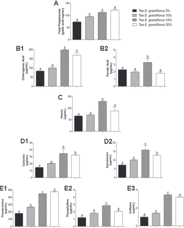
In our study, we found that the
E. grandiflorus
tea is rich in phenolic compounds (Figure 1).
This finding concurs with the study of Flor et
al. (2010), who showed a substantial presence
of polyphenols in an extract of
E. grandiflorus
,
which included flavonoids and their secondary
metabolites (flavones, flavonols and flavanones).
Garcia et al. (2010) identified the presence of
flavonoids, diterpenes and isovitexin in extracts
of
E. grandiflorus
. These compounds were also
observed in green, black (Camellia sinensis (L.) O.
Kuntze) and mate (
Ilex paraguariensis A. St.-Hil
)
tea (Barg et al. 2014). When evaluating different
tea preparations, we found that the total polyphenol
content increased as per the concentration, with
the 20% concentration being richest in phenolic
compounds (Figure 1A). The levels of catechin
were higher in 15 and 20% tea and did not differ
between tea preparations (Figure 1D1). However,
the amounts of chlorogenic acid (Figure 1B1),
ferulic acid (Figure 1B2), vitexin (Figure 1C),
epicatechin (Figure 1D2), caffeine (Figure 1E3)
and theophylline (Figure 1E2) were highest in 15%
tea, and decreased in 20% tea.
sinensis (L.) O. Kuntze) (Van Breda et al. 2013).
Catechin and epicatechin have been detected in
green and black tea (Camellia sinensis (L.) O.
Kuntze), while vitexin has been found in leaves
of yellow passion fruit (
Passiflora edulis Sims f.
flavicarpa Deneger
) (Peres et al. 2011).
In evaluating in vitro antioxidant activity, both
preparations of tea, 0.4% (22.4 ± 1.7%) and 0.8%
(41.3 ± 1.1%) were able to scavenge DPPH, with the
higher concentration showing significantly higher
scavenging capacity (p <0.05). The same was found
for the plant
Limnocharis flava (L.) Buchenau
,
also belonging to the family Alismataceae, which
contained polyphenols and ascorbic acid and
showed the ability to scavenge DPPH (Sakong et
al. 2011). In another study, the antioxidant activity
of green tea (Camellia sinensis (L.) O. Kuntze) was
demonstrated using the DPPH assay, and it also
showed the presence of polyphenols and catechins
(Barg et al. 2014). These results suggest a relationship
between the presence of these compounds and the
antioxidant activity demonstrated.
The use of plants for medicinal purposes is one
of the oldest forms of medical practice to treat, cure
and prevent diseases (Veiga et al. 2005). The plant
E. grandiflorus,
"leather hat," is traditionally used
to fight a variety of illnesses (Rieder et al. 2011).
In our study, we found that
E. grandiflorus
tea had
beneficial effects against oxidative damage induced
in rat liver, while other studies have also shown
some benefits of using the plant
E. grandiflorus
.
According to Portella et al. 2012, the leaves
of Echinodorus macrophyllus exert a
nephroprotec-tive effect in Wistar rats (Portella et al. 2012).
Brugiolo et al. (2011) showed that the extract of E.
grandiflorus
was able to modulate allergic pulmonary
inflammation, and it may also be useful as a potential
therapeutic agent for asthma (Lessa et al. 2008).
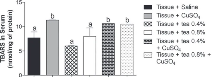
When evaluating the ability of tea to prevent
the induction of lipid peroxidation in human serum
(Figure 2), it was observed that the peroxidation
inducer, CuSO
4, raised the levels of TBARS, and
that both preparations of tea tested were ineffective
in reducing these levels. This is in line with the
findings of Nasser et al. 2011, who observed no
statistical difference in lipid peroxidation in human
serum, before and after the addition of orange
juice (Nasser et al. 2011). However, another study
Figure 2 - Effect of tea of Echinodorus grandiflorus (leather hat) on thiobarbituric acid-reactive substances (TBARS) in human serum. Data are expressed as mean ± standard deviation and were statistically analyzed by analysis of variance (ANOVA) followed by Tukey post-test. Different letters indicate significant differences between groups, with p <0.05.
showed that grape juice protected against CuSO
4-induced lipid peroxidation in serum, except for
conventional white grape juice (Dani et al. 2007). In
this study the authors attributed the ability to inhibit
lipid peroxidation to the content of polyphenols and
ascorbic acid, since the grape juice that showed
no ability to prevent the damage contained lower
amounts of these compounds. These results suggest
that these antioxidant compounds, when present in
greater amounts, provide protection against lipid
peroxidation.
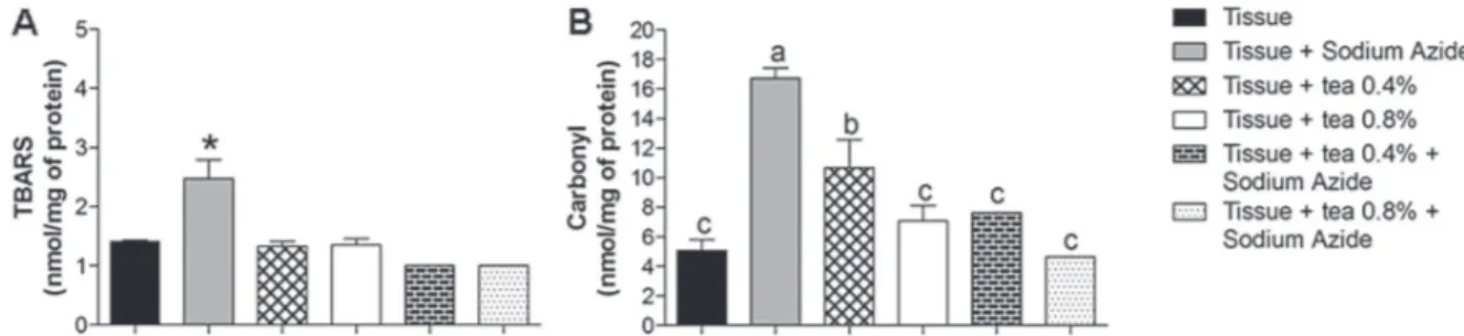
(Figure 3B) assays. It was observed that sodium
azide caused oxidative damage to the tissue due to
its capacity to inhibit the electron transport chain at
complex IV (Champe et al. 2011).
Based on TBARS and carbonyl assays, it
was observed that sodium azide damage tissue
lipids and proteins. Both preparations of tea tested
were able to inhibit lipid peroxidation and protein
Figure 3 - Effect of tea of Echinodorus grandiflorus (leather hat) on thiobarbituric acid-reactive substances (TBARS) (Fig. 3A) and protein carbonyl (Fig. 3B) levels in rat liver. Data are expressed as mean ± standard deviation and were statistically analyzed by analysis of variance (ANOVA) followed by Tukey post-test. Different letters indicate significant differences between the groups. *p<0.05, statistically different from the other groups.
damage, demonstrating protection against sodium
azide-induced damage (p<0.05). Dani et al.
(2008) and Schmitz et al. (2009), working with
rat liver, found that green tea extract was also
able to reduce TBARS and that grape juice was
effective in reducing carbonyl levels, respectively.
Some studies have revealed the presence of
phenolic compounds and more specifically those
of catechin compounds (Dani et al. 2008, Barg et
al. 2014). A similar trend was reported by Hong
et al. (2006) who evaluated the treatment of rat
liver with an extract of Alisma orientalis (family
Alismataceae). It was found that the plant was
useful in the prevention of oxidative stress by
decreasing lipid peroxidation, as seen with other
plants of the same family.
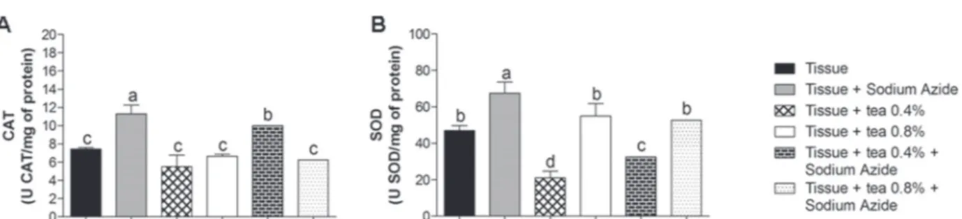
To check the activity of antioxidant enzymes,
superoxide dismutase (SOD) and catalase (CAT)
activities were evaluated in rat liver (Figure 4). These
enzymes are included in the enzymatic defense system.
SOD converts superoxide ion (O
2-) to hydrogen
peroxide (H
2O
2), while CAT converts H
2O
2to water
and oxygen by preventing the formation of hydroxyl
radical (OH
-.) which is highly toxic (Ho et al. 2001,
Halliwell 2006). In our study, we found that sodium
azide induced an increase in CAT (Figure 4A) and
SOD (Figure 4B) activity in the tissue. Meanwhile,
both concentrations of tea tested were able to restore
activity close to those of the control levels, since the
tea helped in attenuate the induced damage. This
corroborated the earlier findings by Hong et al. (2006)
and Dani et al. (2008), who determined the effects of
Alisma orientalis extract and grape juice on rat liver,
respectively. These studies demonstrated similar
effects on SOD and CAT activity and also observed
that the juice and extract were able to reverse the
tissue damage induced.
damage and lipid peroxidation by
E. grandiflorus
tea
as well. Some studies have confirmed the potent
activity of polyphenolic compounds countering lipid
peroxidation and scavenging various radicals,
inclu-ding H
2O
2. It has been suggested that plant phenols
have multifunctional biochemical activities including:
(i) electrophilic radical trapping, (ii) inhibition of
nitrosation, (iii) arachidonic metabolism modulation
and (iv) alteration of carcinogen metabolism (Tseng
et al. 1996, Sroka and Cirowski 2003).
The antioxidant activity of polyphenols depends
on the bioavailability of these compounds. Several
studies have suggested that some flavonoids present in
the diet can have positive effects on the human body,
regardless of their poor absorption. The flavonoids
found in blood and specific organs that result from
digestive or liver activity may differ from native
substances in terms of biological activity (Manach et
al. 2004, Georgiev et al. 2014). The bioavailability of
flavonoids is dependent on their structural properties,
and they are poorly absorbed because they occur
naturally in highly hydrophilic glycosylated forms.
Thus, only aglycones can effectively pass through the
gut wall. It has been suggested that some species of
colon microflora can hydrolyze flavonoid glycosides
to their corresponding aglycones, although the
microbial enzymes may also degrade the entire
compound as well (Bentz 2009, Georgiev et al. 2014).
In general, teas are extensively studied and
they have scientifically proven benefits. However,
studies suggest that phenolic compounds present
in various teas can also act as prooxidants in the
presence of redox-active chemicals and lead to the
formation of reactive oxygen species, which can
cause damage to DNA, lipids and other biological
molecules (Decker 1997, Jain et al. 2013). The
balance between prooxidant and antioxidant effects
of flavonoids is crucial to evaluate the toxicity of
teas. With regards to this toxicity, there have been
reports of some severe allergic reactions (Jain et
al. 2013) and nervous irritability, convulsions,
tachycardia, extrasystoles, and gastric irritation
(due to the presence of xanthine) (Brem et al. 1977,
Jain et al. 2013). However, to date, we do not know
enough about the toxic effects of
E. grandiflorus
tea.
CONCLUSION
On the basis of the findings of this study, it can be
concluded that
E. grandiflorus
tea had antioxidant
activity in the liver, where it was able to reverse the
damage caused by lipid peroxidation and protein
oxidation, besides normalizing the activity of the
antioxidant enzymes SOD and CAT. Furthermore, it is
suggested that this tea can help prevent liver damage
caused by free radicals. This is important because many
synthetic drugs used in treatment are often insufficient
and may have serious adverse effects (Arhoghro
et al. 2009). However, further studies are needed to
provide more knowledge about the composition and
therapeutic actions of
E. grandiflorus
tea.
ACKNOWLEDGMENTS
We are grateful to Centro Universitário
Meto-dista, IPA (Porto Alegre, RS, Brazil), Fundação de
Amparo à Pesquisa do Estado do Rio Grande do Sul
(FAPERGS), Conselho Nacional de Desenvolvimento
Científico e Tecnológico (CNPq) and Coordenação
de Aperfeiçoamento de Pessoal de Nível Superior
(CAPES) for their help and financial support
during this research.
RESUMO
O estresse oxidativo tem sido referido como um dos fatores responsáveis pelo desencadeamento de hepatopatias, sendo necessárias novas formas de tratamento. O presente estudo teve como objetivo avaliar a capacidade antioxidante in vitro do chá de
Echinodorus grandiforus “Chapéu de couro” em fígado de ratos. Nas diferentes preparações de chá foram avaliadas a composição fenólica, a atividade antioxidante pelo ensaio do DPPH e a capacidade de inibição de peroxidação lipídica induzida por sulfato de cobre. A atividade antioxidante foi avaliada em tecidos hepáticos tratados com azida sódica na presença ou ausência de chá, por meio do ensaio de peroxidação lipídica (TBARS), oxidação proteica (Carbonil) e atividade das enzimas antioxidantes catalase (CAT) e superóxido dismutase (SOD). Os resultados demonstram que as diferentes concentrações de chá são fontes importantes de polifenóis, destacando o conteúdo de teobromina, catequina e vitexina. Além disto, os resultados indicam que o chá apresenta atividade antioxidante pela sua capacidade em varrer o radical DPPH. As diferentes preparações de chá foram capazes de impedir os danos a lipídios e proteínas induzidos pela azida sódica, bem como auxiliar no restabelecimento da atividade de enzimas CAT e SOD. Deste modo, pode-se verificar que o chá E. grandiflorus apresentou atividade antioxidante no soro e no fígado, sendo capaz de prevenir danos oxidativos gerados pela azida sódica.
Palavras-chave: estresse oxidativo, compostos fenólicos, espécies reativas, azida sódica.
REFERENCES
AEBI H. 1984. Catalase in vitro. Methods Enzymol 105: 121-126.
ARHOGHRO E, EKPO K, ANOSIKE E AND IBEH G. 2009. Effect of
aqueous extract of bitter leaf (Vernonia Amygdalina Del) on carbon tetrachloride (CCl4) induced liver damage in
albino Wistar rats. Eur J Sci Res 26: 122-130.
BANNISTER J AND CALABRESE L. 1987. Assays for SOD. Methods Biochem. Anal 353: 279-312.
BARG M ET AL. 2014. Evaluation of the protective of Ilex paraguariensis and Camellia sinensis extracts on the prevention of oxidative damage caused by ultraviolet radiation. Environ Toxicol Pharmacol 37: 195-201. BENTZ AB. 2009. A review of quercetin: Chemistry, antioxidant
properties, and bioavailability. J Young Investig Available online: http://www.jyi.org/research/re.php?id=3416 (accessed on 16 March 2014).
BHANDARI M AND KAWABATA J. 2004. Organic acid, phenolic content and antioxidant activity of wild yam (Dioscorea
spp.) tubers of Nepal. Food Chem 88: 163-168.
BREM SS, GULLINO PM AND MEDINA D. 1977. Angiogenesis: A marker for neoplastic transformation of mammary papillary hyperplasia. Science 195: 880-882.
BRUGIOLO A ET AL. 2011. Effects of aqueous extract of
Echinodorus grandiflorus on the immune response in ovalbumin-induced pulmonary allergy. Ann Allergy Asthma Immunol 106: 481-488.
BRUGIOLO S, PETERS V, PIMENTA D, AARESTRUP B, BRUGIOLO
A, RIBEIRO D AND GUERRA M. 2010. Reproductive toxicity of Echinodorus grandiflorus in pregnant rats. J Toxicol Sci 35: 911-922.
CAROCHO M AND FERREIRA ICFR. 2013. A review on antioxidants, prooxidants and related controversy: Natural and synthetic compounds, screening and analysis metho-dologies and future perspectives. Food Chem Toxicol 51:15-25.
CEMEK M, YILMAZ F, BÜYÜKOKUROGLU M, BÜYÜKBEN A,
AYMELEK F AND AYAZ A. 2012. Serum and Liver Tissue Bio-Element Levels, and Antioxidant Enzyme Activities in Carbon Tetrachloride-Induced Hepatotoxicity: Protective Effects of Royal Jelly. J Med Food 1: 1-7.
CHAMPE P, HARVEY R AND FERRIER D. 2011. Biochemistry, 5th ed., Philadelphia: Lippincott Williams & Wilkins, 520 p. COSTA M, TANAKA C, IMAMURA P AND MARSAIOLI A.
1999. Isolation and synthesis of a new clerodane from Echinodorus grandiflorus. Phytochemistry 50: 117-122. DANI C, OLIBONI L, PASQUALI M, OLIVEIRA M, UMEZU F,
SALVADOR M, MOREIRA J AND HENRIQUES J. 2008. Intake of Purple Grape Juice as a Hepatoprotective Agent in Wistar Rats. J Med Food 11: 127-132.
DANI C, OLIBONI L, VANDERLINDE R, BONATTO D, SALVADOR M
DECKER EA. 1997. Phenolics: prooxidants or antioxidants? Nutr Rev 55: 396-407.
DURAK I, AVCI A, KAÇMAZ M, BÜYÜKKOÇAK S, CIMEN M, ELGÜN S AND OZTÜRK H. 1999. Comparison of antioxidant potentials of red wine, white wine, grape juice and alcohol. Curr Med Res Opin 15: 316-320.
FILLIP R, LÓPEZ P, GILBERTI J, COUSSIO J AND FERRARO G.
2001. Phenolic compounds in seven South American Ilex species. Fitoterapia 72: 774-778.
FLOR R, CAMPOS M, SOLANO A, JOKL L AND DANTAS-BARROS A.
2010. Drying of Echinodorus macrophyllus and autoclaving and lyophilization of the fluid-extract: effects on the pharmacochemical composition. J Pharmacog 21: 518-524. GARCIA E, OLIVEIRA M, GODIN A, FERREIRA W, BASTOS L,
COELHO M AND BRAGA F. 2010. Antiedematogenic activity and phytochemical composition of preparations from Echinodorus grandiflorus leaves. Phytomedicine 18: 80-86. GEORGIEV V, ANANGA A AND TSOLOVA V. 2014. Recent
Ad-vances and Uses of Grapes Flavonoids and Nutraceuticals. Nutrients 6: 391-415.
HA H, SHIN H, FEITELSON M AND YU D. 2010. Oxidative stress and antioxidants in hepatic pathogenesis. World J Gastroenterol 16: 6035-6043.
HALLIWELL B. 2006. Reactive Species and Antioxidants. Redox Biology Is a Fundamental Theme of Aerobic Life Plant Physiol 141: 312-322.
HALLIWELL B. 2007. Dietary polyphenols: Good, bad, or indifferent for your health? Cardiovasc Res 73: 341-347. HO J, ZHENG S, COMHAIR S, FARVER C AND ERZURUM S.
2001. Differential Expression of Manganese Superoxide
Dismutase and Catalase in Lung Cancer. Cancer Res 61: 8578-8585.
HONG X, TANG H, WU L AND LI L. 2006. Protective effects of the
Alisma orientalis extract on the experimental nonalcoholic fatty liver disease. J Pharm Pharmacol 58: 1391-1398. JAIN A, MANGHANI C, NIGAM D AND RANI V. 2013. Tea and human
health: The DARK SHADOWS. Toxicol Lett 220: 82-87. KHLEBNIKOV AI, SCHEPETKIN IA, DOMINA NG, KIRPOTINA LN
AND QUINN MT. 2007. Improved quantitative structure– activity relationship models to predict antioxidant activity of flavonoids in chemical, enzymatic, and cellular systems. Bioorg Med Chem 15: 1749-1770.
KOURY J AND DONANGELO C. 2003. Zinc, oxidative stress and physical activity. Rev Nutr 16: 433-441.
LESSA M, ARAÚJO C, KAPLAN M, PIMENTA D, FIGUEIREDO M
AND TIBIRIÇÁ E. 2008. Antihypertensive effects of crude extracts from leaves of Echinodorus GRANDIFLORUS. Fundam Clin Pharmacol 22: 161-168.
LEVINE R, GARLAND D, OLIVER C, AMICI A, CLIMENT I, LENZ A,
AHN B, SHALTIEL S AND STADTMAN E. 1990. Determination of carbonyl content in oxidatively modified proteins. Methods Enzymol 18: 464-478.
LOWRY O, ROSEBROUGH N, FARR A AND RANDALL R. 1951.
Protein measurement with the folin phenol reagent. J Biol Chem 193: 265-267.
MANACH C, SCALBERT A, MORAND C, RÉMÉSY C AND JIMÉNEZ L. 2004. Polyphenols: Food sources and bioavailability. Am J Clin Nutr 79: 727-747.
NASSER A, DOURADO G, MANJATE D, CARLOS I AND CESAR T. 2011. Evaluation of oxidative stress in the blood of
habitual consumers of orange juice. Rev Ciênc Farm Básica Apl 32: 275-279.
NAVARRO V AND SENIOR J. 2006. Drug related hepatotoxicity. N Engl J Med 354: 731-739.
PERES RG, TONIN FG, TAVARES MF AND RODRIGUEZ-AMAYA
DB. 2011. Determination of catechins in green tea infusions by reduced flow micellar electrokinetic chromatography. Food Chem 127: 651-655.
PIETTA P. 2000. Flavonoids as antioxidants. J Nat Prod 63: 1035-1042.
PIMENTA DS, FIGUEIREDO MR AND KAPLAN MAC. 2006. Essential oil from two populations of Echi no dorus grandiflorus (Cham. & Schltdl.) Micheli (Chapéu de couro). An Acad Bras Cienc 78: 623-628.
PINTO A, REGO G, SIQUEIRA A, CARDOSO C, REIS P, MARQUES E, COELHO M AND SABINO K. 2007. Immunosuppressive effects of Echinodorus macrophyllus aqueous extract. J Ethnopharmacol 111: 435-439.
PORTELLA V, COSENZA G, DINIZ L, PACHECO L, CASSALI G,
CALIARI M, BRANDÃO M AND VIEIRA M. 2012. Nephro-protective Effect of Echinodorus macrophyllus Micheli on Gentamicin-Induced Nephrotoxicity in Rats. Nephron Extra 2: 177-183.
RIEDER A, FIGUEIREDO GC AND BONILLA MG. 2011. Plant known as “Leather hat” (Echinodorus spp. (Alimastaceae) and its medicinal use in southwestern Mato Grosso, Brazil. Planta Med 77(12): PF 86; 1327.
SAKONG P, KHAMPITAK T, CHA’ON U, PINITSOONTORN C,
SRIBOONLUE P, YONGVANIT P AND BOONSIRI P. 2011.
Antioxidant activity and bioactive phytochemical contents of traditional medicinal plants in northeast Thailand. J Med Plants Res 5: 6822-6831.
SAUCIER C, MIRABEL M, DAVIUD F, LONGIERAS A AND
GLORIES T. 2001. Rapid fractionation of grape seeds proanthocyanidins. J Agricult Food Chem 49: 5732-5735. SCHMITZ W, CECCHINI R, ESTEVÃO D AND SARIDAKIS H.
2009. Hepatic protection effect of the alcoholic extract from Camellia sinensis (L.) Kuntze (green tea) in Wistar rats treated with diethylnitrosamine. Rev Bras Farmacog 19: 702-709.
SHAHIDI F AND NACZK M. 1995. Food phenolics: sources, che-mistry, effects, applications, 2nd ed., Lancaster: Technomic Publishing Company, 331 p.
SINGLETON V, ORTHOFER R AND LAMUELA-RAVENTOS R.
1999. Analysis of total phenols and other oxidation substrates and antioxidants by means of Folin–Ciocalteau reagent. In: PACKER L (Ed), Methods in Enzymology, Oxidant and Antioxidant (Part A), Boston: Academic Press, Massachussetts, USA, p. 159-178.
SOUZA G, HAAS A, VON POSER G, SCHAPOVAL E AND ELISABETSKY
E. 2004. Ethnopharmacological studies of antimicrobial
SPADA P, DANI C, BORTOLINI G, FUNCHAL C, HENRIQUES J
AND SALVADOR M. 2009. Frozen Fruit Pulp of Euterpe oleraceae Mart. (Acai) Prevents Hydrogen Peroxide-Induced Damage in the Cerebral Cortex, Cerebellum, and Hippocampus of Rats. J Med Food 12: 1084-1088. SROKA Z AND CIROWSKI W. 2003. Hydrogen peroxide
scavenging, antioxidant and anti-radical activity of some phenolic acids. Food Chem Toxicol 41: 753-758.
TSENG T, WANG C, KAO E AND CHU H. 1996. Hibiscus
protocatechuic acid protects against oxidative damage induced by tert-butylhydroperoxide in rat primary hepatocytes. Chem Biol Interact 101: 137-148.
TIBIRIÇÁ E, ALMEIDA A, CAILLLEAUX S, PIMENTA D, KAPLAN
M, LESSA M AND FIGUEIREDO M. 2007. Pharmacological me chanisms involved in the vasodilator effects of extracts from Echinodorus grandiflorus. J Ethnopharmacol 111: 50-55.
VAN BREDA SV, VAN DER MERWE CF, ROBBERTSE H AND
APOSTOLIDES Z. 2013. Immunohistochemical localization of caffeine in young Camellia sinensis (L.) O. Kuntze (tea) leaves. Planta 237: 849-858.
VEIGA V, PINTO A AND MACIEL M. 2005. Medicinal plants: safe cure? Quim Nova 28: 519-528.
WELTMAN M, FARRELL G, HALL P, INGELMAN-SUNDBERG M
AND LIDDLE C. 1998. Hepatic cytochrome P450 2E1 is
increased in patients with nonalcoholic steatohepatitis. Hepatology 27: 128-133.
WILLS E. 1996. Mechanism of lipid peroxide formation in animal tissues. Biochem J 99: 667-676.
YAMAGUCHI T, TAKAMURA H, MATOBA T AND TERAO J. 1999.