255 DOI:10.1590/1982-0224-20130219
Gross morphology of the brain of
Pseudopimelodus bufonius
(Valenciennes, 1840) (Siluriformes: Pseudopimelodidae)
VitorPimentaAbrahão andOscarAkioShibatta
ThegrossmorphologyofthebrainofthepseudopimelodidPseudopimelodus bufoniusisdescribedandcomparedwith congeners.Observationsweremadeonremovedbrainsaftereliminationofbonesfromthetopoftheskullandseveringof thecranialnervesandthespinalcord.Ninemorphometriccharactersassociatedwiththemajorsubdivisionsofthebrain wereidentified,sevenofwhichrevealedsignificantdifferencesamongthespeciesexamined.Thecorpus cerebelliinall examinedspeciesofthegenusisthelargeststructureofthebrain.ThebehaviorofthespeciesofPseudopimelodusisstill unknown,butinotherteleoststhatconditionistypicallycorrelatedwithahigherdegreeofmotorcoordination.Relativesize proportionsofthetectum opticum,eminentia granularis,lobus facialisandlobus vagi,mightberelatedtocarnivoryandan enhancedcapacityforfoodselection.
AmorfologiaexternadoencéfalodePseudopimelodus bufonius édescritaecomparadacomseuscongêneres.Asanálises foramfeitasnocérebroremovidoapósaeliminaçãodosossosdotopodacabeçaesecçãodosnervoscranianosecordão espinhal.Novecaracteresmorfométricosforamobtidosdasprincipaissubdivisõesdoencéfalo,dosquaisemseteocorreram diferenças significativas entre as espécies. Em todas as espécies examinadas do gênero ocorpus cerebelli é a maior estruturadoencéfalo.OcomportamentodasespéciesdePseudopimelodus aindaédesconhecido,masemoutrosteleósteos estacaracterísticaénormalmentecorrelacionadacomumaboacoordenaçãomotora.Alémdisso,asproporçõesrelativas dotectum opticum,eminentia granularis,lobus facialiselobus vagipodemserrelacionadasahábitoscarnívoroseboa capacidadedeselecionaralimentos.
Keywords: Behavior,Catfishes,ComparativeMorphology,Neotropical fishes,Neuroanatomy.
ProgramadePós-GraduaçãoemCiênciasBiológicas,DepartamentodeBiologiaAnimaleVegetal,UniversidadeEstadualdeLondrina, CaixaPostal10.011,86057-970Londrina,PR,Brazil.(VPA)vitorabrahao32@gmail.com(correspondingauthor),(OAS)shibatta@uel.br
Introduction
The neurological system of fi hes has been greatly neglected in systematics studies. Within Teleostei, the major lineages are primarily supported by osteological andmolecularsynapomorphies.Itisestimatedthatalmost three-quarters of the total morphological information relatedtophylogeneticreconstructionisrelatedtoskeletal features (Wiley & Johnson, 2010). Datovo & Vari (2014) in a comprehensive review of morphological features used in fi h systematics, found a similar predominance of osteologicalcharacters(74%)inrelationtoothersetsofdata. Accordingtothoseauthors,6%correspondedtomyology, 5%tosplanchnology,14%toothermorphologicalcharacters (e.g. associated structures of external morphology and reproductivesystem),andonly1%toneurology.
Thebrainofvertebratesisbasicallydividedintoregions thatcontrolspecificfunctionsrelatedtosenseorgans,motor coordination, behavior and metabolic regulation (Butler & Hodos, 2005). The size of a particular structure in the brain typically indicates the predominance of its function and/ortheaccuracyofthesensetowhichitisrelated.Evans (1931)examinedthebrainsofcyprinids,toevaluatefeeding behavior and diet composition with regard to the size of
gustative lobes (lobus facialis andlobus vagi). In a series ofstudies,Eastman&Lannoo(1995,2001,2003a,2003b, 2004, 2007, 2008, 2011) found phylogenetic, ecologic and behaviorcorrelationswithformatandsizeofbrainstructures inAntarcticicefishes(Nototheniiformes).Studiesonbrain evolutionintheTeleosteihavealsobeenconductedbyItoet al.(2007)onthedevelopmentofmainregionsofthebrain, andNorthcutt(2004,2008)onthedevelopmentofgustative lobes and of forebrain. More recently, some authors have focused on the association of development and behavioral responses over the brain structures (Gondaet al., 2011; Kotrschalet al., 2012;Lecchiniet al.,2014).
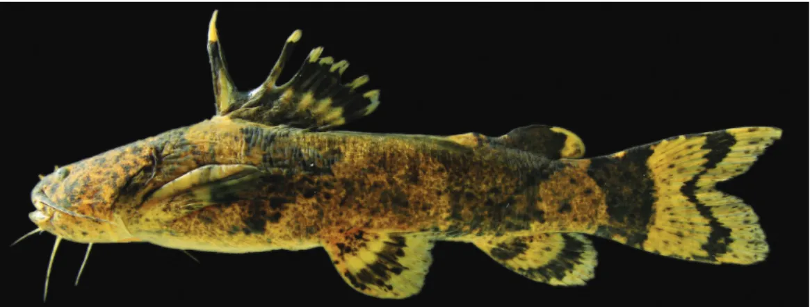
Fig. 1.Pseudopimelodus bufonius,MZUSP94855,113.3mmSL;Brazil,MatoGrosso,Paranatinga,rioCuluene,tributary oftherioXingu.PhotobyJoséL.O.Birindelli.
Material and Methods
Six specimens of P. bufonius (97.8-118.2 mm SL), three specimens ofP. mangurus (88.5-175.4 mm SL), and one specimen ofP. charus (123.7 mm SL) were dissected. Dissections were performed in the following sequence (see Abrahão&Pupo,2014,formoredetailandillustrations):the skincoveringtheskullbonessurroundingthesupraoccipital process was removed, typically scraping it off with a mini spatula. To avoid damaging brain subdivisions and nerves duringdissection,itwasnecessarytoinfertheirpositionsin relation to cranial bones (Fig. 2). Incisions were made with a dental drill powered by a suspended electric motor in the suturesbetweenthepterotic,sphenotic,andfrontal,forming a circle around the supraoccipital process, which was then removed carefully using forceps. Cross-sections were made with a scalpel on themedulla spinalis,nervus vagus (nX), nervus glossopharyngeus(nIX),nervus octavus(nVIII),nervus facialis(nVII),nervus abducens(nVI),nervus trigeminus(nV), nervus trochlearis(nIV),nervus oculomotorius(nIII),nervus linea lateralis anterior (nlla),nervus linea lateralis posterior (nllp),nervus opticus(nII)andnervustractus olfactorius(ntol) (Figs.2-5).Toremovethebrainandtheassociatedolfactory bulbandnervus olfactorius (nI),incisionsweremadeinthe frontal bone and mesethmoid to reach the olfactory organ, removing it while avoiding incisions that could damage the olfactorytract(Fig.2).Thenomenclatureofthebrainregions follows Meek & Nieuwenhuys (1998) and Butler & Hodos (2005). The brains were kept in 4% formalin buffered with CaCO3,afterremovalfromtheneurocranium.Allspecimens werepreservedin70%ethanolafterfixationin4%formalin.
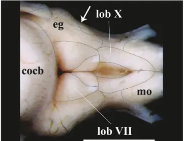
Fig. 3. Diagramatic representation of measurements of major subdivisions of the brain ofPseudopimelodus bufonius, MZUEL5744,116.4mmSL,indorsalandventralviews.Cocb:corpus cerebelli;eg:eminentia granularis;lih:lobus inferior hypothalami;lobVII:lobus facialis;lobX:lobus vagi;telen:telencephalon;tect:tectum opticum.Scalebar=2mm.
Fig. 5. Brain of Pseudopimelodus bufonius, MZUEL 5744,118.2mmSL,in(a)dorsal,(b)lateraland(c)ventral views, and (d) dorsal and (e) ventral views of olfactory organ.bol:bulbus olfactorius;ch:chiasma opticum;cocb: corpus cerebelli; eg: eminentia granularis; lih: lobus inferiorhypothalami;hyp:hypophysis;hyt:hypothalamus; lobVII: lobus facialis; lobX: lobus vagi; mo: medulla oblongata; NII:nervus opticus; NV:nervus trigeminus; NVII: nervus facialis; NVIII: nervus octavus; NIX: nervus glossopharyngeus;NX:nervus vagus;of:olfactory organ; sv: saccus vasculosus; trc:truncus cerebri; telen: telencephalon; tect: tectum opticum; tol:nervus tractus olfactorius.Scalebar=2mm.
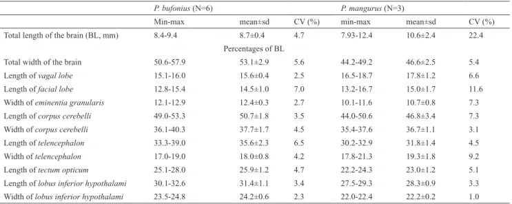
Adigitalcameraattachedtoastereomicroscopewasused tocaptureimagesofthebraintopography.Thefollowing measurementsweretakenusingthesoftwareAxioVision Relv.4.8(Zeiss,2009):vagal lobe length,facial lobe length, eminentia granularis width,corpus cerebelli length,corpus cerebelli width,telencephalon length,telencephalon width, tectum opticum length,lobus inferior hypothalami length, andlobus inferior hypothalami width.Table1showsthese measurementsinproportiontothetotalbrainlength(BL), measuredfromtheanteriorportionofthetelencephalonto
Fig. 6. Posterior portion of rhombencephalon of Pseudopimelodus bufonius, MZUEL 5744, 118.2 mm SL, dorsal view. cocb:corpus cerebelli; eg:eminentia granularis; lobVII:lobus facialis; lobX:lobus vagi; mo: medulla oblongata. Arrow indicates the separation point between anterior and posterior bulges of theeminentia granularis.Scalebar=2mm.
Results
Thebrainiscompletelylocatedbeneaththesupraoccipital processandabovetheparasphenoidandprooticinspeciesof Pseudopimelodus,withexceptionofthebulbus olfactorius and part of the anterior portion of thetelencephalon (Fig. 2). Brain width in relation to BL is significantly larger (p=0.01) inP. bufonius (mean=53.1%) than in the other examinedspecies,whereasinP. mangurusitisthesmallest (mean=46.6%;Table1;Fig.4).Descriptionsbelowarebased onP. bufonius,withcommentsonexaminedcongeners. Rhombencephalon. In all examined species of Pseudopimelodus,themedulla oblongata islocatedabove toefferentprojectionsofnervus vagustomedulla spinalis, andalsolocateddorsallyinrelationtothetruncus cerebri, and posterolaterally to the lobus vagi. The medulla oblongata is composed of two ovoid-shaped structures contacting themedulla spinalis. The posterior portion of those structures is slightly smaller than their anterior counterparts,eachofwhichisconspicuouslyintumescent (Figs.5-6,8).
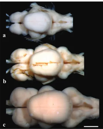
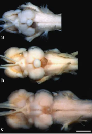
Fig. 8.Brainindorsalview:(a)Pseudopimelodus bufonius, MZUEL5744,118.2mmSL,(b)P. charus,MZUEL6488, 123.7mmSL,(c)P. mangurus,MZUEL2795,175.7mm SL.Scalebar=2mm.
Thelobus vagi is located at the dorsal portion of the rhombencephalon.Indorsalview,itisimmediatelyposterior tothelobus facialis,andanteromediallylocatedinrelation
tothemedulla oblongata.Thelobus vagi is composedof two V-shaped cylindrical paired lobes, whose posterior portioncontacteachother,forminganacutetip(Figs.5-6, 8).Thelengthofthelobus vagiisshorterthanthelength ofthetectum opticum,butislongerthanthelengthsofthe lobus facialis andtheeminentia granularis.Thelobus vagi lengthinrelationtoBLissignificantlylonger(p=0.005)in P. mangurus(Table1,Fig.4).
The lobus facialis is located at the dorsal portion of therhombencephalon, posterior to thecorpus cerebelli, anteriortothelobus vagi,andmediallylocatedinrelation totheeminentia granularis.Asmallportionoftheanterior areaofthelobus facialis isundertheposteriorportionof thecorpus cerebelli. The lobes of thelobus facialis are longitudinally elongated, each approximately rhomboid-shaped, but with rounded edges. A conspicuous bulge is anterolaterally anchored to each lobe. The lobes of the lobus facialisareparallel,butdonotcontactoneanother, thoughbothcontactthecorpus cerebellianteriorly(Figs.5, 6,8).Thelengthofthelobus facialis isshorterlengthwise thanthelobus vagiandtectum opticum,andproportionally longer than the length of theeminentia granularis. The lengthoflobus facialis inrelationtotheBLislongerinP. mangurus (Table1,Fig.4).
Theeminentia granularisisprominentintheexamined species ofPseudopimelodus (Figs. 4-5), and is located at thedorsalportionoftherhombencephalon,posterolaterally tothecorpus cerebelliandlateraltothelobus facialis.The structurehastwoconspicuousroundedbulges,theanterior onemoreprominentthantheposterior.Theanteriorbulge is positioned at the posterolateral portion of thecorpus cerebelli, extending up to approximately, but a little less than half the length of thelobus facialis. The posterior bulgeextendstotheanteriorareaofthelobus vagi(Figs. 5, 6, 8). In terms of lengths, the eminentia granularis is longerthanthemedulla oblongataandshorterthanboththe lobus facialisand lobus vagi.Thelengthoftheeminentia granularis inrelationtoBLissignificantlylonger(p=0.004) inP. bufonius(Table1,Fig.4).
Fig. 9. Braininlateralview:(a)Pseudopimelodus bufonius, MZUEL5744,118.2mmSL,(b)P. charus, MZUEL6488, 123.7mmSL,(c)P. mangurus, MZUEL2795,175.7mm SL.Scalebar=2mm.(ThehypophysisofP. mangurusisnot shownbecauseitwasdamagedduringdissections).
Thenervusglossopharyngeus and thenervus vagus emerge from the lateral wall of therhombencephalon, at approximately the mid-point of thelobus vagi, exiting the efferent projections of the braincase through a foramen on theexoccipital.Thenervus octavus,thenervus linea lateralis anterior and thenervus linea lateralis posterior emerge from the ventrolateral wall of therhombencephalon, at approximatelythemidpointoftheeminentia granularis.The nervusfacialis andthenervus trigeminusemergefromthe ventrolateralportionoftherhombencephalon,anteriortothe nervus octavus,andtheirefferentprojectionsofthebraincase throughforaminaontheparasphenoid,prootic,sphenoticand pterosphenoid.Theanteriorramusofthenervus trigeminus andthenervus linea lateralis anterior haveefferentprojections from the braincase through a single foramen between the orbitosphenoidandpterosphenoid(Fig.2).
Mesencephalon.Thetectum opticumislocatedatthedorsal portion of thetegumentum mesencephali, anterolateral to thecorpus cerebelli in dorsal view, and posterior to the telencephalon in lateral view. Thetectum opticum has twobilaterally-roundedstructures,whichcontactboththe corpus cerebelli andthetelencephalon (Figs.5,8,9).The lengthofthetectum opticumislongerthanthatofthelobus vagi,lobus facialisandeminentia granularis.Thelengthof thetectum opticum inrelationtoBLissignificantlylonger (p=0.01)inP. bufonius(Table1,Fig.4).
Thenervus opticus emergesfromthetectum opticum on themesencephalon,andefferentprojectionsexitintheregion immediately anterior to thelobus inferior hypothalamic, wherethefibersofthisnervecrossthemidlineofthebrain, forming thechiasma opticum. Thesefibers contact each otheronlyinthebaseofthechiasma opticum.Theefferent projectionsofthenervus opticus exitthebraincasethrough aforamenlocatedbetweenthefrontal,orbitosphenoidand pterosphenoid(Fig.2).
Lengthof 12.8-15.4 14.5±1.0 7.0 13.2-16.7 15.0±1.7 11.6
Widthofeminentia granularis 12.1-12.9 12.4±0.3 2.7 10.1-11.6 10.7±0.8 7.3
Lengthofcorpus cerebelli 49.0-53.3 50.7±1.8 3.5 44.0-50.6 46.8±3.4 7.3
Widthofcorpus cerebelli 36.1-40.3 37.7±1.7 4.5 35.4-37.6 36.7±1.1 3.1
Lengthoftelencephalon 33.3-39.0 35.6±2.3 6.5 30.2-32.9 31.8±1.4 4.5
Widthoftelencephalon 17.0-19.0 18.0±0.8 4.2 17.8-21.3 19.3±1.8 9.2
Lengthoftectum opticum 25.1-28.0 25.9±1.2 4.7 22.2-24.3 23.0±1.2 5.1
Lengthoflobus inferior hypothalami 30.1-32.6 31.4±1.1 3.4 27.5-29.3 28.3±0.9 3.3
Truncus cerebri.Thetruncus cerebriisventrallylocated inthebrain,andiscomprisedbytherhombencephalonand mesencephalon. It is more precisely located in the region betweentheposteriorportionofthemesencephalon andthe anterior portion ofmedulla spinalis. Most cranial nerves exit the efferent projections from the truncus cerebri, exceptforthenervus olfactorius and nervus opticus.The cranialnervesleavingthetruncus cerebriinclude:nervus oculomotorius, nervus trochlearis, nervus trigeminus, nervus abducens,nervus facialis,nervus octavus,nervus glossopharyngeus,nervus vagus,nervus linea lateralis anterior,andnervus linea lateralis posterior.Proportional length variations of thetruncus cerebri among species examinedwerenotfound(Figs.5,10).
Fig. 10.Braininventralview:(a)Pseudopimelodus bufonius, MZUEL5744,118.2mmSL,(b)P. charus,MZUEL6488, 123.7mmSL,(c)P. mangurus,MZUEL2795,175.7mm SL.Scalebar=2mm.(ThehypophysisofP. mangurusisnot shownbecauseitwasdamagedduringdissections).
Diencephalon. The lobus inferior hypothalami is
located at the ventral portion of thediencephalon, in a region posterior to thechiasma opticum, ventral to the truncus cerebriandthetectum opticum,posteriortothe telencephalon,andlateraltothehypothalamus.Thelobus inferior hypothalami is semi-circular, with the anterior
portion slightly smaller than the posterior portion. The hypophysis is circular, and anchored medially on the hypothalamus.Thehypothalamus islocatedattheventral portionofthediencephalon,posteriorandabovethelobus inferior hypothalami. Thehypothalamus is also semi-circular,withthemedialportionsstraight(Figs.5,7,10). InallexaminedspeciesofPseudopimelodus,thelength ofthelobus inferior hypothalami islongerthanthatofthe tectum opticum, but significantly longer (p=0.006) inP. bufoniusinrelationtotheBL(Table1,Fig.4).
Telencephalon.InallexaminedspeciesofPseudopimelodus, the telencephalon is anterior to the tectum opticum and posterior to thebulbus olfactorius in lateral view. Less than half the length of the posterior portion of thetelencephalon is positioned ventrally to thecorpus cerebelli in dorsal and lateral views. Thetelencephalon is longitudinally elongated and somewhat cylindrical, withboththeanteriorandposteriormarginsrounded.The anterior margin of thetelencephalon is slightly smaller than the posterior one inP. bufonius (Figs. 5, 8, 9, 10). Thelengthofthetelencephalonislongerthanthelobus inferior hypothalamiandthetectum opticum.Thelength of thetelencephalon in relation to BL is significantly (p=0.04)longerinP. bufonius(Table1,Fig.4).
Thebulbus olfactorius is stalked and is positioned at the anterior portion of the brain. The structure is ventral to the nasal bone, located near the articulation betweenthelateralethmoidandthevomer(Fig.2).The bulbus olfactorius is rounded. It is connected to the olfactory epithelium via thenervus olfactorius, and to thetelencephalonviathenervustractus olfactorius.The olfactory epithelium is rounded, with the anterior edge slightsmallerthantheposteriorone.Itislaterallycurved with a large amount of lamellae on each side, with the appearanceofafeather(Fig.5).
Discussion
Rhyacichthys aspro (Valenciennes, 1837) (Perciformes: Rhyacichthyidae), which also lives in rapids of mountain rivers with rocky bottoms. In both cases, it is presumed thatgreataccuracyintermsofmovementisrequired,and therelativelylargecorpus cerebellimayberelatedtothis. Despite the lack of information on the role of thecorpus cerebelli infish, it is generally accepted that this part of thebrainisessentialinsensorimotorintegration(Meek& Nieuwenhuys, 1998; Nieuwenhuyset al., 1998; Butler & Hodos, 2005). Sharks and teleosts with strong-swimming andwiderangeofhuntingmaneuvershaveincreasinginthe relative size, volume and foliation of thecorpus cerebelli (Lisney&Collin,2006).
InallspeciesofPseudopimelodusexaminedthe tectum opticum is also large, whereas theeminentia granularis, lobus facialis, andlobus vagiarerelativelysmall.Together, those conditions may indicate a rather elaborate feeding behavior.Thecorpus cerebelli receivessensoryinputfrom various regions of the central nervous system, including thelateralline,auditory,visual,vestibular,somatosensory, and mainly sensorimotor systems, which provide spatial orientation, proprioception, and coordination of eye movements.Inthecorpus cerebelli thesensoryinputfrom thoseregionsofthecentralnervoussystemareprocessedin ordertoprovidebalanceandmotorcoordinationofthebody (Kotrschalet al.,1998;Butler&Hodos,2005).Thetectum opticum, in turn, receives sensory information from the retinathroughtheopticnerve,andisalsoresponsiblefor generatingsignalsthatprovidescoordinationofthemotor response(Meek&Nieuwenhuys,1998).Moreover,italso receivesinformationfromtheauditory,andsomatosensory systems,enablingthestructuretocreateretinotopicmapsof theenvironment(Butler&Hodos,2005).Thecombinationof themotorcoordinationofthebodyandalsothecoordination oftheeyespromoteresponsesfromthecorpus cerebelli and thetectum opticumthatprovidestimulusdirectingthefish tocaptureprey(Butler&Hodos,2005).
Thelobus facialis and thelobus vagi are associated to gustative perception, receiving sensory information fromtastebudsinthebarbelsandmouth,andfromtaste budspresentinthepharynxandgillarches,respectively (Meek&Nieuwenhuys,1998).Theeminentia granularis is associated with mechanosensory perception that receives sensory input perceived by the lateral line mechanoreceptors (Finger, 1983; Meek & Nieuwenhuys, 1998). Evans (1931, 1940) recognized functional groups of species of British cyprinoids based on morphological
pattern was observed. Similar results were found on species of the Gadidae (Kotrschalet al., 1998). Studies onthefeedinghabitsofpseudopimelodids(Shibatta1998) indicatethatP. mangurusiscarnivorous.Thisobservation issupportedbythebrainmorphology,giventhatspeciesof Pseudopimelodus haverelativelylargecorpus cerebelli and tectum opticum,andsmalllobus facialis andlobus vagi.
Studiesonbrainarchitectureallowsanincreaseinthe numberofinformativecharactersforsystematics,andalso helpinunderstandingthebehaviorandhabitsof fishes.In addition,itisnoteworthythatthereisagreathorizonfor studies on brain anatomy considering the huge diversity ofteleosts.
Material Examined.Batrochoglanis melanurus:Brazil: MZUEL 3670, 1, 51.2 mm SL, Mato Grosso, Nobres, córrego Cancela, tributaryofrioCuiabá.Batrochoglanis raninus:Brazil: MZUEL 6035, 1, 76.7 mm SL, aquarium specimen. MZUSP 23407 2 of 24,51.4-76.6mmSL,Amazonas,FonteBoa,igarapéTucuxi,rio Solimões basin.Batrochoglanis villosus: Brazil: MZUEL 6037, 1,143.2mmSL,Maranhão,Turiaçu,rioTuriaçu.Cephalosilurus apurensis:Venezuela: MZUEL 6493, 1, 195.6 mm SL, Apure, Cano Bacaral.Cephalosilurus fowleri: Brazil: MZUEL 6040, 1, 275.0 mm SL, Minas Gerais, Três Marias, rio São Francisco. Lophiosilurus alexandri: Brazil: MZUEL 5377, 1, 58.7 mm SL, MinasGerais,TrêsMarias,rioSãoFrancisco.MZUSP96276,1 of 7, 73.1 mm SL, Três Marias, rio São Francisco.Microglanis cottoides: Brazil: MZUEL6033,3,40.1-48.8mmSL,RioGrande doSul,CanudosdoVale,rioForquetinha.29°22’4’’S52°03’19’’W. Microglanis parahybae: Brazil: MZUEL6393,2,39.1-42.0mmSL, RiodeJaneiro,SãoFidelis,rioDoisRios,tributaryofrioParaíbado Sul.Microglanis aff.poecilus: Brazil: INPA6828,1,20.9mmSL, Pará,Trairão,rioJamanxim.Pseudopimelodus bufonius:Brazil: MZUEL 5744, 6, 97.8-118.2 mm SL, Mato Grosso, Paranatinga, rio Culuene, tributary of rio Xingu.Pseudopimelodus charus: Brazil:MZUEL6488,1,123.7mmSL,MinasGerais,SãoRoque deMinas,rioSãoFrancisco.Pseudopimelodus mangurus:Brazil: MZUEL2795,2,138.9-175.4mmSL,Paraná,DiamantedoNorte, rioParanapanema.MZUSP24449,1,88.5mmSL,MatoGrossodo Sul,IlhaSolteira,rioParaná.
Acknowledgements
de Zoologia of the Universidade de São Paulo (MZUSP), andC.Dillman-SmithsonianInstitution-forreadingand commenting the manuscript, and M. de Pinna (MZUSP) forloanandpermissiontodissectspecimens.Wearealso thankfultoUniversidadeEstadualdeLondrinaandCNPq forfinancial support. OAS is a productivity researcher grantedbyFundaçãoAraucária(prot.36.379,414/2013). References Abrahão,V.P.&F.M.R.S.Pupo.2014.Técnicadedissecçãodo neurocrâniodeSiluriformesparaestudodoencéfalo.Boletim SociedadeBrasileiradeIctiologia,112:21-26. Atema,J.1971.Structuresandfunctionsofthesenseoftastein thecatfish(Ictalurus natalis).BrainBehaviorandEvolution, 4:273-294.
Bauchot, R., J. E. Randall, J. M. Ridet&M. L. Bauchot. 1989. Encephalizationintropicalteleostfishesandcomparisonwith theirmodeoflife.JournalfurHirnforschung,30:645-669. Birindelli, J. L. O. & O. A. Shibatta. 2011. Morphology of
the gas bladder in bumblebee catfishes (Siluriformes, Pseudopimelodidae).JournalofMorphology,272:890-896. Butler, A. B. & W. Hodos. 2005. Comparative vertebrate
neuroanatomy:evolutionandadaptation.2nded.Hoboken,N. J.,Wiley-Interscience.715p.
CarlZeiss.2009.TakeoffguideAxioVision.Tutorialtogetstarted with the AxiVision Imaging System (Based on released 4.7.2 december 2008). Jena, De, Carl Zeiss Microimaging GmBH. 35p. Available from: www.usask.ca/biology/scopes/ axivision%204-7-2%20Takeoff%guide.pdf.
Datovo, A. & R. P. Vari. 2014. The adductor mandibulae muscle complex in lower teleosteanfishes (Osteichthyes: Actinopterygii): comparative anatomy, synonymy, and phylogeneticimplications.ZoologicalJournaloftheLinnean Society,171:554-622.
Eastman, J. T. & M. J. Lannoo. 1995. Diversification of brain morphology in Antarctic Notothenioidfishes: basic descriptions andecologicalconsiderations.JournalofMorphology,223:47-83. Eastman, J. T. & M. J. Lannoo. 2001. Anatomy and histology of the brain and sense organs of the Antarctic eel cod Muraenolepis microps (Gadiformes; Muraenolepididae). JournalofMorphology,250:34-50.
Eastman, J. T. & M. J. Lannoo. 2003a. Diversification of brain and sense organ morphology in Antarctic dragonfishes (Perciformes: Notothenioidei: Bathydraconidae). Journal of Morphology,258:130-150.
Eastman, J. T. & M. J. Lannoo. 2003b. Anatomy and histology of the brain and sense organs of the antarctic plunderfish Dolloidraco longedorsalis (Perciformes: Notothenioidei: Artedidraconidae),withcommentsonthebrainmorphology of other artedidraconids and closely related harpagiferids. JournalofMorphology,255:358-377.
Eastman, J. T. & M. J. Lannoo. 2004. Brain and sense organ anatomyandhistologyinhemoglobinlessAntarcticicefishes (Perciformes: Notothenioidei: Channichthyidae). Journal of Morphology,260:117-140.
Eastman, J. T. & M. J. Lannoo. 2007. Brain and sense organ anatomy and histology of two species of phyletically basal non-Antarctic thornfishes of the Antarctic suborder Notothenioidei (Perciformes: Bovichtidae). Journal of Morphology,268:485-503.
Eastman, J. T. & M. J. Lannoo. 2008. Brain and sense organ anatomy and histology of the Falkland Islands Mullet, Eleginops maclovinus(Eleginopidae),thesistergroupofthe AntarcticNotothenioidfishes(Perciformes:Notothenioidei). JournalofMorphology,269:84-103.
Eastman,J.T.&M.J.Lannoo.2011.Divergenceofbrainandretinal anatomyandhistologyinpelagicAntarcticnotothenioidfishes of the sister taxaDissostichus andPleuragramma. Journal of Morphology,272:419-441.
Evans, H. M. 1931. A comparative study of the brains in British Cyprinoids in relation to their habits of feeding, with special referencetotheanatomyofthemedulla oblongata.Proceedings oftheRoyalSocietyB:BiologicalSciences,108:233-257. Evans,H.M.1940.Brainandbodyoffish:astudyofbrainpattern
inrelationtohuntingandfeedinginfish.London,Technical Press164p.
Finger, T. E. 1983. Organization of the teleost cerebellum. Pp. 261-284. In: Northcutt, R. G. & R. E. Davis (Eds.). Fish neurobiology.AnnArbor,UniversityofMichiganPress. Gonda,A.,G.Herczeg&J.Merilä.2011.Populationvariationin brainsizeofnine-spinedsticklebacks(Pungitius pungitius)– localadaptationorenvironmentallyinducedvariation?BMC EvolutionaryBiology,11:75(1-11p.). Ihaka,R.&R.Gentleman.1996.R:Alanguagefordataanalysis and graphics. Journal of Computational and Graphical Statistics,5:299-314.
Ito, H., Y. Ishikawa, M. Yoshimoto & N. Yamamoto. 2007. Diversityofbrainmorphologyinteleosts:brainandecological niche.Brain,BehaviorandEvolution,69:76-86.
Kotrschal,K.,M.J.vanStaaden&R.Huber.1998.Fishbrains: evolution and environmental relationships. Reviews in Fish BiologyandFisheries,8:373-408.
Kotrschal,A.,L.F.Sundström,D.Brelin,R.H.Devlin&N.Kolm. 2012.InsidetheheadsofDavidandGoliath:environmental effects on brain morphology among wild and growth-enhancedcohosalmonOncorhynchus kisutch.JournalofFish Biology,81:987-1002.
Lannoo,M.J.&J.T.Eastman.2000.Nervousandsensorysystem correlatesofanepibenthicevolutionaryradiationinAntarctic Notothenioid fishes, genus Trematomus (Perciformes; Nototheniidae). JournalofMorphology, 245:67-79.
Lecchini,D.,G.Lecellier,R.G.Lanyon,S.Holles,B.Poucet& E.Duran.2014.Variationinbrainorganizationofcoralreef fishlarvaeaccordingtolifehistorytraits.Brain,Behaviorand Evolution,83:17-30.
Lisney, T. J. & S. P. Collin. 2006. Brain morphology in large pelagicfishes: a comparison between sharks and teleosts. JournalofFishBiology,68:532-554.
Lundberg,J.G.1982.Thecomparativeanatomyofthetoothless blindcat, Trogloglanis pattersoni Eigenmann, with a phylogeneticanalysisoftheictaluridcatfishes.Miscelaneous Publications, Useum of Zoology, University of Michigan, 163:1-85.
Lundberg, J. G., A. H. Bornbusch & F. Mago-Leccia. 1991. Gladioglanis conquistador n. sp. from Ecuador with diagnoses of the subfamilies Rhamdiinae Bleeker and Pseudopimelodidae N. Subf. (Siluriformes: Pimelodidae). Copeia,1991:190-209.
filogenéticas.UnpublishedMSc.Dissertation,Universidade FederaldoRiodeJaneiro,RiodeJaneiro.97p.
Rosa,A.C.,F.O.Martins&F.Langeani.2014.Miniaturization inOtothyris Myers,1927(Loricariidae:Hypoptopomatinae). NeotropicalIchthyology,12:53-60.
Ruiz, W. B. G. & O. A. Shibatta. 2011. Two new species of Microglanis (Siluriformes: Pseudopimelodidae) from the upper-middlerioAraguaiabasin,CentralBrazil.Neotropical Ichthyology, 9:697-707.
Shibatta, O. A. 1998. Sistemática e evolução da família Pseudopimelodidae (Ostariophysi, Siluriformes), com a revisãotaxonômicadogêneroPseudopimelodus.Unpublished Ph.D.Dissertation,UniversidadeFederaldeSãoCarlos,São Carlos,357p.
Shibatta, O. A. 2003a. Phylogeny and classification of “Pimelodidae”.Pp.385-400.In:Arratia,G.,B.G.Kapoor,M. Chardon&R.Diogo.Catfishes.Enfield,SciencePublishers.
electrosensorylaterallinesysteminbullheadcatfishIctalurus nebulosus.JournalofComparativeNeurology,217:1-16. Wiley,E.O.&G.D.Johnson.2010.Ateleostclassificationbased
onmonophyleticgroups.Pp.123-182.In:Nelson,J.S.,H.-P. SchultzeM.V.H.Wilson&G.ArratiaFuentes.(Eds.).Origin and phylogenetic interrelationships of teleosts: honoring GloriaArratia:proceedingsoftheInternationalSymposyum at the ASIH Annual Meeting in St. Louis, Missouri, 2007. München,VerlagDr.FriedrichPfeil.
SubmittedDecember03,2013 AcceptedMarch10,2015byLuizMalabarba PublishedJune30,2015
ERRATA
Page 256, caption of Fig. 1:
Where read: MZUSP 94855 ... Paranatinga, rio Culuene, tributary of the rio Xingu. Photo by José L. O. Birindelli. Should read: MZUSP 97220 ... rio Curuá, Iriri-Xingu drainage, near town of Castelo dos Sonhos. Photo by Mark Sabaj-Pérez.
Page 263, Acknowledgements:
Include in the last line: To Mark Sabaj-Pérez for the photograph of Pseudopimelodus bufonius.