Detection and Isolation of Swine Influenza A
Virus in Spiked Oral Fluid and Samples from
Individually Housed, Experimentally Infected
Pigs: Potential Role of Porcine Oral Fluid in
Active Influenza A Virus Surveillance in Swine
Inge Decorte1, Mieke Steensels2, Bénédicte Lambrecht2, Ann Brigitte Cay1☯, Nick De Regge1☯*
1Operational Direction Viral Diseases, Enzootic and (re)emerging diseases, CODA-CERVA, Ukkel, Belgium,2Operational Direction Viral Diseases, Avian virology and immunology, CODA-CERVA, Ukkel, Belgium
☯These authors contributed equally to this work. *[email protected]
Abstract
Background
The lack of seasonality of swine influenza A virus (swIAV) in combination with the capacity of swine to harbor a large number of co-circulating IAV lineages, resulting in the risk for the emergence of influenza viruses with pandemic potential, stress the importance of swIAV surveillance. To date, active surveillance of swIAV worldwide is barely done because of the short detection period in nasal swab samples. Therefore, more sensitive diagnostic meth-ods to monitor circulating virus strains are requisite.
Methods
qRT-PCR and virus isolations were performed on oral fluid and nasal swabs collected from individually housed pigs that were infected sequentially with H1N1 and H3N2 swIAV strains. The same methods were also applied to oral fluid samples spiked with H1N1 to study the influence of conservation time and temperature on swIAV infectivity and detectability in por-cine oral fluid.
Results
All swIAV infected animals were found qRT-PCR positive in both nasal swabs and oral fluid. However, swIAV could be detected for a longer period in oral fluid than in nasal swabs. Despite the high detectability of swIAV in oral fluid, virus isolation from oral fluid collected from infected pigs was rare. These results are supported by laboratory studies showing that the PCR detectability of swIAV remains unaltered during a 24 h incubation period in oral OPEN ACCESS
Citation:Decorte I, Steensels M, Lambrecht B, Cay AB, De Regge N (2015) Detection and Isolation of Swine Influenza A Virus in Spiked Oral Fluid and Samples from Individually Housed, Experimentally Infected Pigs: Potential Role of Porcine Oral Fluid in Active Influenza A Virus Surveillance in Swine. PLoS ONE 10(10): e0139586. doi:10.1371/journal. pone.0139586
Editor:James P. Stewart, University of Liverpool, UNITED KINGDOM
Received:July 24, 2014
Accepted:September 15, 2015
Published:October 2, 2015
Copyright:© 2015 Decorte et al. This is an open access article distributed under the terms of the Creative Commons Attribution License, which permits unrestricted use, distribution, and reproduction in any medium, provided the original author and source are credited.
Data Availability Statement:All relevant data are within the paper and its supporting information file.
Funding:This work was supported by the Federal Public Service of Health, Food Chain Safety and Environment, Belgium [RF 10/6235].
fluid, while swIAV infectivity drops dramatically immediately upon contact with oral fluid (3 log titer reduction) and gets lost after 24 h conservation in oral fluid at ambient temperature.
Conclusions
Our data indicate that porcine oral fluid has the potential to replace nasal swabs for molecu-lar diagnostic purposes. The difficulty to isolate swIAV from oral fluid could pose a drawback for its use in active surveillance programs.
Introduction
H1N1, H1N2 and H3N2 IAV subtypes have become enzootic in swine in many countries worldwide, causing explosive outbreaks of respiratory disease characterized by pyrexia, anorexia, lethargy, dry coughing, sneezing, rhinorrhea and often growth retardation. The clini-cal signs associated with IAV infection of pigs cause substantial economic losses due to the increased time needed to attain slaughter weight [1], making the disease a key concern for the swine industry.
Pigs are important hosts in the ecology of IAV since they are susceptible to infection with influenza viruses from both avian and human origin [2,3]. Co-circulation of different influenza strains in the same production batch can lead to reassortment of the genome segments, increas-ing the diversity of the circulatincreas-ing influenza strains [4–11]. These reassortant viruses can dem-onstrate phenotypically particular characteristics, which might result in facilitated inter-species transmission [12]. Since 2009, multiple independent introductions of the pandemic H1N1/09 virus (pH1N1) into swine have led to several reassortant H1N1, H1N2 and H3N2 viruses in swine worldwide [6,13–19]. Some of these reassortant viruses became established and contin-ued to circulate within the affected production sytems [7,14]. Although recent evidence shows that IAV transmission from humans to pigs occurs more frequent than swine-to-human IAV transmission [3], the recent human cases caused by novel reassortants of pH1N1 and swine H3N2 viruses are of particular concern [20–23]. The continuous circulation of pH1N1 in swine increases the chance of further reassortment what could result in a novel reassortant virus with the potential to cause infection and efficient transmission among humans [7].
experimentally and naturally infected pigs [28]. That study showed that pen-based oral fluid samples were RT-PCR positive for swIAV till 6 days post infection (dpi), but unfortunately, no samples at later time points were collected. Another study on pen-based oral fluid samples by Goodell et al. [29] showed that the probability of detecting SIV in oral fluids and nasal swabs by rRT-PCR was equivalent till 6 dpi and then higher in pen-based oral fluid samples till 16 dpi. Since no information is available from individually housed animals, it was our objective to compare the detection of swIAV strains in oral fluid and nasal swabs collected during experi-mental infection of individually penned animals by qRT-PCR and virus isolation on embryo-nated chicken eggs and Madin-Darby canine kidney (MDCK) cells. Further laboratory studies were also conducted to study the influence of conservation time and temperature on swIAV infectivity and PCR detectability in porcine oral fluid.
Material and Method
Virus titration
Sw/Gent/28/10 (H1N1) (4thand 5thpassage) and Sw/Gent/172/08 (H3N2) (4thpassage), two strains representative of swIAV in Belgium, were propagated in the allantoic cavity of 10-day-old embryonated chicken eggs (ECE). The virus stocks were titrated in ECE. Briefly, eggs were inoculated with 100μL of 10-fold virus stock dilutions made in PBS supplemented with
antibi-otics (107U/L penicillin, 10 g/L streptomicin, 0.25 g/L gentamicin). For each dilution, 5 eggs were inoculated. After 7 days of incubation at 37°C, allantoic fluid was tested for hemaggluti-nating activity with 0.5% chicken erythrocytes. Titers were calculated following the method of Reed and Muench. The H1N1 stocks contained 1 x 108.8EID50/ mL (4thpassage) and 1 x 109.7
EID50/ mL (5thpassage). The H3N2 stock contained 1 x 109.5EID50/ mL.
Effect of oral fluid on virus infectivity
To determine the potential effect of conservation time and temperature on infectivity of swIAV present in oral fluid, oral fluid collected by ropes from swIAV negative pigs (deter-mined by qRT-PCR, ELISA and hemagglutination inhibition (HI) assays) via the same method as described further was spiked (9:1) with the H1N1 virus stock (5thpassage) and conserved either at 4°C or at room temperature (22°C ± 2°C). At 0, 0.5, 2, 6 and 24 h after spiking, ali-quots of 2 mL were taken and 222μL PBS supplemented with antibiotics (108U/L penicillin,
100 g/L streptomicin, 2.5 g/L gentamicin) was added (9:1) and incubated 1 h at room tempera-ture. Afterwards 10-fold dilutions were made in PBS supplemented with antibiotics (107U/L penicillin, 10 g/L streptomicin, 0.25 g/L gentamicin), and per dilution, 5 eggs were inoculated with 100μl each. The titration was further performed as described above. As a control, a
homol-ogous experiment was performed in which the H1N1 virus stock was spiked in PBS instead of oral fluid.
Effect of oral fluid on virus detectability via qRT-PCR
To study the potential effect of oral fluid on swIAV detectability via quantitative reverse tran-scription real-time PCR (qRT-PCR), the H1N1 stock (5thpassage) was spiked in oral fluid col-lected from swIAV negative pigs to a final concentration of either 1 x 108.7EID50/ mL or 1 x
104.7EID50/ mL. The spiked samples were conserved either at 4°C or at room temperature
Animals, inoculation and sample collection
Sixteen Belgian Landrace piglets were purchased at the age of 8 weeks from a commercial swine herd known to be free of PRRS virus (TaqMan NA and EU PRRSV Reagents, Life Tech-nologies) and negative for PRRSV-specific antibodies (PRRS X3 Ab Test, Idexx). Upon entry in the air-filtered level–2 biosecurity facilities (CODA-CERVA Machelen), piglets were ran-domly assigned to the control and infection group and housed individually on slatted floors. Prior to the start of the experiment, pigs were confirmed seronegative to swIAV as determined by a commercial ELISA (Influenza A Ab Test, Idexx). After one week of acclimatization, ten piglets were manually restrained and 0.5 mL of 108.8EID50/mL of the H1N1 strain (sw/Gent/
28/10, 4thpassage) was administered in each nostril by aerosol inoculation with a small plastic nebulizer (length: 4 cm; spray opening: 1 mm). The remaining six pigs were left uninoculated and served as negative control animals. Three weeks later, the inoculated group was infected with the H3N2 strain (sw/Gent/172/08) by aerosol inoculation of 1 mL of 109.5EID50/mL (0.5
mL per nostril). To analyze virus excretion, nasal swabs were collected from all pigs five days before inoculation, as well as on 0, 1, 2, 3, 5, 7, 10, 14 and 21 days post infection (dpi) with the H1N1 strain and at 0, 1, 2, 3 and 5 dpi with the H3N2 strain. Nasal swab samples were sus-pended in 1 mL modified Eagle medium (MEM) (Life technologies), mixed vigorously at 4°C for 1h and stored at -80°C until further use. Oral fluid samples were collected at the same time points following the method described by Prickett et al. [30] with the exception that our sam-ples were collected with 1 meter long polyester ropes (diameter: 16 mm; colour: white with blue pattern; Barrois-Cebef, Brussels) and not with cotton ropes. Oral fluid samples were immediately chilled on ice, centrifuged at 1800 xgfor 10 minutes and stored as aliquots at -80°C until use. At 5 dpi with the H3N2 strain all animals were euthanized by electrocution fol-lowed by exsanguination.
This study was performed in accordance with EU and Belgian regulations on animal welfare in experimentation. The protocol was approved by the joined Ethical and Biosecurity commit-tee of the Belgian Institute of Public Health and CODA-CERVA (procedure agreement no. 120112–01).
Quantitative reverse transcription real-time PCR
For the detection of swIAV, qRT-PCR was performed using an M gene-targeted commercial RT-PCR kit, following the kit protocol. Briefly, RNA from nasal swabs was extracted using the MagMAX Pathogen RNA/DNA kit (Life Technologies) according to the manufacturer’s proto-col. RNA from oral fluid was extracted as previously described [31]. Following extraction, the RNA was amplified with the Vetmax Gold swIAV detection kit (Life Technologies) in a 25μL
reaction mixture using 8μL of extracted RNA. All PCRs were run on a LightCycler 480
Real-time PCR system (Roche). In each run, a 10-fold dilution series of the positive control (10.000 copies/μl) present in the PCR kit and negative control samples were tested with the unknowns.
Samples with a Ct<38 were considered positive. For the oral fluid samples collected during
the in vivo experiment, the obtained Ct values were converted into copy numbers/mL using a linear regression that was fitted to the Ct values obtained for the dilution series of the positive control and taking the dilution factors introduced during sample preparation and RNA extrac-tion into account.
Virus isolation
Nasal swab samples and oral fluid samples with a Ct value<30 were analyzed by virus
(108U/L penicillin, 100 g/L streptomicin, 2.5 g/L gentamicin) was added to the samples (1:9; 100μL supplemented PBS + 900μL oral fluid) and incubated 1h at room temperature. 200μL
of the supernatant was inoculated in quadruplicate into the amniotic cavity of 9 to 10-day-old ECE and incubated at 37°C for 5 days. Eggs were monitored daily for mortality and at day 5 virus growth was detected by using a hemagglutination assay on the allantoic fluids. HA-posi-tives were subsequently subtyped by HI assays.
For isolation of swIAV in MDCK cells from nasal swabs, samples were diluted 3:1 in MEM (Sigma-Aldrich) supplemented with antibiotics and antimycotics (200 IE/mL penicillin, 100μg/mL gentamicin, 0.5μg/mL amphotericin B) (= complete MEM) (180μL sample
+ 60μL complete MEM). Oral fluid was 1:3 diluted in complete MEM (60μL oral fluid
+ 180μL complete MEM). Diluted samples were held for 60 min at 21°C before inoculating the
cells. Confluent monolayers of MDCK cells were prepared in 24-well plates, washed three times with complete MEM and each well was inoculated with 200μL of the diluted samples.
After a 2h absorption period at 37°C in a 5% CO2humidified incubator, cells were washed with
complete MEM and 1 mL of cell culture maintenance medium (complete MEM supplemented with 0.5μg/mL TPCK-treated trypsin (Sigma)) was added. The cell cultures were observed
daily for the appearance of cytopathic effect (CPE) for 7 days. After 7 days, the supernatant was removed and all samples were tested by qRT-PCR.
Statistical analyses
2-sided Fisher’s exact tests were used to evaluate differences in swIAV detection ratios in nasal swabs and oral fluid by qRT-PCR at each time point. Independent-samples t-tests were con-ducted to compare i) the number of oral fluid samples collected from swIAV-infected and mock infected piglets and ii) the log copy numbers in nasal swabs and oral fluids at different time points post infection. Data were analyzed using SPSS Statistics V22.0 (IBM) software and P values<0.05 were considered to be significant.
Results
Sample collection
During the study period, nasal swabs (n = 224) were collected from all pigs at each indicated time point. Despite our efforts to collect oral fluid samples from all piglets at each time point, only 108 oral fluid samples were obtained (Table 1) since piglets were repeatedly not interested to chew on the presented rope. The mean number of samples collected per piglet did not signif-icantly differ between swIAV-infected and mock infected piglets (meaninfected, mock infected= 6.7,
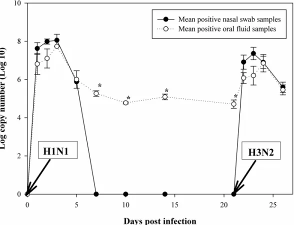
6.8; P = 0.95), indicating that swIAV infection did not influence the biting behavior of the pigs. Nevertheless, individual oral fluid samples from infected piglets were most difficult to obtain at 3 dpi with H1N1 (1 sample from 10 pigs) and H3N2 (3 samples from 10 pigs) strains (Table 2), respectively. This time point corresponded to the peaks of virus excretion (Fig 1).
Detection of swine influenza A virus by quantitative reverse transcription
real-time PCR in samples from experimentally infected piglets
2/8 (25%) of the oral fluid samples were positive at 7, 10, 14 and 21 dpi, respectively. After inoculation with the H3N2 strain, 21 days post H1N1 infection, viral RNA was detected in 24/ 30 (80%) of the nasal swabs collected between 1–3 dpi and in 2/10 (20%) nasal swabs collected at 5 dpi. Detectability in oral fluid went from 83.3% (5/6) at 1 dpi with the H3N2 strain to 100% (9/9) at 2 and 3 dpi and to 80% (4/5) at 5 dpi, respectively. Fisher’s exact tests showed that significantly more oral fluid samples were H1N1 positive by qRT-PCR than nasal swab
Table 1. Successful oral fluid collections by ropes from individually housed piglets aged between 8 and 13 weeks.
swIAV-infected pigs Mock-infected control pigs
1 2 3 4 5 6 7 8 9 10 1 2 3 4 5 6
6* 7 7 4 7 10 6 1 13 6 11 1 1 13 4 11
*The results represent the number of successful collections on 14 different sampling days (1 collection attempt/day)
doi:10.1371/journal.pone.0139586.t001
Table 2. Detectability of swine influenza A virus RNA by qRT-PCR and virus isolation in nasal swab samples (A) and oral fluid samples (B) of pigs sequentially infected with swine influenza A strains sw/Gent/28/10 (H1N1) at day 0 and sw/Gent/172/08 (H3N2) at day 21.
Virus strain H1N1 (dpi) 0 1 2 3 5 7 10 14 21 22 23 24 26
H3N2 (dpi) 0 1 2 3 5
A Pig 1 - + + +†,1,2 - - - - - +†,2 +†,2 +†,2
-Pig 2 - + + + - - - + + +
-Pig 3 - + +†,2 +†,1,2 - - - - - - + +
-Pig 4 - +†,1,2 +†,2 +†,1,2 + - - - - + +†,2 + +
Pig 5 - + + + - - -
-Pig 6 - +†,2 +†,2 +†,1,2 - - - - - + + +
-Pig 7 - +†,2 + + - - - - - + +†,2 + +
Pig 8 - + +†,1,2 +†,1,2 - - - + + +
-Pig 9 - +†,1,2 +†,2 +†,2 - - - - - + - -
-Pig 10 - + + + - - - + + +
-Positive detections 0/10 10/10 10/10 10/10 1/10 0/10 0/10 0/10 0/10 8/10 8/10 8/10 2/10
B Pig 1 ns +†,2 +†
ns + + ns + + ns ns ns ns
Pig 2 ns + + ns - ns - ns - +† +† ns ns
Pig 3 - +†
+†
ns ns + ns + + ns ns ns ns
Pig 4 ns ns ns ns ns ns ns ns ns + +†
+ +
Pig 5 ns ns + ns + ns + - - - + ns ns
Pig 6 - +†,2 +† ns +† + ns ns - + + ns +
Pig 7 - ns ns ns ns ns - - - + ns ns +
Pig 8 ns ns ns ns ns ns ns - ns ns ns ns ns
Pig 9 - +†,2 + +†
+ + + + - + + +
-Pig 10 ns ns ns ns ns + ns - - ns + + +
Positive detections 0/4 5/5 6/6 1/1 4/5 5/5 2/4 3/7 2/8 5/6 6/6 3/3 4/5 Fisher’s exact test (P) Ϯ Ϯ Ϯ Ϯ 0.02‡
0.00‡
0.07 0.05 0.18 1.00 0.50 1.00 0.09
ns: No sample available; +: PCR pos; -: PCR neg
†
: Samples with a Ct value<30 submitted for virus isolation on embryonated chicken eggs (EVE) and MDCK cells
‡: Significant difference between swIAV detection rates in nasal swab samples and oralfluid at that particular time point (p<0,05) Ϯ: No measure of association is computed because both variables are constant
1
: Samples analyzed by virus isolation on ECE with positive result
2
: Samples analyzed by virus isolation on MDCK with positive result
samples at 5 and 7 dpi. At 10 and 14 dpi, the detection rate between nasal swabs and oral fluid was not significantly different. However, P values of 0.07 and 0.05 at those respective time points suggest that swIAV might be longer detectable in oral fluid than in nasal swabs.
Not significantly different amounts of swIAV RNA were found in nasal swabs and oral fluid during the first five dpi for both strains, while significant higher copy numbers were found in oral fluid from 7 dpi onwards (p<0.05) (Fig 1;S1 Table).
Detection of swine influenza A virus by virus isolation in samples from
experimentally infected piglets
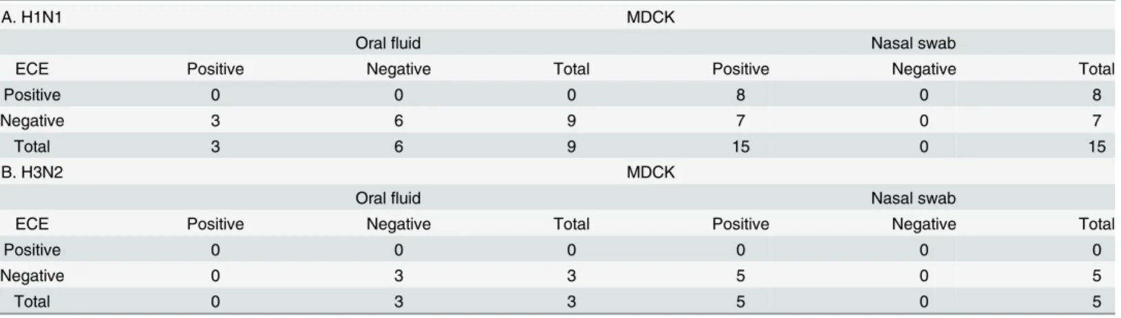
swIAV could be isolated from 8/15 (53.3%) and 15/15 (100%) nasal swabs collected after the H1N1 infection on ECE and MDCK cells, respectively (Table 3). In contrast, the H1N1 strain could not be isolated from oral fluid samples on eggs (0/9) and only from 3/9 (33.3%) oral fluid samples on MDCK cells. All three positive oral fluid samples were collected at 1 dpi. When virus isolation was attempted from nasal swabs and oral fluid collected from H3N2 infected pigs, swIAV was isolated from all nasal swabs with a Ct value<30 on MDCK cells (5/5) but
not on ECE (0/5). All isolations from oral fluid (3) remained negative, both on MDCK cells and on ECE.
Overall, swIAV isolation was more successful on MDCK cells (23/32) compared to ECE (8/32) and nasal swabs were more suitable for virus isolation than oral fluid.
Fig 1. Mean log copy number/mL detected in nasal swab samples and oral fluid samples of pigs sequentially infected with swine influenza A virus strains sw/Gent/28/10 (H1N1) and sw/Gent/172/08 (H3N2).Mean log copy number/mL of viral RNA detected by real-time PCR in nasal swab samples (black circles) and oral fluid samples (white circles) collected from individually housed pigs sequentially infected with the sw/Gent/28/10 (H1N1) strain at day 0 and the sw/Gent/172/08 (H3N2) strain at day 21. Data show the mean (±standard error of the mean) of positive samples. Asterisk denotes a significant difference, as determined by the independent samples T-test (p<0.05).
Influence of conservation time and temperature on swine influenza A
virus infectivity and qRT-PCR detectability in porcine oral fluid
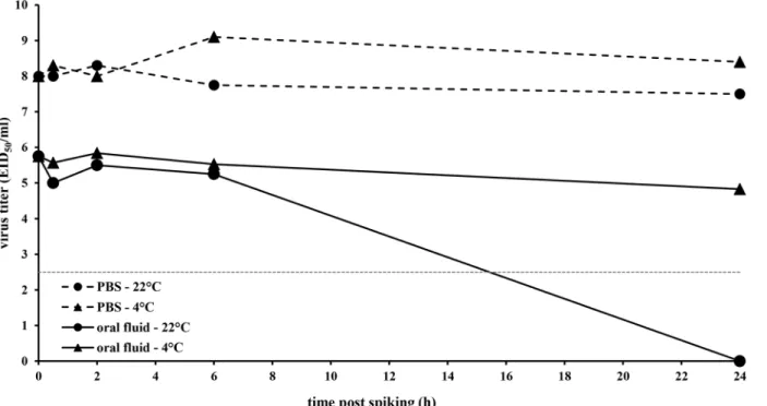
Control experiments whereby swIAV was spiked in PBS to an initial virus concentration of 1 x 108.7EID50/ mL showed that swIAV was stable in PBS during 24 h (Fig 2). No obvious loss in
virus infectivity as measured by virus titration in ECE occurred during this time period, neither when conserved at 4°C (titer 24 h after spiking: 1 x 108.4EID50/ mL), nor at room temperature
(titer 24 h after spiking: 1 x 107.5EID50/ mL). In contrast, spiking the same amount of swIAV
in oral fluid immediately resulted in a thousand fold reduction in virus titer (titer of 1 x 105.75 EID50/ mL at 0 h post spiking). When the spiked oral fluid was further conserved at 4°C, the
remaining amount of infectious virus seemed to remain stable during a 24 h period since viral titers of 1 x 105.84, 1 x 105.53and 1 x 104.83EID50/ mL were found at 2, 6 and 24 h post spiking,
respectively. Incubation of swIAV in oral fluid at room temperature did not result in a further reduction of virus infectivity during the first 6 h post spiking (1 x 105.25EID50/ mL).
Thereaf-ter, however, swIAV infectivity further decreased and dropped below the limit of detection (1 x 102.5EID50/ mL) at 24 h post spiking.
qRT-PCR analysis of oral fluid samples that were spiked with swIAV to concentrations of 1 x 108.7and 1 x 104.7EID50/ mL showed that the observed drop in infectivity after spiking of
swIAV in oral fluid did not coincide with considerable changes in the detectability of viral nucleic acids (Fig 3). The Ct values obtained immediately after spiking of swIAV in PBS and oral fluid were slightly higher in PBS than in oral fluid (indicating a higher retrieved number of viral nucleic acids from oral fluid than from PBS) for both the high (21.3 vs 20.5) and low (35.3 vs 33.6) spiked dose. It can therefore be concluded that spiking of swIAV in oral fluid did not coincide with an immediate reduction of viral nucleic acids, as was observed for the virus infec-tivity. Also further conservation of the spiked oral fluid samples did not result in important changes in the detectable amount of swIAV nucleic acids. The mean Ct values obtained at the different time points till 24 h after spiking did not differ more than 0.5 Ct, irrespective whether the samples were conserved at 4°C or at room temperature and this for both the high and low starting concentration. Similar results were obtained in the control experiments whereby swIAV was spiked in PBS.
Discussion
Collection of a sufficient amount of oral fluid is the first requisite to be able to progress to downstream diagnostics. Prickett et al. [30] described a method to efficiently collect pen-based
Table 3. Comparison of swine influenza A virus isolation from oral fluid samples and nasal swab samples collected from pigs sequentially infected with (A) swine influenza A virus strains sw/Gent/28/10 (H1N1) at day 0 and (B) sw/Gent/172/08 (H3N2) at day 21 in embryonated chicken eggs (ECE) and in Madin-Darby canine kidney (MDCK) cell culture.
A. H1N1 MDCK
Oralfluid Nasal swab
ECE Positive Negative Total Positive Negative Total
Positive 0 0 0 8 0 8
Negative 3 6 9 7 0 7
Total 3 6 9 15 0 15
B. H3N2 MDCK
Oralfluid Nasal swab
ECE Positive Negative Total Positive Negative Total
Positive 0 0 0 0 0 0
Negative 0 3 3 5 0 5
Total 0 3 3 5 0 5
oral fluid samples from pigs, whereby ropes are hung in a pen. The exploratory behaviour of pigs makes them chew on the rope and moisten it with oral fluid that can easily be collected afterwards. The oral fluid collections in this study showed however to be more difficult and dependent on individual animal behaviour. As recently described for a similar study [33], the observed low success rate of oral fluid collection was probably related to the young age of the pigs used and to their individual housing conditions. This is an important aspect to take into consideration when planning future experiments with individually housed piglets.
The laboratory diagnosis of influenza virus infection has typically relied upon the detection of the virus in nasal swabs. Serology to detect antibodies is of low value for swIAV surveillance because vaccination against swIAV is based on inactivated H1N1 and H3N2 vaccines and cur-rent serologic tests do not diffecur-rentiate between vaccinated and infected animals [34]. Only in holdings where no swIAV vaccination is practiced, testing of paired sera (acute and conva-lescent serum) might be useful. Therefore, virological assays are currently preferred over serol-ogy for surveillance. To allow virus detection, nasal swabs need to be collected during the acute phase of infection which is limited in time, mostly between 1 to 4–5 dpi [27]. The results of this study are in agreement with these findings since for both the H1N1 and H3N2 strain used, virus could be detected in nasal swabs from 1 till 5 dpi.
Recent publications have suggested the utility of pen-based oral fluid samples for the viro-logical diagnosis of swIAV by qRT-PCR [28–29,35]. It was described that the probability to detect swIAV in oral fluids and nasal swabs by qRT-PCR was equivalent till 6 dpi and then higher in pen-based oral fluid samples till the end of the experiment at 16 dpi. This is in agree-ment with our results showing that in experiagree-mentally infected and individually penned animals swIAV could be detected by qRT-PCR for a longer time period and with a higher detection
Fig 2. Influence of conservation time and temperature on swine influenza A virus infectivity in porcine oral fluid as determined by virus titration on embryonated chicken eggs.H1N1 virus stock was spiked in porcine oral fluid or PBS to a final concentration of 1 x 108.7EID50 / mL and conserved either at 4°C or at room temperature (22°C±2°C). At 0, 0.5, 2, 6 and 24h after spiking, aliquots of 2 mL were taken and incubated with PBS supplemented with antibiotics (9:1) for 1h at room temperature. Afterwards 10-fold dilutions were made and per dilution, 5 eggs were inoculated with 100μl each. Titers were
calculated using the method of Reed and Muench.
rate in porcine oral fluid compared to nasal swabs. Especially noteworthy was the detection of swIAV RNA till at least 21 dpi in 25% of the oral fluid samples, while all nasal swabs already became negative at 7 dpi.
The virus detected in nasal swabs most probably originates from local virus replication, which normally occurs up to 5 days after intranasal inoculation. H1N1 swIAV is only detect-able by virus isolation till 6 dpi in nasopharynx, tonsils, trachea, and some lung parts [36]. The reported presence of only limited numbers of single swIAV positive cells in nasal mucosa and nasopharynx after H1N1 swIAV infection in pigs [36] can help to explain the fast drop in qRT-PCR positive nasal swabs after local virus replication has stopped. The influenza RNA detected up till 21 dpi in oral fluids does probably not originate from such local virus replica-tion in the nasal mucosa, but might rather be explained by the detecreplica-tion of viral RNA in expec-torated sputum. Such sputum contains cellular debris from the lower respiratory tract (e.g. trachea, bronchi, aveoli, bronchiole) [37] which is known, just like the interstitial and alveolar macrophages, to contain high amounts of viral RNA and antigen in naturally infected pigs [36,
38]. The long turnover time of these cells that are potential sources of viral RNA in
Fig 3. Influence of conservation time and temperature on qRT-PCR detectability of swine influenza A virus in porcine oral fluid.H1N1 virus stock was spiked in porcine oral fluid or PBS to final concentrations of either 1 x 108.7EID
50/ mL or 1 x 104.7EID50/ mL. The spiked samples were conserved either
at 4°C or at room temperature (22°C±2°C) and at 0, 0.5, 2, 6 and 24 h after spiking, aliquots were collected and stored at -80°C. After RNA extraction using the MagMAX Pathogen RNA/DNA kit (Life Technologies), swIAV RNA was amplified with the Vetmax Gold swIAV detection kit (Life Technologies). Mean Ct values (±standard error) of three independent replicates are shown.
expectorated sputum—one to three weeks for epithelial cells in trachea, large bronchi and small bronchi in adult mice and 35 days for the alveolar macrophages [39,40]–seems in line with the prolonged swIAV detection in oral fluid. Also our finding that the level of viral nucleic acids remained unaltered during a 24 h incubation period in oral fluid helps to the explain why swIAV can be detected for a prolonged period in oral fluid.
The possibility to detect the H3N2 strain in both nasal swabs and oral fluid by qRT-PCR after a previous infection with a H1N1 strain was another important observation. This might be explained by the fact that although cross-protective immunity has been described for swIAV [41], heterosubtypic immunity induced by natural infection is mostly weak [42]. As a result, intranasal inoculation with H1N1 induces only partial protection against subsequent infection with other influenza A virus subtypes, like the H3N2 strain.
Besides the importance to detect swIAV RNA in diagnostic samples for surveillance pur-poses, it might also be important to isolate the virus from the diagnostic sample for further characterization. Our results show that swIAV isolation was more successful on MDCK cells than on ECE. These findings suggest that although ECE are still considered the golden standard for isolation of swIAV from infected animals [43], MDCK cells might be superior to ECE for the detection of certain subtypes/strains of swIAV. In this context, it has already been reported that the success rate of one of both isolation systems is mainly dependent upon the virus sub-type [44]. Furthermore, our results indicate that swIAV isolation is more efficient from nasal swab samples than from oral fluid samples, even if they are collected from the same pig at the same time point. Similar observations have been reported before [28,29] and suggest that oral fluid has a negative impact on swIAV infectivity. This is supported by our laboratory experi-ments, showing that spiking of swIAV in oral fluid results in an immediate thousand fold reduction in virus titer, thereby reducing the chance on a positive virus isolation from an oral fluid sample during diagnosis. Furthermore, continued conservation of the spiked oral fluid samples at room temperature further decreased the virus infectivity till it was completely lost after 24 h. This rapid loss of infectivity in oral fluid seems to be in line with the observation that swIAV was only successfully isolated from oral fluid samples collected at 1 day post infec-tion with the H1N1 strain. With regard to diagnosis, these results indicate that freshly collected oral fluid samples should be tested in the briefest delay to maximize virus detection and strongly argue to cool oral fluid samples as fast as possible upon collection and conserve them at 4°C when virus isolation is envisioned.
The causative factor for the reduction of swIAV infectivity in oral fluid has not yet been irre-futably identified. Goodell et al. [29] suggested that the lower isolation rates from oral fluid samples compared to nasal swab samples might be caused by the presence of anti-influenza antibodies in the oral fluid samples. This seems to be in agreement with our incapability to iso-late swIAV from oral fluid collected after the H3N2 infection performed 21 days after the primo infection with H1N1. It should however be kept in mind that only three oral fluid sam-ples with Ct<30 were available for virus isolation. Detmer et al. [28] suggested that
excluded for the moment is the possibility that simply no infectious virus was present in those samples and that the positive qRT-PCR results originate from viral RNA in expectorated spu-tum as discussed in a previous paragraph.
Finally, it should be emphasized that oral fluid was collected with polyester ropes in this study while most studies rely on cotton ropes to collect oral fluid from pigs. Although it has been shown that rope material influences downstream antibody and hormone detection [50–
52], multiple studies have shown that polyester is a suitable material to collect samples for downstream virus detection by PCR or isolation. Decorte et al. [33] showed that there was no difference in the detection of PRRSV virus by PCR when oral fluid was collected from pigs either with cotton or polyester ropes. Other studies showed that polyester swabs were suitable for swIAV sample collection from pigs for downstream PCR detection [53] and that polyester swabs were equally suitable as cotton swabs for downstream pseudorabies virus and bovine herpes virus 1 isolation [54–55]. Furthermore, the obtained results that swIAV could be detected both in nasal swabs and polyester rope collected oral fluid from all pigs from the same time point (1 dpi) confirm the suitability of polyester ropes for swIAV sample collection.
Although further research is advisory to evaluate the influence of virus strain or subtype and inoculation dose, our data indicate that porcine oral fluid samples collected with ropes hold potential for diagnostic purposes seen the possibility to detect swIAV RNA for a longer period than in nasal swabs. The difficulty to isolate swIAV from these oral fluid samples could however pose a drawback and has to be studied more intensively.
Supporting Information
S1 Table. Cycle threshold (Ct) values obtained for the detection of swine influenza A virus.
The Vetmax Gold swIAV kit (Life Technologies) was used for the detection of swine influenza A virus RNA by qRT-PCR in nasal swab samples (A) and oral fluid samples (B) of pigs sequen-tially infected with swine influenza A strains sw/Gent/28/10 (H1N1) at day 0 and sw/Gent/ 172/08 (H3N2) at day 21.
(DOCX)
Acknowledgments
The authors would like to acknowledge Prof. Kristien Van Reeth who kindly donated the swine influenza virus strains. We also thank the veterinarians Willem Van Campe and Laurent Mos-tin and the animal care takers of CODA-CERVA Machelen for the assistance in carrying out the biological sample collection. We also thank Virginie Colasse, Catherine Rasseneur and Marc Boschmans for excellent technical assistance.
Author Contributions
Conceived and designed the experiments: MS BL ABC NDR. Performed the experiments: ID NDR. Analyzed the data: ID MS BL ABC NDR. Wrote the paper: ID MS BL ABC NDR.
References
1. Fouchier RA, Osterhaus AD, Brown IH. Animal influenza virus surveillance. Vaccine. 2003; 21: 1754– 1757. PMID:12686089
2. Crisci E, Mussa T, Fraile L, Montoya M. Review: Influenza virus in pigs. Mol Immunol. 2013; 55: 200– 211. doi:10.1016/j.molimm.2013.02.008PMID:23523121
4. Zhou NN, Senne DA, Landgraf JS, Swenson SL, Erickson G, Rossow K, et al. Genetic reassortment of avian, swine, and human influenza A viruses in American pigs. J Virol. 1999; 73: 8851–8856. PMID: 10482643
5. Zell R, Motzke S, Krumbholz A, Wutzler P, Herwig V, Durrwald R. Novel reassortant of swine influenza H1N2 virus in Germany. J Gen Virol. 2008; 89: 271–276. PMID:18089751
6. Starick E, Lange E, Fereidouni S, Bunzenthal C, Höveler R, Kuczka A, et al. Reassorted pandemic (H1N1) 2009 influenza A virus discovered from pigs in Germany. J Gen Virol. 2011; 92: 1184–1188. doi:10.1099/vir.0.028662-0PMID:21307227
7. Liu QF, Ma JJ, Liu HX, Qi W, Anderson J, Henry SC, et al. Emergence of novel reassortant H3N2 swine influenza viruses with the 2009 pandemic H1N1 genes in the United States. Arch Virol. 2012; 157: 555–562. doi:10.1007/s00705-011-1203-9PMID:22198410
8. Karasin AI, Schutten MM, Cooper LA, Smith CB, Subbarao K, Anderson GA, et al. Genetic characteri-zation of H3N2 influenza viruses isolated from pigs in North America, 1977–1999: evidence for wholly human and reassortant virus genotypes. Virus Res. 2000; 68: 71–85. PMID:10930664
9. Fan XH, Zhu HC, Zhou BP, Smith DK, Chen X, LAM TT, et al. Emergence and Dissemination of a Swine H3N2 Reassortant Influenza Virus with 2009 Pandemic H1N1 Genes in Pigs in China. J Virol. 2012; 86: 2375–2378. doi:10.1128/JVI.06824-11PMID:22171260
10. Claas ECJ, Kawaoka Y, Dejong JC, Masurel N, Webster RG. Infection of Children with Avian-Human Reassortant Influenza-Virus from Pigs in Europe. Virology. 1994; 204: 453–457. PMID:8091678 11. Castrucci MR, Donatelli I, Sidoli L, Barigazzi G, Kawaoka Y, Webster RG. Genetic Reassortment
between Avian and Human Influenza-a Viruses in Italian Pigs. Virology. 1993; 193: 503–506. PMID: 8438586
12. Marshall N, Priyamvada L, Ende Z, Steel J, Lowen AC. Influenza Virus Reassortment Occurs with High Frequency in the Absence of Segment Mismatch. PLoS pathogens. 2013; 9: e1003421. doi:10.1371/ journal.ppat.1003421PMID:23785286
13. Welsh MD, Baird PM, Guelbenzu-Gonzalo MP, Hanna A, Reid SM, Essen S, et al. Initial incursion of pandemic (H1N1) 2009 influenza A virus into European pigs. Vet Rec. 2010; 166: 642–645. doi:10. 1136/vr.4851PMID:20495164
14. Vijaykrishna D, Poon LL, Zhu HC, Ma SK, Li OT, Cheung CL, et al. Reassortment of pandemic H1N1/ 2009 influenza A virus in swine. Science. 2010; 328: 1529. doi:10.1126/science.1189132PMID: 20558710
15. Song MS, Lee JH, Pascua PN, Baek YH, Kwon HI, Park KJ, et al. Evidence of human-to-swine trans-mission of the pandemic (H1N1) 2009 influenza virus in South Korea. J Clin Microbiol. 2010; 48: 3204– 3211. doi:10.1128/JCM.00053-10PMID:20610681
16. Nelson MI, Vincent AL, Kitikoon P, Holmes EC, Gramer MR. Evolution of novel reassortant A/H3N2 influenza viruses in North American swine and humans, 2009–2011. J Virol. 2012; 86: 8872–8878. doi: 10.1128/JVI.00259-12PMID:22696653
17. Moreno A, Di Trani L, Faccini S, Vaccari G, Nigrelli D, Boniotti MB, et al. Novel H1N2 swine influenza reassortant strain in pigs derived from the pandemic H1N1/2009 virus. Vet Microbiol. 2011; 149: 472– 477. doi:10.1016/j.vetmic.2010.12.011PMID:21208754
18. Kitikoon P, Vincent AL, Gauger PC, Schlink SN, Bayles DO, Gramer MR, et al. Pathogenicity and trans-mission in pigs of the novel A(H3N2)v influenza virus isolated from humans and characterization of swine H3N2 viruses isolated in 2010–2011. J Virol. 2012; 86: 6804–6814. doi:10.1128/JVI.00197-12 PMID:22491461
19. Ducatez MF, Hause B, Stigger-Rosser E, Darnell D, Corzo C, Juleen K, et al. Multiple reassortment between pandemic (H1N1) 2009 and endemic influenza viruses in pigs, United States. Emerg Infect Dis. 2011; 17: 1624–1629. doi:10.3201/eid1709.110338PMID:21892996
20. Lindstrom S, Garten R, Balish A, Shu B, Emery S, Berman L, et al. Human Infections with Novel Reas-sortant Influenza A(H3N2)v Viruses, United States, 2011. Emerg Infect Dis. 2012; 18: 834–837. doi: 10.3201/eid1805.111922PMID:22516540
21. Cox CM, Neises D, Garten RJ, Bryant B, Hesse RA, Anderson GA, et al. Swine Influenza Virus A (H3N2) Infection in Human, Kansas, USA, 2009. Emerg Infect Dis. 2011; 17: 1143–1144. doi:10.3201/ eid/1706.101488PMID:21749798
22. Wong KK, Greenbaum A, Moll ME, Lando J, Moore EL, Ganatra R, et al. Outbreak of influenza A (H3N2) variant virus infection among attendees of an agricultural fair, Pennsylvania, USA, 2011. Emerg Infect Dis. 2012; 18: 1937–1944. doi:10.3201/eid1812.121097PMID:23171635
24. Corzo CA. Active Surveillance for Influenza A Virus among Swine, Midwestern United States, 2009– 2011. Emerg Inf Dis. 2013; 19: 954–960.
25. Kyriakis CS, Brown IH, Foni E, Kuntz-Simon G, Maldonado J, Madec F, et al. Virological Surveillance and Preliminary Antigenic Characterization of Influenza Viruses in Pigs in Five European Countries from 2006 to 2008. Zoonoses Public Health. 2011; 58: 93–101. doi:10.1111/j.1863-2378.2009.01301. xPMID:20042068
26. Mak PWY, Jayawardena S, Poon LLM. The Evolving Threat of Influenza Viruses of Animal Origin and the Challenges in Developing Appropriate Diagnostics. Clin Chem. 2012; 58: 1527–1533. doi:10. 1373/clinchem.2012.182626PMID:22968105
27. Janke B. Clinicopathological Features of Swine Influenza. Curr Top Microbiol Immunol. 2013; 370: 69– 83. doi:10.1007/82_2013_308PMID:23404579
28. Detmer SE, Patnayak DP, Jiang Y, Gramer MR, Goyal SM. Detection of Influenza A virus in porcine oral fluid samples. J Vet Diag Invest. 2011; 23: 241–247.
29. Goodell CK, Prickett J, Kittawornrat A, Zhou F, Rauh R, Nelson W, et al. Probability of detecting influ-enza A virus subtypes H1N1 and H3N2 in individual pig nasal swabs and pen-based oral fluid speci-mens over time. Vet Microbiol. 2013; 166: 450–460. doi:10.1016/j.vetmic.2013.06.029PMID: 23910522
30. Prickett J, Simer R, Christopher-Hennings J, Yoon KJ, Evans RB, Zimmerman JJ. Detection of Porcine reproductive and respiratory syndrome virus infection in porcine oral fluid samples: a longitudinal study under experimental conditions. J Veterinary Diag Invest. 2008; 20: 156–163.
31. Decorte I, Van der Stede Y, Nauwynck H, De Regge N, Cay AB. Effect of saliva stabilisers on detection of porcine reproductive and respiratory syndrome virus in oral fluid by quantitative reverse transcriptase real-time PCR. Vet J. 2013; 197: 224–228. doi:10.1016/j.tvjl.2013.02.001PMID:23489844
32. Webster RG, Krauss S. WHO manual on animal influenza diagnosis and surveillance. World Health Organization, Department of Communicable Disease Surveillance and Response. 2002.
33. Decorte I, Van Campe W, Mostin L, Cay AB, De Regge N. Diagnosis of the Lelystad strain of Porcine reproductive and respiratory syndrome virus infection in individually housed pigs: comparison between serum and oral fluid samples for viral nucleic acid and antibody detection. J Vet Diag Invest. 2015; 27: 47–54.
34. Detmer S, Gramer M, Goyal S, Torremorell M, Torrison J. Diagnostics and surveillance for swine influ-enza. Curr Top Microbiol Immunol 370: 85–112. doi:10.1007/82_2012_220PMID:22566130 35. Romagosa A, Gramer M, Joo HS, Torremorell M. Sensitivity of oral fluids for detecting influenza A virus
in populations of vaccinated and non-vaccinated pigs. Influenza Other Respir Viruses. 2012; 6: 110– 118. doi:10.1111/j.1750-2659.2011.00276.xPMID:21777397
36. Van Reeth K, Braeckmans D, Cox E, Van Borm S, van den Berg T, Goddeeris B, et al. Prior infection with an H1N1 swine influenza virus partially protects pigs against a low pathogenic H5N1 avian influ-enza virus. Vaccine. 2002; 27: 6330–6339.
37. Rubin BK. Physiology of airway mucus clearance. Resp Care. 2002; 47: 761.
38. Jung T, Choi C, Chae C. Localization of swine influenza virus in naturally infected pigs. Vet Pathol Online. 2002; 39: 10–16.
39. Shorter RG, Titus JL, Divertie MB. Cell turnover in the respiratory tract. CHEST J. 1964; 46: 138–142. 40. Spencer H, Shorter R. Cell turnover in pulmonary tissues. Nature. 1962; 194: 880.
41. Reeth KV, Brown I, Essen S, Pensaert M. Genetic relationships, serological reaction and cross-protection between H1N2 and other influenza A virus subtypes endemic in European pigs. Virus Res. 2004; 103: 115–124. PMID:15163499
42. Tamura SI, Tanimoto T, Kurata T. Mechanisms of broad cross-protection provided by influenza virus infection and their application to vaccines. Jpn J Infect Dis. 2005; 58: 195–207. PMID:16116250 43. Lombardo T, Dotti S, Renzi S, Ferrari M. Susceptibility of different cell lines to Avian and Swine
Influ-enza viruses. J Virol Meth. 2012; 185:82–88.
44. Chiapponi C, Zanni I, Garbarino C, Barigazzi G, Foni E. Comparison of the usefulness of the CACO–2 cell line with standard substrates for isolation of swine influenza A viruses. J Virol Meth. 2010; 163: 162–165.
45. Fábián TK, Hermann P, Beck A, Fejérdy P, Fábián G. Salivary defense proteins: their network and role in innate and acquired oral immunity. Int J Mol Sci. 2012; 13: 4295–420. doi:10.3390/ijms13044295 PMID:22605979
47. Eaton MD, Scala AR. Reversible effect of hypotonic solutions on growth of influenza virus in tissue cul-tures. Proc Soc Exp Biol Med. 1956; 92:289–297. PMID:13350324
48. Tolskaya EA, Agol VI, Voroshilova MK, Lipskaya GY. The osmotic pressure of the maintenance medium and reproduction of poliovirus. Virology. 1966; 29: 613–621. PMID:18611523
49. Park NJ, Li Y, Yu T, Brinkman BMN, Wong DT. Characterization of RNA in saliva. Clin Chem. 2006; 52: 988–994. PMID:16601067
50. Decorte I, Van Breedam W, Van der Stede Y, Nauwynck HJ, De Regge N, Cay AB. Detection of total and PRRSV-specific antibodies in oral fluids collected with different rope types from PRRSV-vacci-nated and experimentally infected pigs. BMC Vet Res. 2014; 17: 134.
51. Olsen C, Karriker L, Wang C, Binjawadagi B, Renukaradhya G, Kittawornrat A, et al. Effect of collection material and sample processing on pig oral fluid testing results. Vet J. 2013; 198: 158–163. doi:10. 1016/j.tvjl.2013.06.014PMID:24011474
52. Hansen AM, Garde AH, Persson R. Measurement of salivary cortisol—effects of replacing polyester with cotton and switching antibody. Scand J Clin Lab Invest. 2008; 68: 826–829. doi:10.1080/ 00365510802056207PMID:18609107
53. Edwards JL, Nelson SW, Workman JD, Slemons RD, Szablewski CM, Nolting JM, et al. Utility of snout wipe samples for influenza A virus surveillance in exhibition swine populations. Influenza Other Respir Viruses. 2014; 8: 574–579. doi:10.1111/irv.12270PMID:25043408
54. Hanson BR, Schipper IA. Effects of swab materials on infectious bovine rhinotracheitis virus. Am J Vet Res. 1976; 37: 707–708. PMID:180853