EPILEPTIFORM ABNORMALITIES AND
QUANTITATIVE EEG IN CHILDREN WITH
ATTENTION-DEFICIT/HYPERACTIVITY DISORDER
Lineu Corrêa Fonseca
1, Glória Maria A.S. Tedrus
1, César de Moraes
2, Amanda de Vicente Machado
3,
Marcela Pupin de Almeida
3, Débora Ortolan Fernandes de Oliveira
4Abstract – There is much controversy about the importance of the electroencephalogram (EEG) in assessing the attention-deficit/hyperactivity disorder (ADHD). The objective of this study was to assess the use of EEG and quantitative EEG (qEEG) in ADHD children. Thirty ADHD children and 30 sex- and age-matched controls with no neurological or psychiatric problems were studied. The EEG was recorded from 15 electrode sites during an eyes-closed resting condition. Epileptiform activity was assessed, as were the absolute and relative powers in the classical bands after application of the Fast Fourier transform. Epileptiform activity was found in 3 (10%) ADHD children. As compared to the controls, the ADHD group showed significantly greater absolute delta and theta powers in a diffuse way, and also greater absolute beta power and smaller relative alpha 1 and beta powers at some electrodes. A logistic multiple regression model, allowed for 83.3% sensibility and specificity in diagnosing ADHD.
KEy worDs: attention-deficit/hyperactivity disorder, EEG, children.
Atividade epileptiforme e eletrencefalograma quantitativo em crianças com transtorno de déficit de atenção/hiperatividade
Resumo – Há controvérsias sobre a importância do eletrencefalogama (EEG) na avaliação do transtorno de déficit de atenção/hiperatividade (TDAH). o objetivo deste estudo foi avaliar, em crianças com TDAH, o EEG digital e quantitativo. Foram estudadas 30 crianças com TDAH e 30 sadias, sem evidências de problemas neurológicos ou psiquiátricos e pareadas por idade e gênero. Foi registrado o EEG em 15 posições de eletrodos, durante repouso e olhos fechados. Foi realizada pesquisa de atividade epileptiforme e feita análise de freqüências nas faixas clássicas, após aplicação da transformada rápida de Fourier. Foi encontrada atividade epileptiforme em 3 (10%) crianças com TDAH. o grupo TDAH teve, em relação ao grupo controle, significativamente, maior potência absoluta delta e teta, de modo difuso, assim como maior potência absoluta beta e menor potência relativa alfa 1 e beta, em alguns eletrodos. Um modelo de regressão múltipla logística possibilitou sensibilidade e especificidade de 83,3% no diagnóstico de TDAH.
PAlAvrAs-cHAvE: transtorno de déficit de atenção/hiperatividade, EEG, infância.
1Professor of Neurology; 2Professor of Psychiatry, Pontifícia Universidade católica de campinas, campinas sP, Brazil (PUc-campinas), scholarship hold-ers; 3PIBIc/cNPq, PUc-campinas; 4FAPIc/reitoria PUc-campinas.
received 10 March 2008. Accepted 4 June 2008.
Dr. Lineu Corrêa Fonseca – Rua Sebastião de Souza 205 / 122 - 13020-020 Campinas SP - Brasil.
The attention-deicit/hyperactivity disorder (ADHD) is one of the commonest behavioral disorders in childhood. Due to advancing knowledge with respect to the preva-lence, natural history, genetics, biology and treatment of ADHD, a greater number of patients are now receiving adequate treatment. The diagnosis of ADHD is based on the DsM Iv1 criteria of which the essential characteristic consists of a persistent pattern of lack of attention and/ or hyperactivity-impulsivity more frequent and serious than that typically observed in individuals with an
scales such as that of conners2 about behavioral as-pects and continued execution tests may be useful in the assessment. However, due to limitations, these tests and scales cannot be considered diagnostic3. There is current concern with respect to exaggeration in the diagnosis of ADHD and consequent exposition of many children to un-necessary medical treatment. Thus the search continues for better accuracy in the diagnosis using objective proce-dures. since ADHD is considered to be the result of a brain dysfunction and the electroencephalogram (EEG) assess-es brain function, it is natural that this method be exam-ined with respect to this clinical condition.
Electroencephalogram and ADHD
EEG studies in children with ADHD are searching for data with respect to various brain function aspects. one of the alterations that can occur in an EEG is that of ep-ileptiform activity (EA), characterized by electrographic elements that correspond to the recording of excessive neuronal discharge and abnormal components of the ba-sic epilepsy mechanism. EA can occur with less frequency in non-epileptic individuals. A greater recording of EA has been described in ADHD children than in normal children4.
Although it only occurs in a small proportion of ADHD children (about 6%), EA could be a factor in the origin of the attention deicit 5,6,7, and thus pharmacotherapy with the objective of reducing EA could eventually produce beneit with respect to this behavior8.
Quantitative electroencephalogram (qEEG) and ADHD
Advances in computer technology and the creation of programs have made it possible to register EEG digi-tally using analogical-digital transformation. since it uses numbers, the digital EEG allows for quantitative analyses (qEEG) such as the composition of the electrical brain ac-tivity frequencies (frequency analysis). segments of the recording free of artifacts are chosen, and the Fast Fouri-er Transform applied, this being a mathematical process that identiies the various frequency bands (delta, theta, alpha and beta) on the qEEG, from the temporal series of the original digital EEG data.
various qEEG studies were carried out with individuals suffering from ADHD, assessing different parameters us-ing a variable number of electrodes in patients with their eyes open and closed, both at rest and when carrying out activities9,10. In studies with ADHD children at rest with their eyes closed, differences have been observed in re-lation to normal controls, such as an increase in the del-ta and thedel-ta powers11, increase in theta12, increase in the-ta and decrease in bethe-ta13,14 and increase in theta and de-crease in alpha and beta15.
qEEG in ADHD diagnosis
various research studies have assessed the value of qEEG in ADHD diagnosis. Monastra et al. (1999, 2001)16,17 afirmed that qEEG data allow for differentiation between ADHD children and normal children with a speciicity of 94% and sensibility of 90%. other studies indicate the val-ue of qEEG in ADHD diagnosis7,18,19, but there is still not suf-icient evidence to use qEEG as a routine diagnostic meth-od20. Although qEEG obtained with children with their eyes open or carrying out tasks were less trustworthy in the test-retest than those registered at rest with the eyes closed21, only one study of the sensibility and speciicity under this functional condition was found19.
Thus the objective of the present research was to study ADHD children using digital and quantitative elec-troencephalograms, determining their diagnostic value at rest with the eyes closed.
METHOD
Thirty schoolchildren in the 8 to 11 year-old age range suffer-ing from the attention-deicit/hyperactivity disorder accordsuffer-ing to the DsM-Iv-Tr1 criteria, referred by the outpatients sections
of the Infancy & Adolescence Psychiatry Department and the Pediatric & Neurological Pediatric Department of (HMcP PUc-campinas), were included in this study.
All the children were free of medication at the time of test-ing, and those taking methylphenidate were taken off this med-ication for at least 12 h prior to the assessment.
The following procedures were carried out: medical history, psychiatric evaluation and traditional neurological examination; conner’s Parent and Teacher rating scales; wechsler Intelligence scale for children (wIsc-III); digital and quantitative EEG.
The dEEG was recorded with a resolution of 12 bits, 0.5 and 35 Hz ilters and 200 samples per second, using the Braintech 3.0 equipment (EMsA Equipamentos Médicos). Impedance was
maintained below 10 kΩ. The exam was carried out with the
child in the dorsal decumbent position in an ambient of silence with reduced luminosity. The electrodes were placed according to the International 10-20 system, with the use of an addition-al two electrodes placed 1 cm below (left side) and above (right side) the external angle of the eyelid, with the objective of eval-uating eye movements. The inter-connected ear lobe electrodes served as the reference. recording was carried out during three periods, alternating 2 minutes rest with the eyes closed with two minutes with the eyes open. The type, location and side of ep-ileptiform activity were assessed.
follow-ing frequency bands: delta (up to 3.9 Hz), theta (4.29 to 7.8 Hz), alpha (8.2 to 12.5 Hz) and beta (above 12.89 Hz). To obtain the normal distribution, the values for absolute power (X) were sub-stituted by their logarithms, y=log (X), and the relative power values (r) transformed by logit, y=log(r/1-r).
Data analysis
Thirty children made up the control group (cG) of “healthy” children, paired with the study group according to age, gender and the scholastic level of their parents. These children had no history of neurological (for example personal antecedents or close relatives suffering from epileptic its, head injury with loss of consciousness, encephalitis or reduced mental capacity) or psychiatric problems, showed normal neurological and intel-lectual development, normal neurological and
electroenceph-alographic examinations, an absence of cognitive deicit in the raven progressive matrixes test, had never repeated a school year and presented performance compatible with their age and school grade in the school Performance Test.
A comparison was made between the study group and the control group with respect to the absolute and relative powers in the delta, theta, alpha and beta bands of the qEEG (T-test).
The Ethics in research committee of FcM-PUc-campinas, organ recognized by the Brazilian National commission on Eth-ics in research (coNEP/Ms) approved the project.
RESULTS
Table 1 shows the distribution of the 30 ADHD chil-dren, which was identical to that of the control group with respect to age and gender.
In the wIsc-III evaluation, all the children showed an IQ above 70 and the means and standard deviations were as follows: total IQ, 94.5±19.9; verbal IQ, 96.3±19.3; perfor-mance IQ, 93.8±17.9.
Digital electroencephalogram
Epileptiform activity was registered in 3 (10.0%) of the ADHD children, in small numbers and short duration.
The characteristics of the EA in these 3 cases were, respectively: 1 – spikes showing their main projections in the left and median parietal regions, either spontaneous or evoked by tapping the right foot; 2 – generalized spike-wave complexes at 3-4 Hz, lasting for 1 second; 3 – spikes Table 1. Distribution of the 30 ADHD children, and similarly of
the control group, according to age and gender.
Age (years)
Boys Girls Total
Nº % Nº % Nº %
7 3 12.5 0 0 3 10.0
8 8 33.3 1 16.6 9 30.0
9 8 33.3 2 33.3 10 33.3
10 3 12.5 2 33.3 5 16.6
11 2 8.3 1 16.6 3 10.0
Total 24 80 6 20 30 100
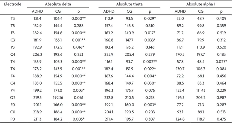
Table 2. Mean values for the absolute delta, theta and alpha 1 powers of the ADHD and control (CG) groups of children, and the value for p in the respective comparisons.
Electrode Absolute delta Absolute theta Absolute alpha 1
ADHD cG p ADHD cG p ADHD cG p
T3 131.4 106.4 0.000** 110.9 93.5 0.029* 52.0 48.7 0.409
T5 152.9 144.4 0.288 157.4 145.8 0.310 89.2 99.8 0.359
F3 182.4 154.6 0.000** 163.2 140.9 0.017* 71.2 66.9 0.519
c3 181.9 155.1 0.001** 166.8 147.7 0.033* 86.7 79.9 0.312
P3 192.9 172.5 0.016* 192.4 176.2 0.146 117.1 110.9 0.520
o1 206.2 192.6 0.253 225.9 205.4 0.279 170.5 197.7 0.183
T4 135.9 105.3 0.000** 116.1 93.7 0.002** 57.8 48.4 0.027*
T6 178.2 143.9 0.001** 182.4 151.9 0.022* 130.7 106.7 0.084
F4 188.9 154.9 0.000** 167.6 144.4 0.004* 72.2 68.1 0.456
c4 183.0 155.5 0.000** 168.4 149.7 0.030* 88.5 83.3 0.464
P4 199.2 171.0 0.003* 196.3 175.7 0.076 123.4 111.43 0.229
o2 219.5 192.16 0.061 232.8 210.5 0.218 195.3 203.2 0.987
F0 201.1 166.0 0.000** 192.1 160.0 0.003* 77.2 71.3 0.287
c0 218.9 186.4 0.000** 204.1 190.5 0.203 93.1 89.1 0.513
P0 211.3 184.2 0.005* 211.4 195.7 0.307 124.8 118.7 0.475
in the right frontal-temporal region. These children did not suffer from epileptic its.
Results of the qEEG: comparison between the ADHD and control groups
Tables 2 and 3 show the values obtained for the abso-lute powers for the ADHD and control groups, and also the values for p in their comparison (T-test).
Note that the absolute delta and theta powers were signiicantly greater for the ADHD group at the majority of electrodes as compared to the control group (Table 2).
The absolute alpha 1 (Table 2), alpha 2 and beta (Ta-ble 3) powers were greater for the ADHD group, but only reached a signiicant level at a few electrodes.
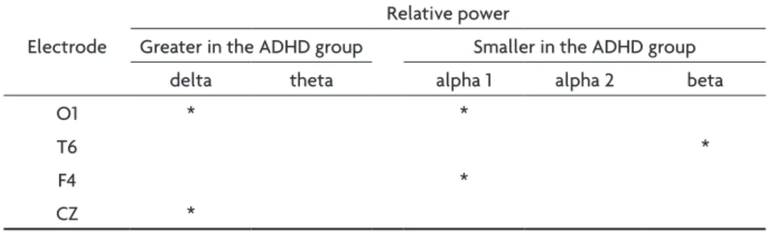
with respect to the relative powers, at the majority of the electrodes the means were greater for the relative
delta and theta powers, and lower for the alpha 1, alpha 2 and beta powers in the ADHD group as compared to the control group, but statistical signiicance was only found for some of the delta (o1, c0), alpha 1 (F4, o1) and beta (T6) electrode positions (Table 4).
Table 3. Mean values for the absolute alpha 2 and beta powers of the ADHD and control (CG) groups of children, and the value for p in the respective comparisons.
Electrode Absolute Alpha 2 Absolute Beta
ADHD cG p ADHD cG p
T3 45.6 41.6 0.246 87.8 84.0 0.307
T5 63.9 64.3 0.956 94.4 93.7 0.869
F3 59.1 52.1 0.147 104.4 92.3 0.061
c3 68.3 60.1 0.126 99.0 86.6 0.030*
P3 79.6 70.2 0.130 109.2 100.5 0.156
o1 102.0 105.3 0.873 120.6 122.6 0.793
T4 51.3 41.4 0.016* 91.2 83.2 0.114
T6 87.1 68.5 0.082 106.3 96.0 0.101
F4 60.1 59.0 0.205 112.7 94.9 0.014*
c4 70.5 60.3 0.097 101.8 90.0 0.029*
P4 82.0 70.5 0.077 110.9 99.9 0.065
o2 110.5 103.8 0.689 125.4 126.4 0.994
F0 62.6 54.4 0.047* 116.7 95.5 0.004*
c0 66.1 60.7 0.215 111.3 94.7 0.012*
P0 75.8 68.2 0.135 108.4 95.6 0.039*
*T-test, *p<0.05
Table 4. Electrode sites and frequency ranges showing signiicant differences between the ADHD and control groups.
relative power
Electrode Greater in the ADHD group smaller in the ADHD group
delta theta alpha 1 alpha 2 beta
o1 * *
T6 *
F4 *
cZ *
T-test, *p<0.05
Table 5. Classiication between the ADHD and control groups according to the absolute delta T4 and absolute theta F0 and C0 powers (logistic multiple regression).
Forecast by the model
observed ADHD control
ADHD 25 5
control 5 25
The qEEG and discrimination between the ADHD and the controls
A multiple logistic regression analysis allowed for the correct classification of 83.3% of the cases (Table 5) as from the data for the absolute F7 and T4 delta powers. The sensibility and speciicity were 83.3% in the classii-cation of the ADHD group.
DISCUSSION
Epileptiform activity on the EEG – The inding of epilepti-form activity (EA) in 3 (10%) of the ADHD children is simi-lar to the values of 6.1% and 5.6% found in the literature4,5 and higher than that found in healthy children (2%-3%)22,23. The EA could be a factor generating the attention-def-icit5-7 and one of the mechanisms could be the occurrence of transitory cognitive impairment during the EA24.
In a recent paper on the evaluation of children with rolandic epilepsy and EA, transitory cognitive impairment was only shown in a small percentage of the children, and in these cases there were no cognitive or behavioral im-pairments25. These indings suggest that transitory cogni-tive impairment is not an important factor in the genesis of behavioral alterations in children with ADHD.
The clinical use of routine EEG in children with ADHD seems to be limited and its recommendation would de-pend on the suspicion of epileptic manifestations.
qEEG in the comparison between the ADHD and control groups –
In the present research the absolute theta power was shown to increase in a diffuse way, but preserving the pos-terior regions in agreement with the literature13,14,26.
The inding of a diffuse increase in the delta power, as found in the present study, has been less frequently pointed out11.
It is possible that the relatively low socio-economic level of various children in the present study could have been a causal factor in the increase in delta activity, simi-lar to that described by Harmony et al. (1990)27 in healthy children with low socio-cultural stimulation.
The increase in the relative delta power in the left occip-ital region is in agreement with the increases in delta power in posterior regions described by some other authors15,26,28.
The smaller relative alpha and beta powers observed in the present research have also been described previ-ously15,26,28.
It is known that in the development of a healthy child, there is a tendency for the absolute powers in the delta and theta bands to decrease with age, and the relative alpha power to increase23.
Based on these data, one of the ADHD models based on the qEEG is the maturational lag model of ADHD. on
the other hand, since alterations in EEG are frequently very stable and considering that different sub-groups ex-ist within the ADHD, the hypothesis of a developmental deviation model has been raised28.
Nevertheless, these models do not appear to ade-quately explain the complexity of ADHD18.
qEEG in the diagnosis of ADHD – In the present research, the children were assessed at rest with their eyes closed, since this is a simpler situation showing trustworthiness in the test-retest21.
The model was reached with the absolute delta T4 and theta powers in F0 and c0. Alterations in c0 and F0 in ADHD have already been pointed out in the literature16.
The inding of 83.3% for both sensibility and speciicity in the present research was similar to the values found by Magee et al. (2005)19 in a study using similar procedures, of 89.0% and 79.6%, respectively.
In studies using other methods for the evaluation of qEEG, the values for sensibility and speciicity found were 80.9%-74.00% by Mann et al.12, 86.0%-98.0 by Monastra et al.16; and 83.1%-88.2% by chabot & serfontain13.
recent research has shown discordant results in the comparison between qEEG and psychiatric evaluation data and rating scales, with high29 or low30 values for sen-sibility and speciicity.
one of the aspects limiting transposition of the sen-sibility and speciicity values found in research studies is that the parameters of the qEEG may be particular for that research (ADHD versus normal) and not apply to new groups of patients for whom a differential diagnosis of different clinical conditions is carried out20.
Another question refers to the negative predictive val-ue, where a qEEG within normal parameters can, in about 20% of cases, correspond to the ADHD that will be diag-nosed by other methods.
Nevertheless the EEG analysis has provided highly signiicant indings in children with ADHD, and new ap-proaches to this procedure could provide additional ele-ments to reinforce its diagnostic contribution.
AcknowlEDGEmEnts – The authors are grateful to Professor
Elisa-beth Marinelli de camargo Pacheco for supervising the psychologi-cal aspects of the present research.
REFERENCES
1. American Psychiatric Association. Manual diagnóstico e estatístico de transtornos mentais, 4th edition. Porto Alegre: Artmed, 2003:112-119. 2. Dias MR, Barbosa GA, Gaião AA. Adaptação do Questionário
Abre-viado de Conners para Professores: uma avaliação psicométricra. R Psiquiatr RS 1997;19:202-210.
3. Cantwell DP. Attention deicit disorder: a review of the past ten years.
4. Richer LP, Shevell MI, Rosenblatt BR. Epileptiform abnormalities in
children with attention deicit hyperactivity disorder. Pediatr Neurol
2002;26:125-129.
5. Holtmann M, Becker K, Kentner-Figura B, Schmidt MH. Increased fre-quency of rolandic spikes in ADHD Children. Epilepsia 2003; 44:1241-1244.
6. Becker K, Sinzing JK, Holtmann M. Attention deicits and subclinical
epileptiform discharges: are EEG diagnostics in ADHD optional or
es-sential. Dev Med Child Neurol 2004;46:501-502.
7. Boutros N, Fraenkel L, Feingold A. A four-step approach for develop
-ing diagnostic tests in psychiatry: EEG in ADHD as a test case. J Neu
-ropsychiatr Clin Neurosci 2005;17:455-464.
8. Laporte N, Sebire G, Gillerot Y, et al. Cognitive epilepsy: ADHD relat
-ed to focal discharges. P-ediatr Neurol 2002;27:307-311.
9. Chabot RJ, Michele FD, Prichep L, John ER. The clinical role of comput-erized EEG in the evaluation and attention disorders in children and
adolescents. J Neuropsychiat Clin Neurosci 2001;13:171-186.
10. Hermens DF, Soei EX, Clarke SD, Kohn MR, Gordon E, Williams LM. Resting EEG theta activity predicts cognitive performance in
attention-deicit hyperactivity disorder. Pediatr Neurol 2005;32:248-256. 11. Matsuura M, Okubo Y, Toru M, et al. A cross-national EEG study of chil
-dren with emotional and behavioral problems: a WHO collaborative
study in the Western Paciic Region. Biol Psychiatry 1003;34:973-991
12. Mann C, Lubar J, Zimmerman A, Miller C, Muenchen R.
Quantita-tive analysis of EEG in boys with attention deicit hyperactivity disor
-der: controlled study with clinical implications. Pediatr Neurol 1992;
8:3036.
13. Chabot RJ, Serfontein G. Quantitative electroencephalographic proiles of children with attention deicit disorder. Biol Psychiatry 1996;40:951-963.
14. Bresnahan SM, Anderson JW, Barry RJ. Age-related changes in
quanti-tative EEG in attention-deicit/hyperactivity disorder. Biol Psychiatry
1999;46:1690-1697.
15. Clarke AR, Barry RJ, Carthy RM, et al. Quantitative EEG in low-IQ
chil-dren with attention-deicit hyperactivity disorder. Clin Neurophysiol
2006;117:1708-1714.
16. Monastra VJ, Linden M, Green G, et al. Assessing attention deicit hy -peractivity disorder via quantitative electroencephalography: an initial
validation study. Neuropsychology 1999;13:424.
17. Monastra VJ, Linden M, Lubar JF. The development of a quantitative
electroencephalographic scanning process for attention deicit- hyperac
-tivity disorder: reliability and validity studies. Neurophysiology 2001;
15;136-144.
18. Barry RJ, Clarke AR, Johnstone SJ. A review of electrophysiology in
at-tention-deicit/hyperactivity disorder: I. Qualitative and quantitative electroencephalography. Clin Neurophysiol 2003;114:171-183.
19. Magee CA, Clarke AR, Barry RJ, McCarthy R, Selikowitz M. Examining the diagnostic utility of EEG power measures in children with attention
deicit/hyperactivity disorder. Clin Neurophysiol 2005;116:1033-1040. 20. Loo SK, Barkley RA. Clinical utility of EEG in attention deicit hyper
-activity disorder. Appl Neuropsychol 2005;12:64-76.
21. John E, Ahn H, Princhep I, Trepetin M, Brown D, Kaye H. Developmen-tal equations of the electroencephalogram. Science 1980;2210:1255-1258. 22. Eeg-Olofsson O, Peterson I, Sellden U. The development of the eletro-encaphalogram in normal children and adolescentsfrom the age of 1 through 21 years. Acta Paeditr Scand 1970;208(Suppl):S20-S81. 23. Fonseca LC, Tedrus GMAS Pontas evocadas por estímulos
somatos-sensitivos e atividade epileptiforme no eletrencefalograma em
crian-ças normais. Arq Neuropsiquiatr 2003;61:793-795.
24. Binnie CD. Cognitive impairment during epileptiform EEG discharg-es. Epilepsia 2002;43(Suppl 8).
25. Fonseca LC, Tedrus GMAS, Pacheco EMC. Epileptiform EEG discharg-es in benign childhood epilepsy with centrotemporal spikdischarg-es: reactivity and transitory cognitive impairment. Epilepsy Behav 2007;11:65-70.
26. Lazzaro I, Gordon E, Li W, et al. Quantiied EEG activity in adolescent attention deicit hyperacticity. Clin Electroencephalogr 1998;29:37-42.
27. Harmony T, Marosi E, Diaz de León AE , Becker J, Fernández T. Effect of sex, psychosocial disadvantages and biological risk factors on EEG
maturation. Electroencephalogr Clin Neurophysiol 1990;75:482-491. 28. Clarke AR, Barry RJ, McCarthy R, Selikowitz M. EEG-deined subtypes
of children with attention-deicit/hyperactivity disorder. Clin Neuro -physiol 2001;112:2098-2105.
29. Quintana H, Snyder SM, Purnell W, Aponte C, Sita J. Comparison of a standard psychiatric evaluation to rating scales and EEG in the
differ-ential diagnosis of attention-deicit/hyperactivity disorder. Psychiatry
Res 2007152:211-222.
30. Coolidge FL, Starkey MT. Comparison of a parent-rated DSM-IV
mea-sure of attention-deicit/hyperactivity disorder and quantitative EEG parameters in an outpatient sample of children. J Clin Neurophysiol