1Professora-Adjunta de Neurologia / Escola de Medicina e Cirurgia (UNIRIO), Consultora Laboratório Neurolife, Responsável pelo Laboratório de Líquido Cefalorraquidiano (LCR) / Serviço de Patologia Clínica, Hospital Universitário Clementino Fraga Filho / Universidade Federal do Rio de Janeiro RJ, Brazil (HUCFF/UFRJ); 2Neurologista, Diretor-Médico do Laboratório Neurolife, Rio de Janeiro, Brazil.
Received 16 January 2006, received in final form 21 September 2006. Accepted 20 November 2006.
Dra. Marzia Puccioni-Sohler - Praia do Flamengo 66 / Bl. B 219/220 - 22210 903 Rio de Janeiro RJ - Brasil. E-mail: [email protected]
FACTORS ASSOCIATED TO THE POSITIVE
CEREBROSPINAL FLUID CULTURE IN
TUBERCULOUS MENINGITIS
Marzia Puccioni-Sohler
1, Carlos Otávio Brandão
2ABSTRACT - Central nervous system involvement is the most common neurological complication in the course of tuberculosis. The lack of rapid and sensitive tests delays the early diagnosis. Here, we retrospec-tively reviewed the cerebrospinal fluid (CSF) examination of 30 patients with tuberculous meningitis con-firmed by bacteriological tests (culture and/or polymerase chain reaction). The purpose of the present study was to determine the CSF parameters associated to the positive CSF culture for Mycobacterium tubercu-losis in tuberculous meningitis. We found higher frequency of positive CSF culture in patients infected with HIV as well in patients with high number of neutrophils and high protein content (characteristic in the ear-ly or acute-stage patients), which suggests that the positive culture found in these patients may be asso-ciated with the presence of high bacillary load in CSF occurring in these stages.
KEY WORDS: tuberculous meningitis, cerebrospinal fluid, HIV, Mycobacterium tuberculosis, culture.
Fatores associados à positividade da cultura do líquido cefalorraquidiano na meningite tuber-culosa
RESUMO - A meningite tuberculosa é a complicação neurológica mais freqüente no curso da tuberculose. Entretanto, a carência de testes rápidos e sensíveis dificulta o diagnóstico precoce, contribuindo para o elevado índice de letalidade desta condição. Na presente análise, é feita revisão dos achados do líquido cefalorraquidiano (LCR) de 30 pacientes com o diagnóstico de meningite tuberculosa confirmado pelo exame bacteriológico. O objetivo do estudo consiste em caracterizar os parâmetros associados à positivi-dade da cultura para Mycobacterium tuberculosis no LCR. Observamos maior freqüência de cultura posi-tiva entre os pacientes infectados pelo HIV e naqueles que apresentam aumento de neutrófilos e da con-centração de proteína no LCR. Nossos achados se justificam pelo fato de que na co-infecção com o HIV ocorre maior carga bacilífera em comparação aos pacientes não co-infectados. A presença de neutrofilor-raquia e hiperproteinorneutrofilor-raquia são marcadores de inflamação aguda, onde se supõe existir também maior concentração de bactérias no LCR.
PALAVRAS-CHAVE: meningite tuberculosa, líquido cefalorraquidiano, HIV, cultura, Mycobacterium tuber-culosis.
Tuberculous (TB) meningitis is a challenge for pub-lic health authorities around the world. In countries with high levels of poverty, the situation is even more serious. Around 30 percent of cases with TB menin-gitis result in death1. Mortality is even greater in
indi-viduals also infected by HIV2,3.
The high mortality rate associated to TB menin-gitis is related especially to its late diagnosis. It is a result of: the similarity between its clinical manifes-tations to those of chronic infections of the central nervous system (CNS), such as neurocysticercosis, neu-robrucellosis and cryptococcal meningitis4,5; the lack
of quick and sensitive diagnostic tests; and the
resist-ance to antituberculosis agents. The neurological complications that occur in survivors are common and include: decreased intellectual capacity, psychi-atric disorders, recurring seizures, visual and oculo-motor disorders, deafness and hemiparesis4.
would make the laboratorial diagnosis of the disease even more difficult.
METHOD
Cases with a diagnosis defined as TB meningitis were identified by reviewing 13,000 medical records of patients who were seen at Neurolife CSF Laboratory in the city of Rio de Janeiro, between the period December 1998 to July 2002. The diagnosis of TB meningitis was based on the pres-ence of a medical history compatible with meningitis (head-ache, nausea, vomiting and signs of irritation of the me-ninges) and/or focal neurological signs (seizures, motor deficit, paralysis of cranial nerves) associated with the de-monstration of the Mycobacterium tuberculosis agent4,6 through bacterial culture exam (Lowenstein–Jensen medi-um) or polymerase chain reaction (PCR). The diagnosis of infection by HIV was confirmed with a seropositive demon-stration for HIV (ELISA test confirmed by Western blot). The study was submitted to and approved by the Ethics and Research Committee.
All patients were submitted to a routine exam of the CSF, including: total and specific cytology with cytosedi-mentation using the Suta sedicytosedi-mentation chamber; deter-mination of the protein and glucose concentrations by spec-troscopy; microbiological study (direct test and exam of the culture for common germs, Mycobacterium tuberculosis
(BK), fungi, latex for common germs and fungi); and immu-nological reactions to syphilis (VDRL) and HIV (ELISA). All patients with a negative culture for Mycobacterium tuber-culosis were submitted to a PCR test for the bacterial genome.
Statistical analysis – With the aim of verifying whether there were a significant difference in the cytology and
bio-chemical measurements between patients who were seropositive for HIV and those who were negative as well as between those with a positive and negative culture, we conduced a Mann-Whitney test (a non-parametric test), since the variables did not show normal distribution (Gauss distribution). The criterion for determining significance that was used was a level of 5%, that is, if the value of p
in the statistical test is smaller or equal to 0.05, it is statis-tically significant. The statistics software SAS System proc-essed the statistical analysis.
RESULTS
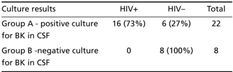
Of the total group, 30 (0.23%) fulfilled the estab-lished criteria for the diagnosis of tuberculous menin-gitis, of which 22 (73%) displayed a positive culture for Mycobacterium tuberculosis (group A) (Table 1). The remaining eight patients (group B), who had dis-played a negative culture, turned out positive for BK in the PCR test. The direct exam (BAAR) was nega-tive for all patients. There was no difference between the average and standard deviation of the age dis-tribution between both groups: 47±15 years of age Table 1. Correlation between HIV infection and positive BK culture in CSF of 30 patients with tuberculous meningitis.
Culture results HIV+ HIV– Total
Group A - positive culture for BK in CSF
16 (73%) 6 (27%) 22
Group B -negative culture for BK in CSF
0 8 (100%) 8
Table 2. CSF findings in 30 patients with tuberculous meningitis, according the bacteriological positive (Pos)/neg-ative (Neg) culture results.
Culture n Median Minim Maximum p value
Total leucocytes/mm3 Pos 22 132 1 1070 0.17
Neg 8 65,5 2 960
Neutrophils (%) Pos 22 72 0 93 0.0007
Neg 8 5,5 0 33
Lymphocytes (%) Pos 22 17 4 90 0.004
Neg 8 69 47 89
Monocytes (%) Pos 22 7 1 15 0.0006
Neg 8 12 10 35
Eosinophils (%) Pos 22 0 0 1 0.066
Neg 8 0,5 0 3
Macrophages (%) Pos 22 0 0 5 0.25
Neg 8 1 0 12
Protein (mg/dL) Pos 22 150 24 840 0.014
Neg 8 63,5 40 200
Glucose (mg/dL) Pos 22 32 5 136 0.17
for group A and 42±27 years of age for group B (p>0.05). Whereas in group A, 16 patients (73%) showed positive serology for HIV, in group B, 100% were seronegative (Table 1). All the patients with positive serology for HIV showed a positive BK cul-ture.
Of the total group, two patients presented nor-mal cytological and biochemical CSF exams: one patient from group B (negative culture/positive PCR) had negative serology for HIV and, the other from group A (positive culture), presented positive serol-ogy for HIV. Most of the patients (28/30) presented neutrophils in the CSF. For the patients in group A, the median proportion of neutrophils in the CSF was 72% (0-93) and for group B, 5.5% (0-33) (p>0.05) (Table 2). The number of leukocytes and the percent-age of monocytes in group A were significantly low-er than those in group B (Table 2).
In relation to the comparison of the findings of the CSF in patients with or without HIV infection, there was no statistical difference in the cytological and biochemical values between both groups (Table 3). Of the total group, around half of the patients with positive serology (9/16) and negative serology (5/14) for HIV showed a proportion of neutrophils in the CSF above 50% in the differential cell count. However, if we consider the six patients who had neg-ative serology for HIV and positive culture for BK in the CSF, five of these patients had more than 50% neutrophils in the CSF.
DISCUSSION
TB meningitis is caused by the Mycobacterium tu-berculosis, an acid-fast microorganism, and, in rare cases, by Mycobacterium bovis. The disease is a result of the bacterial dissemination originating in anoth-er location in the body4. According the Rich’s
descrip-tion, in the initial phase of the disease the formation of meningeal tubercules which are secondary to the spread of the agent in the meninges and in the sub-pial regions of the brain. The bacterial is released in the subarachnoid space right after the break of one or more of these tubercules4. There is also a
hypoth-esis that miliary bacterial dissemination occurs with a direct invasion of the bacillus in the choroid plexus or in the meningeal ducts4.
The clinical manifestations set in slowly in the course of three weeks. Signs of meningeal irritation (stiffness of the neck, Kernig and Brudzinski signs), headache, low fever, anorexia and irritability appear already in the first week. After this initial period, signs of localization and high intracranial pressure appear, followed by paralysis of the III, VI and VII cra-nial nerves and seizures. After this, the patient evolves into a coma state with high fever. If the treatment is not initiated promptly, death occurs after five to eight weeks counting from the beginning of the symp-toms1,4.
An exam of the CSF in TB meningitis evidences an increase in pressure, a slightly turbid aspect with clot formation, pleocytosis of 10–500 leucocytes/mm3,
Table 3. CSF findings according the presence or absence of HIV infection.
HIV n Median Minim Maxim p value
Total leucocytes/mm3 Pos 16 132 1 1070 0.61
Neg 14 106.5 2 960
Neutrophils (%) Pos 16 69.5 0 93 0.12
Neg 14 27.5 0 86
Lymphocytes (%) Pos 16 22.5 6 90 0.26
Neg 14 51 4 89
Monocytes (%) Pos 16 7.5 1 15 0.18
Neg 14 10.5 4 35
Eosinophils (%) Pos 16 0 0 1 0.1
Neg 14 0 0 3
Macrophages (%) Pos 16 0 0 5 0.075
Neg 14 1 0 12
Protein (mg/dL) Pos 16 147 24 840 0.58
Neg 14 115 40 580
Glucose (mg/dL) Pos 16 33.5 5 136 0.67
with a predominance of lymphocytes. The CSF pro-tein concentration is higher than normal (100-500 mg/dL) and increases even more if the flow of the CSF is obstructed7,8. Glucose levels are reduced, with
levels below 40 mg/dL and the concentration of lac-tate is increased (>19 mg/dL). The laclac-tate is not spe-cific for tuberculous meningitis but is a good mark-er of cmark-erebral metabolism in bactmark-erial and fungal me-ningitis. It has no influence of blood concentration, as occurs with the glucose, and is a product of glu-cose metabolism in CSF9. Atypical findings in the CSF
such as a concentration of neutrophils and normal exam results may contribute to making the diagno-sis more difficult7. This is exacerbated due to the low
sensitivity of the direct exam10.
A concentration of neutrophils in the CSF or a nor-mal biochemistry analysis occur in the initial and acute phases of the infection. In a previous study7, a
normal cytological exam of the CSF (<5 cells/mm3with
an absence of neutrophils) was seen in 21.4%, a pre-domination of neutrophils in 39% and, 5.4% had a positive bacilloscopy. In our study, 6% of the patients had a CSF exam with normal cytology and biochem-istry: one case was seropositive for HIV and, had pos-itive BK culture in the CSF and, the other had nega-tive serology for HIV and neganega-tive culture/posinega-tive PCR test for BK. On the other hand, in 94% of the cases, we found neutrophils in the CSF. The highest proportion of neutrophils was predominant in the CSF of patients with a positive culture for BK. There was no difference in the neutrophils count, as in oth-er parametoth-ers of the CSF, except for the positivity of the culture, between patients with positive serology for HIV and those with negative serology for HIV. All cases with HIV infection had positive BK culture in CSF. From the seronegative HIV patients, six had pos-itive CSF culture and, eight were negative. This find-ing showed that, in this study, the highest neutrophils count was associated to the positivity of the BK cul-ture in the CSF, regardless of the presence or absence of infection by HIV. On the other hand, the differen-tiating factor between the patients with positive and negative BK cultures in the CSF was in the predomi-nance in the total number of neutrophils in the CSF, not merely in its presence. It was suggested that pleo-cytosis with a predominance of neutrophils in the CSF could be associated to the increased probability of positivity in the bacteriological diagnosis10.
The Mycobacterium tuberculosis may oftentimes lead to persistent neutrophilic meningitis in patients that are HIV positive. This is characterized by the ini-tial and persistent presence (with a minimum
inter-val of seven days) of neutrophilic pleocytosis (50%), high spinal fluid protein concentration (>40 mg/dL) and low spinal fluid glucose concentration (<2/3 gly-cemia). Hypothetically, in the case of HIV infection, the fundamental immunocellular response to con-trol tuberculosis is not at full capacity and this could allow for the release of chemotaxic factors for neu-trophils and a persistence of the response in the acute phase11. The type of cell response can have a
prog-nostic effect. A higher percentage of neutrophils was associated to higher survival rates12. It has been
sug-gested that neutrophils may have a protective role against Mycobacterium tuberculosis. As ours is a ret-rospective study, it was not possible to evaluate the survival rates of our patients. The slow course of TB meningitis in patients infected with HIV may also be associated to the use of other drugs that interfere with the levels of tuberculostatic agents in circula-tion or the presence of strains that are resistant to the medication11.
The direct exam, using the Ziehl-Neelsen stain (BAAR), should be conducted, despite the fact that the CSF is paucibacillary and that there is low levels of positivity in the test. It is simple to execute and allows for an immediate confirmation of the diagno-sis. Most researchers mention a sensitivity range that
varies between 10% and 40%10,12. However, when
larger volumes of CSF (>5 mL) and/or serial samples are analyzed, the positivity may reach 87%6,13-15. On
the other hand, the sensitivity of the direct exam drops from 52% to 2% after approximately 5-15 days from the beginning of treatment10. In our caseload,
the patients showed negativity in the direct exam, which was probably related to the small volume of the sample that was analyzed and could also be relat-ed to the detailrelat-ed exam above-mentionrelat-ed was not performed.
The isolation of mycobacterias in the Lowenstein-Jensen culture in the CSF represents the gold stan-dard for the diagnosis of TB meningitis. Nevertheless, due to the slow growth (30 to 60 days) this exam is useful only from an epidemiological point of view, not a clinical one. The radiometric method (BACTEC) detects the growth of mycobacteria by measuring the levels of 14CO2released. This method is more sensitive and the bacterial growth can be verified in approximately 14 days8.
The PCR test has variable sensitivity and specifici-ty. In some studies, the sensitivity is low (56-70%), while specificity is high (98-100%)10,12. It is
bacilloscopy of the samples come out negative, as it contributes to the early diagnosis and swift applica-tion of the appropriate treatment.
Previous reports of patients with tuberculous meningitis did not show a significant change in the cytological and biochemical parameters in terms of co-infection with HIV2,3. However, differences have
been reported in bacteriological tests of patients co-infected with HIV in relation to those not co-infected. In the case study by Dubè et al.2, despite not having
statistical value, the direct exam for BK was positive in 27% of patients co-infected and only 6% of those not co-infected with HIV2. Some authors suggest that
the patients co-infected with HIV present more intense bacterial proliferation and greater concen-trations of bacilli in the meninges than do patients that are not co-infected10,16. In our caseload, we
observed a direct correlation between positivity for
Mycobacterium tuberculosis in the bacterial culture and co-infection with HIV.
Chandramuki et al., certified that anti-M. tuber-culosis antibodies were found more frequently in patients clinically diagnosed with a negative culture when compared to those who tested positive to the culture14. Based on these data, the authors
suggest-ed that there might be in inverse relation between the presence of antibodies in the CSF and an increase in the bacillary load for BK. In this same case study, patients who were co-infected with HIV showed a weaker antibody response in the CSF than those who were not co-infected14.
Twaites et al., made a list of some of the factors involved in a greater accuracy of the bacteriological confirmation of the CSF for TB meningitis: at least 6 mL of CSF, direct exam of the smear for at least 30 minutes, the presence of neutrophils, and an increase in lactate in the CSF6.
The adenosine deaminase (ADA) activity in the CSF may be used as a screening test. It is increased in the CSF of TB meningitis. But it is not specific because may be found in high levels in other bacterial menin-gitis, lymphoma, neurobrucellosis and cryptococal meningitis17.
The changes in the CSF respond slowly to treat-ment for TB meningitis (6 to 12 months). Twaites et al, studied pro- and anti-inflammatory cytokines in serial samples of CSF and peripheral blood of 21 patients diagnosed with TB meningitis13. They found
that high concentrations of lactate, o interleukin-8 and gamma-interferon steadily decreased in amount after the beginning of the specific treatment. On the
other hand, the dysfunction of the blood-CSF barri-er and immunoactivation remained constant aftbarri-er 60 days of treatment. Pleocytosis (>5 cells/mm3) was
found in 62% of patients after nine months of treat-ment and high spinal fluid protein concentration (>40 mg/dL) was found in 38% of patients, while the decreased ratio between CSF glucose and that of the blood was found in 6% of cases. The levels of TNF-alpha were persistently high in the CSF in TB menin-gitis, which could represent the primary biological factor responsible for maintaining the inflammato-ry process. The high initial concentration (during the first seven days of treatment) of lactate in the CSF, the low leukocyte count and the glucose concentra-tion were all associated to a higher death rate. Lac-tate production is secondary to the phenomenon of hypoxia, which results from obliterative vasculitis and ischemic infarction which occur in TB meningitis. The high concentration of lactate may represent a good prognostic marker for this disease, due to the evi-dence that it has a correlation with the seriousness of the process.
In the present study, we found that the factors associated to positivity in the CSF culture in TB menin-gitis consisted of: the presence of co-infection with HIV, high spinal fluid protein concentration and larg-er proportions of spinal fluid neutrophil in the CSF (markers of the acute phase), probably associated to a higher bacteremia. According to the literature, oth-er factors associated to a greatoth-er positivity in the bac-teriological diagnosis include a greater volume of CSF and a longer microscope analysis time. It is impor-tant to emphasize that the CSF exam in TB meningi-tis can present, in a small minority of cases, atypical findings such as normal cellular or biochemical results or persistent spinal fluid neutrophil concentrations. These parameters should not be used as exclusion criteria during diagnosis. On the other hand, the inflammatory alterations may persist for a period greater than nine months after the beginning of the treatment for complete normalization10. This study
reinforces the need for exams with greater sensitiv-ity and specificsensitiv-ity and that can allow for an early di-agnosis of TB meningitis, thereby avoiding the delay and lack of diagnosis and reducing lethal outcomes.
Acknowledgment - The authors thank Mrs Rosangela Noe from CIC/HUCFF/UFRJ, Rio de Janeiro, Brazil, for the statistical analysis.
REFERENCES
2. Dube MP, Holtom PD, Larsen RA. Tuberculous meningitis in patients with and without human immunodeficiency virus infection. Am J Med 1992;93:520-524.
3. Schutte CM. Clinical, cerebrospinal fluid and pathological findings and outcomes in HIV-positive and HIV-negative patients with tuberculous meningitis. Infection 2001;29:213-217.
4. Adams RD, Victor M. Neurologia. 5.Ed. México: Nueva Editorial Interamericana 1996.
5. Lan S-H, Chang. W, Lu C, Lui C-C, Chang H-W. Cerebral infarction in chronic meningitis: a comparison of tuberculous meningitis and cryp-tococcal meningitis. Q J Med 2001;94:247-253.
6. Twaites GE, Chau TTH, Farrar JJ. Improving the bacteriological diag-nosis of tuberculous meningitis. J Clin Microbiol 2004;42:378-379. 7. Karstaedt AS, Valtchanova S, Barriere R, Crewe-Brrown HH.
Tubercu-lous meningitis in South African urban adults. Q J Med 1998;91: 743-747.
8. Heringer RR, Fernandes LE, Goncalves RR, Puccioni-Sohler M. Location of the lesion and the cerebrospinal fluid findings in tuberculous menin-gitis: differences in the lumbar, cisternal and ventricular compartiments. Arq Neuropsiquiatr. 2005;63:543-547.
9. Cabeça HLS, Gomes HR, Machado LR, Livramento JA. Dosage of lac-tate in the cerebrospinal fluid in infectious diseases of the central nerv-ous system. Arq Neuropsiquiatr 2001;59:843-848.
10. Twaites GE, Hien TT. Tuberculous meningitis: many questions, too few answers. Lancet Neurol 2005:4:160-170.
11. Marinho SF, Paciullo VHA, Fonseca MO, et al. Meningite neutrofilica persistente em paciente com síndrome de imunodeficência adquirida. Soc Bras Méd Trop 1997;30:241-245.
12. Machado LR, Livramento JA, Bydlowski SP, Bendit I, Bravo LM, Spina-França A. Polymerase chain reaction in the diagnosis of tuberculous meningitis. Arq Neuropsiquiatr 1994;52:445-446.
13. Twaites GE, Simmons CP, Guyrn NTH, et al. Pathophysiology and prognosis in Vietnamese adults with tuberculous meningitis. J Inf Dis 2003;188:1105-1015.
14. Chandramuki A, Lyashchenko K, Kumari HBV, et al. Detection of anti-body to Mycobacterium tuberculosis protein antigens in the cere-brospinal fluid of patients with tuberculous meningitis. J Infect Dis 2002;186:678-683.
15. Fallon RJ, Kennedy DH. Rapid diagnosis of tuberculous meningitis. J Infect 1993;26:226.
16. Katrak SM, Shembalkar PK, Bijwe SR, Bhandarkar. The clinical, radi-ological and pathradi-ological profile of tuberculous meningitis in patients with and without human immunodeficiency vírus infection. J Neurol Sci 2000;181:118-126.