DOI: 10.1590/0004-282X20150001
ARTICLE
Evaluation of the efficacy of sodium valproate
in convulsive status epilepticus following to
ıschemic stroke
Avaliação da eicácia do valproato de sódio no status epilepticus convulsivo devido ao
acidente vascular cerebral isquêmico
Hasan Hüseyin Özdemir1, Bülent Müngen2, Selçuk İlhan3
Stroke has long been recognised as a common cause of epileptic seizures, and indeed is the most common cause of seizures in adults aged ≥ 601. Previous studies have shown that the frequency of seizures in stroke ranges between 2.3% and 19%2,3,4,5.he outcome for patients having early-onset sei-zures is poor, with a high in-hospital mortality rate and a high incidence of status epilepticus (SE)6. herefore, the role of
stroke as an aetiological factor in SE is accepted2. SE is pri-marily observed in patients with a severe stroke. It can be a presenting symptom of acute stroke, and almost half of adult
SE cases are caused by combined acute and remote symp-tomatic cerebrovascular disorders1. SE occurs in 20.7% of pa-tients at stroke onset, in 11.7% of papa-tients at late-onset and at very late-onset in only 2.1% of patients7. In addition, stroke
patients sufering from SE have worse outcomes than pa-tients who do not experience seizures, and mortality rates in stroke patients with SE can also be higher3,4.
here are several challenges when treating stroke patients with SE, including increased mortality, concomitant diseas-es, lack of evidence supporting the use of various treatment
1Dicle University Neurology, Diyarbakır, Turkey;
2Fırat University Neurology, Elazığ, Turkey;
3Fırat University Pharmacology Department, Elazığ, Turkey.
Correspondence: Hasan Hüseyin Özdemir; Dicle University Neurology; 21280 Diyarbakır, Turkey; E-mail: [email protected]
Conlict of interest: There is no conlict of interest to declare.
Received 26 May 2014; Received in inal form 11 November 2014; Accepted 01 December 2014.
ABSTRACT
Objective: Convulsive status epilepticus (CSE) is very rarely observed after ischaemic stroke. Sodium valproate (SV) is one of the agents used in the treatment of CSE, but its role still controversial, and its degree of eficacy in treating CSE that develops following stroke is unclear.
Method: We evaluated 19 patients who were treated with intravenous (IV) SV (20 mg/kg, 2 mg/kg/h-12h) after diazepam. Patients’ modiied Rankin scores (mRS), SE types, and changes in biochemical parameters after treatment were assessed. Results: CSE was successfully treated in 12 (63.15%) patients. Side effects such as hypotension and allergic reactions were observed in two patients. Refractory SE devel-opment was observed in 5 (29.4%) patients with high mRS (> 3). No signiicant deterioration in patients’ laboratory evaluations, conducted before and after status, was observed. Conclusion: SV may be safe and effective in the treatment of CSE observed after ischaemic stroke, especially in patients with low mRS.
Keywords: convulsive status epilepticus, ischaemic stroke, sodium valproate.
RESUMO
Objetivo: Status epilepticus convulsivo (SEC) é muito raramente observado após acidente vascular cerebral isquêmico. Valproato de sódio (VS) é um dos agentes utilizados no tratamento do SEC, mas seu papel ainda é controverso e seu grau de eicácia não é claro no SEC pós acidente vascular. Método: Avaliamos 19 pacientes que foram tratados com AV endovenoso (EV) (20 mg/kg, 2 mg/kg/h-12h) após diaz-epam. Valores da escala modiicada de Rankin (mRS) dos pacientes, tipos de SE e mudanças nos parâmetros bioquímicos foram avaliados.
Resultados: SEC foi tratado com sucesso em 12 pacientes (63,15%). Efeitos colaterais como hipotensão e reações alérgicas foram obser-vados em dois pacientes. Desenvolvimento de SE refratário foi observado em cinco pacientes (29,4%) com altos valores de mRS (> 3). Não houve deterioração signiicativa nas avaliações laboratoriais dos pacientes feitas antes ou depois do status. Conclusão: AV pode ser eicaz no tratamento do SEC observado após acidente vascular cerebral isquêmico, especialmente nos pacientes com baixo mRS.
options and a high rate of adverse efects. herefore, treat-ment of epileptic seizures and SE is very important in pa-tients who have sufered strokes, and such treatment should be rapid-acting and with few side efects.
Convulsive status epilepticus (CSE) is the most common and life-threatening form of SE6. It is observed in 0.2% of
pa-tients after ischaemic stroke, and occurs more often than non-convulsive SE4,8. Several studies have shown that intravenous (IV) sodium valproate (SV) is efective in the treatment of CSE, although the correct dose to administer when treating repeti-tive seizures remains controversial9,10. Previous studies that
have evaluated the eicacy of SV in treating SE have described various causes of SE; there is no clear evidence regarding the SE that develops during the acute period after ischaemic stroke. Moreover, the eicacy of SV in the treatment of CSE that de-velops after stroke has not been fully elucidated. herefore, in this study, we evaluated the eicacy of SV in treating CSE that develops in the acute period following stroke.
METHOD
he study population was derived from the SE Data Bank of Fırat University Hospital, Turkey, and this included all SE patients (n = 151) admitted to the hospital from January 2007 to October 2013. his hospital-based data bank included in-formation regarding the duration and type of SE, underlying factors, such as structural deicits or medical pathologies, and demographic data. We included only patients who had experienced ischaemic stroke, and excluded patients with a history of seizure, pseudoseizures, nonconvulsive SE, sub-arachnoid haemorrhage, head injury, hypertensive and met-abolic encephalopathy, diabetic ketoacidosis, acute/chronic renal and liver failure, malignant hypertension, arteritis, fever and cerebrovenous thrombosis. As a result, we included only 19 of the 151 identiied patients.
All patients underwent a CT scan at admission, a second scan after 24-72 h and further scans when necessary. All pa-tients with ischaemic stroke were investigated using Doppler carotid ultrasonography, transthoracic echocardiography, and 24-hour holter electrocardiography if a cardioembolic stroke was suspected. In all patients, liver function and whole blood tests, plasma urea, creatinine, electrolyte and plasma glucose levels were measured on admission and after 48 h.
An experienced neurologist evaluated neurological dei-cits on admission. he degree of functional disability was graded using a modiied Rankin scale (mRS) before SE, but only one patient’s mRS was graded after SV treatment11. In addition, patients were again evaluated during hospital dis-charge (outcome).
he diagnosis was based on direct observation of sei-zures by the medical staf at the time of hospitalisation, the patient’s history provided by his or her neurologist, reliable
descriptions obtained from ambulance personnel when sei-zures occurred during transportation, or on observations of close family members when seizures occurred at home.
According to the 1981 classiication criteria, CSE was divid-ed into generalizdivid-ed convulsive status epilepticus (GCSE), com-plex partial secondary-generalised convulsive status epilepticus (CPS-GCSE), and tonic status epilepticus (TSE)12. In addition, pa-tients with refractory status epilepticus (RSE) were identiied12.
Diazepam (DZP) was given at a rate of 10 µg/kg/min and was increased by 10 µg/kg/min every 5 min until SE control or a maximum dose of 100 µg/kg/min was reached. After DZP treatment, 20 mg/kg in 100 ml of IV saline-infused SV was administered over 15 min, following which, a load-ing dose/continuous infusion was administered at a rate of 2 mg/kg/h as the maintenance dose for 12 h. After discontin-uation of IV infusion, a maintenance IV dose of 400 mg every 8 h was continued. Refractory status epilepticus (RSE) was accepted as a diagnosis for continuous seizures not respon-sive to DZP and SV.
Adverse local efects (burning, pain and phlebitis at injec-tion site) and systemic efects (low blood pressure, heart rate disorder and respiratory suppression) were also monitored. All patients also received general medical management, in-cluding oxygen, cooling, electrolyte balancing, prevention of complications and other symptom-relief treatment, such as mannitol, acetyl salicylic acid, and clopidogrel. None of the patients were administered t-PA.
EEG recording devices were not appropriate in some circumstances so EEG was performed 6 to 48 hours. All pa-tient’s therapy was started clinically. An EEG was carried out at the end of seizure for 15 patients, during seizure for two patients, and two patients did not undergo an EEG. EEGs were considered abnormal when burst-suppression pattern, focal, lateralised or generalised slowing or epileptiform dis-charges were present.
he stopping of SE table in patients was conirmed by the clinical cessation of seizure activity. In addition, in order to electrographically evaluate nonconvulsive SE, EEG was car-ried out using a device in the intensive care unit.
Statistical analysis
Analysis of data was carried out using SPSS 21.0. For con-tinuous variables such as age and time of start of SE, descrip-tive statistics were calculated and reported as mean ± SD. he paired t-test was used to compare the values of means.
RESULTS
We found that seven patients had GCSE, 10 had CPS-GCSE and two had TSE. he mean age of the patients was 71.05 ± 11.0 (age: 58-90 years). Infarct was observed in the entire hemisphere to A. carotis interna stenose in two pa-tients, at the A. cerebri media (MCA) region in 13 patients, and in the temporal regions in four patients (Table 1).
We observed an allergic reaction during the SV infusion in one patient, and a signiicant decrease in blood pressure in a dif-ferent patient. he infusion was discontinued in these two pa-tients, and they were excluded from the study. Of the patients who completed the study, 10 were female and seven were male.
We found that SV treatment halted SE in 63.15% (12) of the patients. Following the development of ischaemic stroke, the onset of SE was 33.55 ± 26.75 hours. he average of mRS before SE was 3.21 ± 1.22, and outcome mRS was 3.23 ± 1.09. he aver-age time it took for SV to control SE was 17.91 ± 4.98 min.
RSE developed in ive of the patients, and two of them died. Of the ive patients in whom RSE was detected, the Rankin score was 5 in four of the patients and 4 in one pa-tient. Infarct was observed in the left hemisphere of four of ive patients in whom RSE developed, and in the right hemi-sphere of one patient.
EEG indings revealed periodic lateralising epileptiform abnormalities in six patients, focal slowing in ive patients, generalised slowing in four patients and a burst-suppression pattern in two patients.
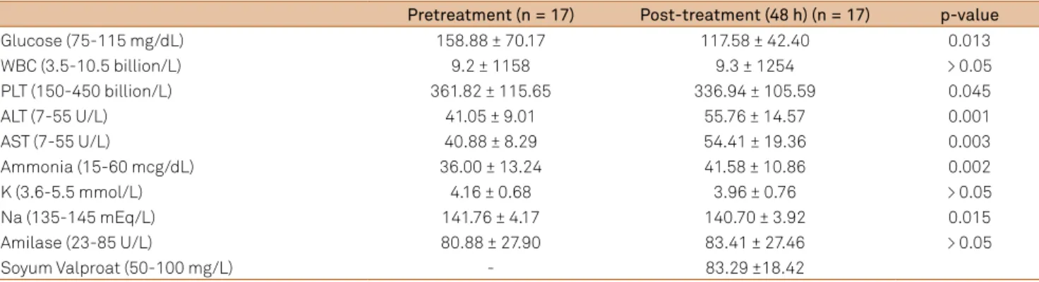
We observed an increase in alanine aminotransferase, aspartate aminotransferase and ammonia values, a decrease in glucose, platelets and sodium values and no statistically signiicant diferences in patients’ white blood cell and amy-lase levels. However, we did not observe deterioration in liv-er and kidney function tests, thrombocytopenia, and hypo-natremia (Table 2).
DISCUSSION
In the present study, SV treatment halted SE in 12 (63.15%) patients. Some previous studies have shown that there is no relationship between the occurrence of SE and gender, age, stroke risk factors, stroke type (ischaemic or haemorrhagic), stroke topography and stroke cause, cortical involvement or size of lesion in patients with stroke14. However, damage to the cerebral cortex is widely considered to be a sine qua non
for the development of epilepsy, and indeed epilepsy is rare in
Table 1. Demographic characteristics of patients.
Patient-Age/ Gender Lesion Before treatment mRS/ Outcome mRS
Risk factors
Status epilepticus starting time (after ischemia)
Type of status epilepticus
Time to seizure controlled (min)
1-85F Left MCA infarction 2/3 DM, HT 48 h CPS-GCSE 15
2-60F Right temporal lobe
infarction
2/2 AF 36 h CPS-GCSE 15
3-58M Left MCA infarction 4/4 AF 72 h GCSE-RSE 30
4-58F Right MCA infarction 3/3 AF 6 h CPS-GCSE 20
5-83M Right MCA infarction 3/4 DM 12 h CPS-GCSE 20
6-75M Right MCA infarction 3/3 DM, HT 96 h TSE 15
7-63F Left temporal lobe
infarction
2/2 AF 24 h CPS-GCSE 15
8-60F Right temporal lobe
infarction
2/2 AF 10 h CPS-GCSE 20
9-75F Right MCA infarction 2/2 DM, HT 72 h CPS-GCSE 20
10-62F Right temporal lobe
infarction
2/2 AF 36 h CPS-GCSE 10
11-73F Right MCA infarction 3/3 DM, HT 6 h TSE 15
12-58M Left MCA infarction 3/3 DM, AF 48 h CPS-GCSE 20
13-81F# Left MCA infarction 5/5 DM, HT 10 h GCSE-RSE 35
14-62F# Right MCA infarction 5/5 AF 36 h CPS-GCSE 30
15-82M# Left MCA infarction 5/5 DM, HT 36 h GCSE-RSE 50
16-90M# Right hemisphere
infarction
5*/ - DM, HT 30 min GCSE-RSE
(ex)
-17-85M# Left hemisphere
infarction
5/ - DM,HT 1 h GCSE-RSE
(ex)
-18-75 F Right MCA infarction
(allergic reactions)
2/3 HT 48 h GCSE 25
19-65 F Right MCA infarction
(hypotension)
3/4 DM 40 h GCSE 30
strokes that occur in the posterior fossa or deep white mat-ter14. Several studies have shown that cortical involvement is a risk factor independent of infarct size; in other studies it was associated with large infarcts15,16. In our study, we
ob-served infarct in an entire hemisphere in two patients, in the region corresponding to the irrigation area of MCA in 13 pa-tients, and in the temporal regions in four patients. Our ind-ings may indicate that a large infarct area, including the tem-poral region, may be important for the development of SE. Likewise, infarct was shown in the left hemisphere in four of ive patients in whom RSE developed, and in the right hemi-sphere in one patient. We more frequently observed RSE in patients with left hemisphere lesions. However, we cannot speculate on the possible causes of this, since many factors contribute to the development of RSE.
Rankin scale is very important in the assessment of dis-ability in patients with stroke11. Our patients’ outcome aver-age mRS had increased in a similar manner to that reported in previous studies3,4. In addition, we found that the patients with high mRS did not adequately respond to IV SV treat-ment, and the development of RSE occurred more frequently in these patients; those patients with low mRS responded far better to this treatment.
RSE occurs in up to 44% of all patients with SE17.he
mor-tality of RSE is at least 16%-23%, and again depends on age and aetiology18. It is treated with coma induction using an-aesthetics such as propofol or barbiturates. RSE developed in ive of our patients, two of whom died.
he National Institutes of Health warn patients tak-ing SV about the risk of serious or life-threatentak-ing damage to the liver and pancreas that is associated with the use of this drug19. Case presentations have shown that SV causes hypotension and allergies, but that it is a safe and efec-tive drug, despite these side efects10,20,21. In our study, two
patients developed hypotension and hypersensitivity reac-tions. However, many complications developed in SE, and
changes in metabolic parameters were also monitored dur-ing the course of stroke. Although changes were statistically observed in some parameters in the assessment carried out 48 h after treatment, thrombocytopenia and hyponatremia with liver dysfunction and liver failure did not develop in any of the patients in our study.
Phenytoin is recommended as the irst-line antiepilep-tic drug for SE, and its metabolism may be afected by other drugs, which increase phenytoin blood levels and toxicity22. Tiamkao et al. showed that the time to seizure control and number of non-dependent patients was increased, and du-ration of hospitalisation and number of deaths was reduced, with SV treatment compared to phenytoin treatment. In our study, the average time at which SV halted SE was similar to that observed in their study. In addition, these authors re-ported that SV is non-inferior to intravenous phenytoin as the irst-line treatment in SE, with no signiicant cardiovas-cular compromises, and that SV may be used in elderly pa-tients with cardiovascular risks or hepatitis10. Moreover,
hav-ing little interaction with other drugs and with linear kinetics, SV has an advantage over phenytoin in this respect. SV may be also similarly be more efective and safe than phenytoin in the elderly with SE.
Our study had some limitations. It was a retrospective study, and the number of patients used was small. In addi-tion, EEG was not performed at the start of seizures, meaning that SE was evaluated more clinically than electrographically.
Development of SE after ischaemic stroke is a very rare condition that may be associated with numerous factors. According to our observations, SV may be efective in the treat-ment of CSE that develops during the acute period after isch-aemic stroke, especially in patients with a low mRS. However, strict monitoring should be conducted with regard to hyper-sensitivity and hypotension. In addition, we observed that SV was less efective in the treatment of RSE, but that RSE was more frequently identiied in patients with a high mRS.
Table 2. Changes in biochemical parameters.
Pretreatment (n = 17) Post-treatment (48 h) (n = 17) p-value
Glucose (75-115 mg/dL) 158.88 ± 70.17 117.58 ± 42.40 0.013
WBC (3.5-10.5 billion/L) 9.2 ± 1158 9.3 ± 1254 > 0.05
PLT (150-450 billion/L) 361.82 ± 115.65 336.94 ± 105.59 0.045
ALT (7-55 U/L) 41.05 ± 9.01 55.76 ± 14.57 0.001
AST (7-55 U/L) 40.88 ± 8.29 54.41 ± 19.36 0.003
Ammonia (15-60 mcg/dL) 36.00 ± 13.24 41.58 ± 10.86 0.002
K (3.6-5.5 mmol/L) 4.16 ± 0.68 3.96 ± 0.76 > 0.05
Na (135-145 mEq/L) 141.76 ± 4.17 140.70 ± 3.92 0.015
Amilase (23-85 U/L) 80.88 ± 27.90 83.41 ± 27.46 > 0.05
Soyum Valproat (50-100 mg/L) - 83.29 ±18.42
References
1. DeLorenzo RJ, Hauser WA, Towne AR, Boggs JG, Pellock JM, Penberthy L, et al. A prospective, population-based epidemiologic study of status epilepticus in Richmond, Virginia. Neurology. 1996;46(4):1029-35. http://dx.doi.org/10.1212/WNL.46.4.1029
2. Lowenstein DH, Alldredge BK. Status epilepticus at an urban public hospital in the 1980s. Neurology. 1993;43(3 Pt 1):483-8. http://dx.doi.org/10.1212/WNL.43.3_Part_1.483
3. De Reuck j, Claeys I, Martens S, Vanwalleghem P, Van Maele G, Phlypo R, et al. Computed tomographic changes of the brain and clinical outcome of patients with seizures and epilepsy after an ischaemic hemispheric stroke. Eur J Neurol. 2006;13(4):402-7. http://dx.doi.org/10.1111/j.1468-1331.2006.01253.x
4. Rumbach L, Sablot, D, Berger E, Tatu L, Vuillier F, Moulin T. Status epilepticus in stroke: report on a hospital-based stroke cohort. Neurology. 2000;54(2):350-4. http://dx.doi.org/10.1212/WNL.54.2.350
5. Menon B, Shorvon SD. Ischaemic stroke in adults and epilepsy. Epilepsy Res. 2009;87(1):1-11.http://dx.doi.org/10.1016/j.eplepsyres.2009.08.007
6. De Reuck J, Van Maele G. Status epilepticus in stroke patients. Eur Neurol. 2009b:62(3):171-5. http://dx.doi.org/10.1159/000227289
7. De Reuck J. Management of stroke-related seizures. Acta Neurol Belg. 2009;109(4):271-6.
8. Bateman BT, Claassen J, Willey JZ, Hirsch LJ, Mayer SA, Sacco RL et al. Convulsive status epilepticus after ischemic stroke and intracerebral hemorrhage: frequency, predictors, and impact on outcome in a large administrative dataset. Neurocrit Care. 2007;7(3):187-93. http://dx.doi.org/10.1007/s12028-007-0056-2
9. Misra UK, Kalita J, Patel R. Sodium valproate vs phenytoin in status epilepticus: a pilot study. Neurology. 2006;67(2):340–2. http://dx.doi.org/10.1212/01.wnl.0000224880.35053.26
10. Tiamkao S, Sawanyawisuth K, Chancharoen A. The eficacy of intravenous sodium valproate and phenytoin as the irst-line treatment in status epilepticus: a comparison study. BMC Neurol. 2013;27;13:98. http://dx.doi.org/10.1186/1471-2377-13-98
11. Wolfe CD, Taub NA, Woodrow EJ, Burney PG. Assessment of scales of disability and handicap for stroke patients. Stroke. 1991;22(10):1242-4. http://dx.doi.org/10.1161/01.STR.22.10.1242
12. Commission on Classiication and Terminology of the International League Against Epilepsy. Proposal for revised clinical and electro-encephalographic classiication of epileptic seizures. Epilepsia. 1981;22(4):489-501. http://dx.doi.org/10.1111/j.1528-1157.1981.tb06159.x
13. Kolominsky-Rabas PL, Weber M, Gefeller O, Neundoerfer B, Heuschmann PU. Epidemiology of ischemic stroke subtypes according to TOAST criteria. Stroke. 2001;32:2735-40. http://dx.doi.org/10.1161/hs1201.100209
14. Velioğlu SK, Özmenoğlu M, Boz C, Alioğlu Z. Status epilepticus after stroke. Stroke. 2001;32(5):1169-72. http://dx.doi.org/10.1161/01.STR.32.5.1169
15. Bladin CF, Alexandrov AV, Bellavance A, Bornstein N, Chambers B, et al. Seizures after stroke: a prospective multicenter study. Arch Neurol. 2000;57(11):1617-22. http://dx.doi.org/10.1001/archneur.57.11.1617
16. Kilpatrick CJ, Davis SM, Tress BM, Rossiter SC, Hopper JL, Vandendriesen ML. Epileptic seizures in acute stroke. Arch Neurol. 1990;47(2):157-60. http://dx.doi.org/10.1001/archneur.1990.00530020053014
17. Yaffe K, Lowenstein DH. Prognostic factors of pentobarbital therapy for refractory generalized status epilepticus. Neurology. 1993;43(5):895-900. http://dx.doi.org/10.1212/WNL.43.5.895
18. Holtkamp M, Othman J, Buchheim K, Meierkord H. Predictors and prognosis of refractory status epilepticus treated in a neurological intensive care unit. J Neurol Neurosurg Psychiatry. 2005;76(4):534-9. http://dx.doi.org/10.1136/jnnp.2004.041947
19. Nanau RM, Neuman MG. Adverse drug reactions induced by valproic acid. Clin Biochem. 2013;46(15):1323-38. http://dx.doi.org/10.1016/j.clinbiochem.2013.06.012
20. Bota RG, Ligasan AP, Najdowski TG, Novac A. Acute hypersensitivity syndrome caused by valproic Acid: a review of the literature and a case report. Perm J 2011;15(2):80-4.
21. Ota KS. Probable valproate sodium-associated
hypotension. Am J Geriatr Pharmacother. 2010;8(3):281-4. http://dx.doi.org/10.1016/j.amjopharm.2010.04.005