DOI: 10.1590/0004-282X20130080
ARTICLE
Walking execution is not affected by divided
attention in patients with multiple sclerosis
with no disability, but there is a motor
planning impairment
A execução da marcha não é afetada pela atenção dividida em pacientes com esclerose
múltipla sem incapacidade, mas existe um comprometimento do planejamento motor
Leandro Alberto Calazans Nogueira1,2, Luciano Teixeira dos Santos3, Pollyane Galinari Sabino1,
Regina Maria Papais Alvarenga1, Luiz Claudio Santos Thuler1,4
ABSTRACT
Purpose: We analysed the cognitive influence on walking in multiple sclerosis (MS) patients, in the absence of clinical disability.
Method: A case-control study was conducted with 12 MS patients with no disability and 12 matched healthy controls. Subjects were referred for completion a timed walk test of 10 m and a 3D-kinematic analysis. Participants were instructed to walk at a comfort-able speed in a dual-task (arithmetic task) condition, and motor planning was measured by mental chronometry. Results: Scores of walking speed and cadence showed no statistically significant differences between the groups in the three conditions. The dual-task condition showed an increase in the double support duration in both groups. Motor imagery analysis showed statistically significant differences between real and imagined walking in patients. Conclusion: MS patients with no disability did not show any influence of divided attention on walking execution. However, motor planning was overestimated as compared with real walking.
Key words: mild cognitive impairment, multiple sclerosis, walking.
RESUMO
O objetivo do estudo foi analisar a influência cognitiva na caminhada de pacientes com esclerose múltipla (EM) sem incapacidade clínica. Foi conduzido um estudo caso-controle com 12 pacientes com EM sem incapacidade com 12 pessoas saudáveis como controles pareados. Os sujeitos fizeram um teste de caminhada de 10 metros , acompanhado de análise cinemática 3D, e foram orientados a caminhar em ve-locidade confortável, realizando dupla-tarefa (tarefa aritmética), e o planejamento motor foi medido pela cronometria mental. Os valores de velocidade da caminhada e da cadência não evidenciaram diferenças estatisticamente significativas entre os grupos nas três condições. A condição de dupla-tarefa demonstrou um aumento na duração do duplo apoio em ambos os grupos. A imagética motora evidenciou difer-enças estatisticamente significativas entre a caminhada real e a imaginada nos pacientes com EM. Pacientes com EM sem incapacidade não apresentaram influência da atenção dividida na execução da caminhada. Entretanto, o planejamento motor esteve superestimado.
Palavras-Chave: comprometimento cognitivo leve, esclerose múltipla, caminhada.
1Programa de Pós-graduação em Neurologia, Universidade Federal do Estado do Rio de Janeiro (UNIRIO), Rio de Janeiro, Brazil; 2Instituto Federal de Educação, Ciência e Tecnologia do Rio de Janeiro (IFRJ), Rio de Janeiro, Brazil;
3Programa de Pós-graduação em Biodinâmica do Movimento Humano, Escola de Educação física e Desportos, Universidade Federal do Rio de Janeiro (UFRJ),
Rio de Janeiro, Brazil;
4Coordenação de Pesquisa Clínica e Incorporação Tecnológica-Instituto Nacional de Câncer José Alencar Gomes da Silva (INCA), Rio de Janeiro, Brazil.
Correspondence: Luiz Claudio Santos Thuler; Rua Mariz e Barros 775; 20270-004 Rio de Janeiro RJ – Brasil; E-mail: [email protected] Support: This work was supported in part by the Coordination for the Improvement of Higher Level Personnel (CAPES) – Brazil. Conflict of interest: There is no conlict of interest to declare.
Diiculty experienced while walking is the most visible sign of the functional impairments caused by multiple sclero-sis (MS)1. Only 21% of the MS patients estimate their walking ability correctly2. he need to concentrate on walking, caused by MS, was the most common problem found in a study in-volving 703 patients1. Walking is a complex sensorimotor task, requiring a dynamic interaction between the spinal lo-comotor pattern generators and the hierarchically organised supraspinal locomotion centres in the brainstem, cerebellum and forebrain3. Walking has traditionally been considered as an automatic or relex-controlled task; however, recent stud-ies have suggested that there are signiicant attention require-ments for postural and balance control4. Epidemiological, cognitive and neuroimaging studies suggest that walking is inluenced by higher order and cortical control mechanisms5. Cognitive deicits can lead to gait instability and an in-crease in gait variability. Cognitive impairment in MS ranges from 40% to 65%, and cognitive dysfunction has been consis-tently demonstrated even in patients with clinically isolated syndromes, with early-stage disease and with low disability levels6. MS pathophysiology involves spinal and supraspinal white matter lesions. Both cross-sectional and longitudinal studies have found that white matter changes are associated with gait disturbance and falls7.
MS patients have both motor and cognitive impairments, making them vulnerable to dual-tasking. Dual-tasks require an individual to have the ability to simultaneously perform two tasks. When two tasks are simultaneously performed, more than the total capacity of the individual is required and the performance of either or both tasks can deteriorate4. Studies that have analysed attention and gait have used the dual-task paradigm, using gait as a primary task and a simul-taneous secondary cognitive task. Gait analysis conducted while dual-tasking was usually based on visual observation in most prior studies, reporting various dual-task-related gait changes such as increase in walking time, number of steps and mediolateral deviation8.
Cognitive aspects of the neural control of action and mo-tor planning in the absence of sensorimomo-tor feedback has been widely studied by motor imagery (MI)9. MI can be de-ined as a dynamic state during which an individual mentally simulates a given action without any motor output10. Brain activation during locomotion-imagined movement has the same pattern but with a lower amplitude as compared with active movement11. Clinically, mental chronometry can be re-liably used for the screening of patients capable of MI or for measuring the temporal congruency between real and imag-ined movements12.
Early stages of MS show compensatory cortical activa-tions, mainly located in regions involved in executive pro-cessing13. he aim of this study was to analyse the cognitive inluence on walking in patients with MS, in the absence of
clinical disability. here is a lack of studies assessing the efect of dual-task on MS patients. To the best of our knowledge, in previously published literature, there is no description on the inluence of MI or motor planning on walking in MS patients.
METHODS
Subjects
A case-control study was conducted with two groups of subjects (MS patients and control patients). Data were collected at the Physical herapy clinic of Gafrée and the Guinle University Hospital (GGUH), Rio de Janeiro, Brazil. Twelve subjects diagnosed with MS (average age: 30.6 years, average height: 168 cm, average weight: 67.17 kg, gender: 9 fe-males and 3 fe-males and average International Physical Activity Questionnaire score: 3221.75) and 12 healthy control sub-jects (average age: 33.2 years, average height: 169 cm, average weight: 68.17 kg, gender: 9 females and 3 males and average International Physical Activity Questionnaire score: 3874.62) participated in this study. No statistically signiicant difer-ences were found between the study groups. he patients were recruited from the outpatient clinic of GGUH, and a control group of healthy subjects was recruited from the staf and student community of the physical therapy department. he inclusion criteria consisted of a diagnosis of MS accord-ing to the criteria established by McDonald et al.14 and no dis-ability on the Expanded Disdis-ability Status Scale (EDSS≤1.5). he exclusion criteria included patients with other forms of idiopathic demyelinating disease, patients currently un-dergoing an MS attack and patients with another associat-ed neurological disease. he control subjects were healthy adults without a clinical diagnosis of MS and no reports of neurological impairments. Subjects from the MS group were matched with subjects from the control group, based primar-ily on gender and age, followed by an agreement between the subject pairs in terms of height, weight and physical activity level. he study was approved by the Human Research Ethics Committees of GGUH, and all subjects provided informed consent prior to their participation in the study.
Procedures
perform 10m-TWT while executing an arithmetic task; mo-tor planning was measured bymental chronometry.
Instruments
Disability
A standardised, bedside neurological examination was performed by a neurologist. he scores obtained in this ex-amination allowed neurological impairment and disability to be established using the EDSS. Clinicians determined a pa-tient’s EDSS level by irst assigning a separate grade for the eight functional systems, including pyramidal, cerebellar, bowel and bladder, cerebral, brain stem, sensory, visual and other functions. A composite of grades was then used to de-termine an individual’s EDSS score, ranging from 0 (normal neurologic exam) to 10.0 (death due to MS)16. he EDSS is the most widely used scale for MS disability and is a commonly used rating system for evaluating the degree of neurological impairment in MS based on neurological indings.
Gait clinical trial
Participants were instructed to walk barefoot at a self-selected, comfortable speed along a 14-m walkway. A ‘dy-namic start’ was used, where the subject may accelerate 2 m before entering the timed 10 m distance and decelerate 2 m afterwards. As long as the subjects were capable of ambu-lating the required 14 m, they were allowed to participate in the test. Timing was marked when the lead foot crossed the starting line and was stopped when the lead foot crossed the inish line. Speed was only calculated for the 10 m distance between the starting and the inish line, to avoid measuring the acceleration and deceleration phases of gait. he second walking trial was recorded to minimise the learning efect. he walking time and the number of steps were registered. he gait speed, cadence, step and stride length were then es-timated. he 10m-TWT is a valid and reliable measure for pa-tients with neurological impairment17.
3D-kinematic analysis
Video analyses were simultaneously performed in the same environment. A three-dimensional analysis was per-formed with four video camera recordings (Kodak Zi10 sam-pled at 60 frames per second). Data were collected across the central 4 m of the walkway to exclude the acceleration and deceleration phases of each trial. To evaluate the hip, knee and ankle kinematics during gait, 15 adhesive markers were attached to the subjects to deine the thigh, shank and foot segments, according to the Helen Hayes protocol previously described for gait analysis18. he same examiner placed the markers. he Human software (HMA Technology) was used for video analysis. For three-dimensional analysis, the soft-ware accepts two-dimensional source digitised data and uses a direct linear transformation to produce the 3D coordinate
ile. A gait cycle was digitised and synchronised for each sub-ject. he 3D-kinematic analysis was used to objectively mea-sure the cadence (steps per minute), swing phase (% gait cy-cle), stance phase (% gait cycy-cle), double support duration (% gait cycle) and step width (cm). he kinematic method is the most accurate method to measure the temporal-spatial gait characteristics.
Dual-task
Subjects were similarly instructed to perform the 10m-TWT while executing an arithmetic task, namely, counting aloud backwards from 100, subtracting by 3, to manipulate the attention demands of subjects during a motor task. One investigator walked beside the patients adjacent to the walk-way to provide support if a loss of balance occurred. Gait speed and cadence were measured by 10m-TWT.
Motor planning
Motor planning was measured by mental chronometry. his strategy is based on the observation that the duration of mentally simulated and executed motor tasks are com-parable. hus, knowing the time length of the physical act, the investigator asked the patient to signal the beginning and termination of the imagined movement. A comparable time period of the imagery and physical performance of the task is considered to be an evidence of the engagement in MI practice of the required task. Subjects were instructed to imagine themselves ( irst-person perspective) walking along the walkway, then, kinaesthetic MI was used. Bakker et al.9 showed that kinaesthetic MI has higher correspondence with gait execution than visual MI. he motor planning results were obtained from the walking imagination time along the 10 m distance. he average walking speed in the physical act and the imagined movement (MI) were compared to analyse the motor planning congruence.
Data analysis
Normal probability plots were inspected for each variable. he data distribution of each variable was verified through the Shapiro–Wilk test. Comparison between the groups was performed using the paired and non-paired Student’s t-test or the Mann–Whitney U test. he χ2 test was used to analyse categorical variables. Signiicance level was established at 5% (p<0.05). All data were analysed using the Statistical Package for the Social Sciences (SPSS, Inc., Chicago, Illinois, US) ver-sion 17.0 software.
RESULTS
Table 2. Comparison of temporal-spatial characteristics in multiple sclerosis patients and healthy control patients in comfortable walking and dual-task conditions.
MS patients (n=12)
Healthy controls (n=12) Comfortable walking
(mean±SD)
Dual-task
(mean±SD) p-value
Comfortable walking (mean±SD)
Dual-task
(mean±SD) p-value
Walking speed (m/s) 1.27±0.24 1.19±0.23 NS 1.21±0.10 1.18±0.13 NS
Cadence (steps/min) 117.21±12.93 118.12±19.84 NS 110.79±5.38 110.00±3.11 NS Double support duration (%) 24.38±3.85 27.96±5.21 <0.01* 25.17±4.12 29.75±1.82 <0.01*
Step width (cm) 13.7±4.43 12.32±4.21 NS 11.81±3.14 13.04 (3.64 NS
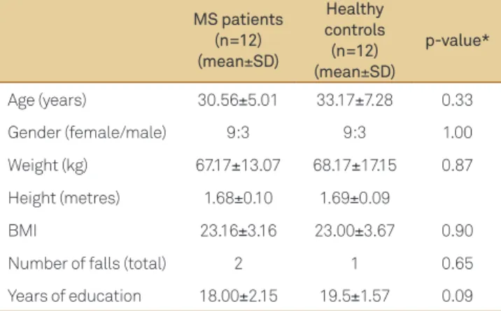
MS: multiple sclerosis; SD: standard deviation; *Signiicance level <0.05; NS: not signiicant. Table 1. Demographic data of the multiple sclerosis patients and
healthy control group.
MS patients (n=12) (mean±SD)
Healthy controls (n=12) (mean±SD)
p-value*
Age (years) 30.56±5.01 33.17±7.28 0.33
Gender (female/male) 9:3 9:3 1.00
Weight (kg) 67.17±13.07 68.17±17.15 0.87
Height (metres) 1.68±0.10 1.69±0.09
BMI 23.16±3.16 23.00±3.67 0.90
Number of falls (total) 2 1 0.65
Years of education 18.00±2.15 19.5±1.57 0.09
BMI: body mass index; MS: multiple sclerosis; SD: standard deviation; *Signii-cance level <0.05.
Fig 1. Comparison of double support duration in multiple sclerosis patients and healthy controls in normal walking and dual-task conditions.
40
* Dual Task *
Multiple Sclerosis Patients Health Controls
Normal Walking
30
20
10
0
Fig 2. Comparison of walking speed in multiple sclerosis patients and healthy controls in normal walking and motor imagery conditions.
* Motor Imagery
Real Walking
Multiple Sclerosis Patients Health Controls 2.5
2.0
1.5
1.0
0.5
0.0 Most of the participants were young adults with female pre-dominance and normal body fat. here were no diferences in the falls history and education level between the groups. Patient demographic data are shown in Table 1.
he scores of walking speed and cadence showed no sta-tistically signiicant diferences between the groups in com-fortable walking, dual-task condition and MI. he dual-task condition showed an increase in double support duration in both groups. However, step width did not show any sig-niicant diferences between comfortable walking and the dual-task condition in both groups. he temporal-spatial walking values are described in Table 2. he comparison of double support duration in MS patients and healthy con-trols in normal walking and dual-task conditions is present-ed in Figure 1.
MI analysis showed statistically signiicant diferences between real and imagined walking in MS patients (MI=1.90 m/s, SD±0.93 vs. normal walking=1.27 m/s, SD±0.24; p=0.02), while healthy controls showed a tendency to higher the walk-ing speed in imagined walkwalk-ing, but this result was not sta-tistically signiicant (MI=1.55 m/s, SD±0.52 vs. normal walk-ing=1.21 m/s, SD±0.11; p=0.05). MS patients overestimated
walking speed by 33%, while matched healthy controls over-estimated walking speed by 22%. Figure 2 shows the compar-ison in walking speed in MS patients and healthy controls in normal walking and MI conditions.
DISCUSSION
A matched healthy control group did not show overestima-tion of motor planning in the same task. MI-based exercises, as a ther apy tool, have been used for Parkinson’s disease and post-stroke patients, with good results. However, for MS pa-tients, it is important to con sider their ability to generate cor-rect motor images. Cognitive impairments are highly preva-lent in MS patients, even in early stages, and in patients with mild disability measured by the EDSS6.
he dual-task condition did not show any diferences in walking speed, cadence and step width. However, double sup-port duration signiicantly increased. A previous study showed that cadence does not difer with dual-task condition’s clinical-ly isolated syndromes, suggestive of MS19. he increase in dou-ble support duration was a compensatory strategy to maintain walking stability. Hamilton et al.20 showed greater decrements in performance under dual-task conditions in cognitive task performance, walking speed and swing-time variability in MS patients. Kalron et al.19 also showed that combined walking and cognitive tasks were expressed in prolonged double sup-port duration, as shown by the present results. hey also de-scribed a reduction in gait speed in the early stages of the dis-ease. Our results showed an increase of 13% in double support duration in dual-task conditions, while Kalron et al.19 showed an increase of 8% in double support duration. Despite the simi-larity of results, the diferences found in both studies may be related to the methods used as Kalron et al.19 used the Word List Generation Test as the executive function, while we used an arithmetic task. Stoquart-Elsankari et al.21 showed that the action slowing of an MS patient was mainly related to the at-tention deicit, even in patients without motor deicit on clini-cal examinations, where divided attention and decisional pro-cesses were preserved. MS patients showed an interference of cognitive task on motor execution although the severity of clinical disability with more impairment, as compared with healthy subjects, still needs to be clariied.
he double support duration was increased in patients and matched healthy controls during the dual-task condi-tion. Slower walking speed5,22 and an increase in stride-time variability are common indings, even in healthy adults per-forming dual-tasks22. Walking speed is the most common inding described in studies with dual-task condition and it is usually slower in many populations. he results of the pres-ent study relect the increase of walking stability under cog-nitive demands. A functional magnetic resonance imaging study, with diferent types of dual-tasks, revealed that corti-cal areas along the inferior frontal sulcus, middle frontal gy-rus and intraparietal sulcus are involved in dual-task perfor-mance, which highlights the role of cognitive function and the frontal lobe on mobility23. he question that should be asked is at which stage of the MS disease process do dual-task conditions afect gait performance; speciically, which gait parameters are afected and to what extent.
MS patients with no disability showed a reduction in mo-tor planning accuracy compared with matched healthy con-trols. A recent study24, which was the irst research that inves-tigated MI in MS patients, focused on upper limb move ment capacity and described signiicant diferences in temporal organisation. Heremans et al.24 used mental chronometry during the Box and Block Test in moderate disability (av-erage EDSS=6.5) in MS patients. he present study showed that MS patients overestimated the imagined movement by 33%, while Heremans et al.24 showed an increase of 14% in the upper limb most afected side. he temporal invariance between executed and imagined movements, which was well documented in young adults, suggests that similar motor representations are shared between covert and overt stages of actions25. Young adults had their MI ability preserved ir-respective of the width of the path, while the elderly group signiicantly overestimated the duration of imagined move-ments with respect to the executed movemove-ments26. MI accura-cy was signiicantly deteriorated in elderly adults; this could be attributed to functional changes in the brain that occur with ageing, inluencing cognitive and motor abilities25. he temporal congruence of real and imagined movements in post-stroke patients remains similar to that of age-matched controls27.
Neuroimaging studies have been described to have re-markable similarities between the real and imagined locomo-tion network. he major activated areas were the motor/pre-motor and multisensory cortices, parahippocampal gyri and midline cerebellum. here were deactivations in multisensory vestibular cortical areas in both conditions28. In MS patients, it has been shown that an ipsilateral sensorimotor cortex deac-tivation with a simple motor task29 and an existence of com-pensatory cortical activations at the earliest stage of MS were mainly located in regions involved in executive processing13,30.
he sample size was a potential limitation of this study. herefore, the possibility of making generalisations from our indings may still be fairly limited. he second possible limi-tation is that we did not ind any gait research in MS patients with no disability (EDSS≤1.5) that analyses the inluence of motor planning with which to compare our data. Despite these limitations, the originality of the research theme and intriguing results provide an impetus for future research. Studies with larger sample sizes that include participants with greater disability from the present study and diferent levels of cognitive impairments should be undertaken to con-irm the present results.
1. Iezzoni LI, Rao SR, Kinkel RP. Patterns of mobility aid use among working-age persons with multiple sclerosis living in the community in the United States. Disabil Health J 2009;2:67-76.
2. Ringel I, Zettl UK. Estimates of the walking distance in multiple sclerosis patients and their effect on the EDSS. J Neurol 2006;253:666-667.
3. Rossignol S, Dubuc R, Gossard JP. Dynamic sensorimotor interactions in locomotion. Physiol Rev 2006;86:89-154.
4. Woollacott M, Shumway-Cook A. Attention and the control of posture and gait: a review of an emerging area of research. Gait Posture 2002;16:1-14.
5. Holtzer R, Mahoney JR, Izzetoglu M, et al. fNIRS study of walking and walking while talking in young and old individuals. J Gerontol A Biol Sci Med Sci 2011;66:879-887.
6. Amato MP, Zipoli V, Portaccio E. Multiple sclerosis-related cognitive changes: a review of cross-sectional and longitudinal studies. J Neurol Sci 2006;245:41-46.
7. Xiong YY, Mok V. Age-related white matter changes. J Aging Res 2011;2011:617927.
8. Beauchet O, Allali G, Annweiler C, et al. Does change in gait while counting backward predict the occurrence of a irst fall in older adults? Gerontology 2008;54:217-223.
9. Bakker M, de Lange FP, Stevens JA, Toni I, Bloem BR. Motor imagery of gait: a quantitative approach. Exp Brain Res 2007;179:497-504.
10. Decety J. The neurophysiological basis of motor imagery. Behav Brain Res 1996;77:45-52.
11. Jahn K, Deutschlander A, Stephan T, Strupp M, Wiesmann M, Brandt T. Brain activation patterns during imagined stance and locomotion in functional magnetic resonance imaging. Neuroimage 2004;22:1722-1731.
12. Malouin F, Richards CL, Durand A, Doyon J. Reliability of mental chronometry for assessing motor imagery ability after stroke. Arch Phys Med Rehabil 2008;89:311-319.
13. Audoin B, Ibarrola D, Ranjeva JP, et al. Compensatory cortical activation observed by fMRI during a cognitive task at the earliest stage of MS. Hum Brain Mapp 2003;20:51-58.
14. McDonald WI, Compston A, Edan G, et al. Recommended diagnostic criteria for multiple sclerosis: guidelines from the International Panel on the diagnosis of multiple sclerosis. Ann Neurol 2001;50:121-127.
15. Borel S, Schneider P, Newman CJ. Video analysis software increases the interrater reliability of video gait assessments in children with cerebral palsy. Gait Posture 2011;33:727-729.
References
16. Kurtzke JF. Rating neurologic impairment in multiple sclerosis: an expanded disability status scale (EDSS). Neurology 1983;33:1444-1452.
17. Rossier P, Wade DT. Validity and reliability comparison of 4 mobility measures in patients presenting with neurologic impairment. Arch Phys Med Rehabil 2001;82:9-13.
18. Vaughan C, Davis B, O’Connor J. Dynamics of human gait. 2nd ed. Cape Town: Kiboho Publishers,1999:p23-30.
19. Kalron A, Dvir Z, Achiron A. Walking while talking–dificulties incurred during the initial stages of multiple sclerosis disease process. Gait Posture 2010;32:332-335.
20. Hamilton F, Rochester L, Paul L, Rafferty D, O’Leary CP, Evans JJ. Walking and talking: an investigation of cognitive-motor dual tasking in multiple sclerosis. Mult Scler 2009;15:1215-1227.
21. Stoquart-Elsankari S, Bottin C, Roussel-Pieronne M, Godefroy O. Motor and cognitive slowing in multiple sclerosis: an attentional deicit? Clin Neurol Neurosurg 2010;112:226-232.
22. Doi T, Asai T, Hirata S, Ando H. Dual-task costs for whole trunk movement during gait. Gait Posture 2010;33:712-714.
23. Szameitat AJ, Schubert T, Muller K, Von Cramon DY. Localization of executive functions in dual-task performance with fMRI. J Cogn Neurosci 2002;14:1184-1199.
24. Heremans E, D’Hooge AM, De Bondt S, Helsen W, Feys P. The relation between cognitive and motor dysfunction and motor imagery ability in patients with multiple sclerosis. Mult Scler 2012;18:1303-1309.
25. Personnier P, Ballay Y, Papaxanthis C. Mentally represented motor actions in normal aging: III. Electromyographic features of imagined arm movements. Behav Brain Res 2010;206:184-191.
26. Personnier P, Kubicki A, Laroche D, Papaxanthis C. Temporal features of imagined locomotion in normal aging. Neurosci Lett 2010;476:146-149.
27. Malouin F, Richards CL, Durand A, Doyon J. Clinical assessment of motor imagery after stroke. Neurorehabil Neural Repair 2008;22:330-340.
28. la Fougere C, Zwergal A, Rominger A, et al. Real versus imagined locomotion: a [18F]-FDG PET-fMRI comparison. Neuroimage 2010;50:1589-1598.
29. Manson SC, Wegner C, Filippi M, et al. Impairment of movement-associated brain deactivation in multiple sclerosis: further evidence for a functional pathology of interhemispheric neuronal inhibition. Exp Brain Res 2008;187:25-31.