ARTICLE
DOI: 10.1590/0004-282X20130084
Hematoma volumes of spontaneous
intracerebral hemorrhage: the ellipse (ABC/2)
method yielded volumes smaller than those
measured using the planimetric method
Volumes de hematomas em hemorragias intracerebrais espontâneas:
o método da elipse (ABC/2) produziu volumes inferiores do que aqueles
determinados pelo método planimétrico
Adriano Keijiro Maeda1, Luiz Roberto Aguiar2, Carolina Martins3,
Gerson Linck Bichinho4, Munir Antônio Gariba4
Cerebrovascular accidents (CVA) include ischemic cerebral attacks, subarachnoid hemorrhage, and spontaneous intracere-bral hemorrhage (ICH). he latter is the deadliest type of CVA, accounting for 10% of total strokes1,2. Stroke is considered the third single cause of mortality in the United States of America, following cardiovascular diseases and neoplasms. In 2005, stroke was the cause of one in every 15 deaths in the USA, and 8 to 15%
of patients with acute ischemic strokes and 37 to 38% of the ce-rebral hemorrhage ones died within 30 days3. In addition, only 20% of patients with ICH recovered functional independence, suggesting a poor prognosis for recovery4.
Intracranial hypertension is considered the main reason for surgical indication in subjects with ICH, especially in cases in which there is a progressive neurological deterioration5,6. he
1MD, Postgraduate Health Technology Program, Pontiical Catholic University of Paraná, Curitiba PR, Brazil;
2MD, MsC, PhD, Postgraduate Health Technology Program, Pontiical Catholic University of Paraná; Neurosurgeon at Hospital Santa Cruz, Curitiba PR, Brazil;
3MD, PhD, Neurosurgeon at Hospital Pelópidas Silveira, Instituto de Medicina Integral Professor Fernando Figueira (IMIP), Recife PE, Brazil;
4MSc, PhD, Postgraduate Health Technology Program, Pontiical Catholic University of Paraná, Curitiba PR, Brazil.
Correspondence: Luiz Roberto Aguiar; Rua Imaculada Conceição 1.155; 80215-901 Curitiba PR - Brasil; E-mail: [email protected] Conflict of interest: There is no conlict of interest to declare.
Received 26 March 2012; Received in inal form 26 March 2013; Accepted 02 April 2013.
ABSTRACT
Objective: To compare two different methods for measuring intracerebral hemorrhage (ICH) volume: the ellipse volume (called ABC/2), and the software-aided planimetric. Methods: Four observers evaluated 20 brain computed tomography (CT) scans with spontaneous ICH. Each professional measured the volume using the ABC/2 and the planimetric methods. The average volumes were obtained, and the intra- and in-ter-rater variability was determined. Results: There is an absolute 2.24 cm3 average difference between both methodologies. Volumes yielded by the ABC/2 method were as much as 14.9% smaller than by the planimetric one. An intra-observer variability rate of 0.46% was found for the planimetric method and 0.18% for the ABC/2. The inter-observer rates were 1.69 and 1.11% respectively. Conclusions: Both methods are reproducible. The ABC/2 yielded hemorrhage volumes as much as 14.9% smaller than those measured using the planimetric methodology.
Key words: cerebral hemorrhage, tomography, evaluation studies.
RESUMO
Objetivo: Comparar dois métodos diferentes para determinar o volume da hemorragia intracerebral: volume da elipse (chamado ABC/2), e método planimétrico auxiliado por computador. Métodos: Quatro diferentes observadores avaliaram as imagens de 20 tomograias cerebrais com diagnóstico de hemorragia intracerebral espontânea. Cada proissional determinou o volume da hemorragia usando os dois métodos. Foram comparadas as médias dos volumes obtidos, bem como suas variabilidades intra e interobservadores. Resultados: Foi observada dife-rença estatisticamente signiicativa entre os volumes calculados por meio dos dois métodos, com uma variação média absoluta de 2,24 cm3 e com volumes até 14,9% menores para o método ABC/2. A média da variabilidade intraobservador foi de 0,46% para o método planimétrico e 0,18% para o ABC/2. As taxas de variabilidade interobservador foram de 1,69 e de 1,11%, respectivamente. Conclusões: Ambos os métodos são reprodutíveis. O volume determinado pelo ABC/2 pode ser até 14,9% menor que aquele determinado pelo método planimétrico.
hemorrhage size, ventricular extension and expansion of the he-matoma, along with altered level of consciousness, are decisive factors in the evolution of patients7,8. An increase in the ICH vol-ume is a major contributor to poor prognosis, especially when it occurs early on, since it indicates neurological deterioration9-14.
Given the importance of measuring accurately the hemor-rhage lesion volume in properly determining patient prognosis, the aim of this study was to compare two methods that are cur-rently used for determining spontaneous hemorrhage volume: the ellipsoid (or ABC/2), and the planimetric.
METHODS
his was a retrospective study, carried out from October 2007 to January 2008, approved by the Ethics and Research Committee of the Pontiical Catholic University of Paraná, in Curitiba, Paraná, Brazil.
he hematoma volumes of spontaneous ICH, using patients in the acute phase, were measured by two diferent methods (ABC/2 and the planimetric method) of computed tomography (CT) scans, obtained by volumetric acquisition in a Somaton® equipment (Siemens, Erlangen, Germany). he ield of view (FOV) was selected at the time of examination according to pa-tient’s characteristics (weight and position) with a pitch of 1:1 and 3 mm thick cuts. Further reconstructions to 1 mm were made to all cuts.
Four independent raters evaluated the CT scans of 20 pa-tients with spontaneous ICH, including ive cases of posterior fossa and 15 of supratentorial hemorrhage. Bleeding was ob-served in 83 CT slices. Two measurements were made for each method (ABC/2 and planimetric) in two independent sessions,
with at least a two-day interval. Volume measurements using the
ABC/2 method were performed using DicomViewer®
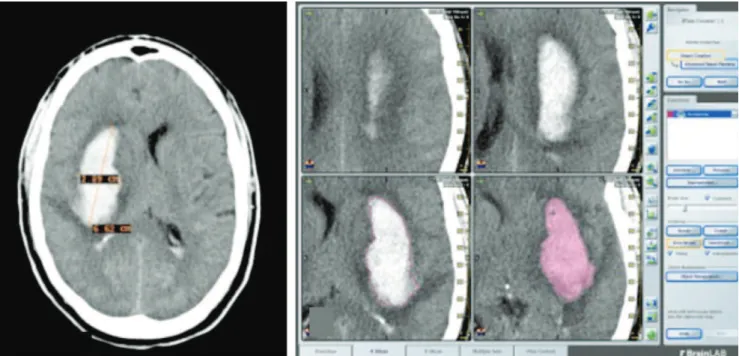
software (DicomWorks, Limoges, France). he images were transferred as DICOM iles, and the tomography slices showing the hemor-rhage were individually assessed. he slice with the largest area of hematoma was chosen for the measurements. Measure A cor-responds to the greatest diameter of hemorrhage; B to the larg-est perpendicular diameter to A; whereas C was the sum of the thickness of slices containing the hemorrhage (Fig 1A). For the sum of the slice thickness (C), one was excluded if the hematoma area was smaller than 25% of the larger obtained place. Half of the slice thickness was taking into consideration if the measured area were between 25 to 75% when compared to the largest one, as proposed by Kothari et al.15. Values for A, B, and C were then multiplied, and the inal result was divided by two, which was
expressed in cm3. Each observer performed two independent
measurements using the ABC/2 method, with at least a two-day interval.
he planimetric measurements were performed using
the planning station of BrainLab®
neuronavigation equip-ment. he DICOM iles of the CT images were transferred to
the workstation using PatXFer®
2.0 and exported to Iplan®
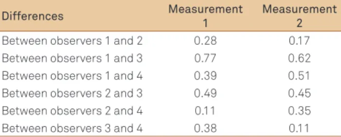
2.6 Cranial software (BrainLab, Munich, Germany), a com-ponent of the utilities’ package used for planning navigation. Hemorrhage edges were individually determined for each slice using the available software tools (brush and smart-brush), as seen in Fig 1B. his software calculates the total hemorrhage volume, expressed in cm3.
The volumes obtained using the two methods were
compared using Student’s t-test for dependent samples,
and the correlation between them was estimated using Pearson’s correlation coeicient and tested for its major signiicance.
Fig 1. (A) Axial computed tomography scan slice displaying the method of linear measurement using DicomWorks software;
he method error was determined, and the results were compared for inter- and intraobserver measurements using the variance model. he intraobserver error was determined by averaging the values obtained from each observer using both measurement methods (ABC/2 and planimetric method). he interobserver error was calculated applying the diferences be-tween the average values for every two observers, for the ABC/2 and planimetric methods.
RESULTS
here was a signiicant diference in the average vol-umes obtained using both methods, the absolute diference
being 2.24 cm3. he average measurement obtained with
ABC/2 method was 14.9% lower than that obtained with the planimetric one. Table 1 and Fig 2 show average, minimum,
Fig 2. Average and standard deviation for planimetric and
ellipsoid method. PLANI 30
28 26 24 22 20 18 16 14 12 10 8 6 4 2 0
ABC/2
Mediana 25%-75% Min-Max
Fig 3. Pearson’s correlation coeficient between planimetric
and ellipsoid method (r=0.984; p<0.001).
60
50
40
30
20
10
0
-10
ABC/2
-5 0 5 10 15 20 25 30 35 40 45 50
PLANI
Method Average (cm3)
Standard
deviation (cm3) p-value*
PLANI 15.04 12.46
ABC/2 12.8 11.37
(PLANI – ABC/2) 2.24 2.38 <0,001
Table 1. Planimetric and ABC/2 method comparison.
*Student’s t-test for paired samples; p<0.05.
and maximum values, which ranged from 25 to 75% of data. However, a good correlation between both methodologies can be observed as showed by Pearson’s correlation coei-cient, in the two methods (r=0.984; p<0.001), as seen in Fig 3. he intraobserver error is listed in Table 2 for ABC/2 method and in Table 3 for the planimetric.
he interobserver error was calculated using the difer-ences between the average values for every two observers. Table 4 shows the results for ABC/2 and Table 5 for the pla-nimetric method.
DISCUSSION
he ABC/2 method provides an estimated value of the spontaneous ICH volume. Although several studies validate this method for clinical purposes16-21, it is considered to be the most efective for measuring ICH that have a rounded or elliptical shape20, as opposed to irregularly-shaped hemor-rhages, which are in fact more common among patients who take anticoagulants19.
In a previous study, Gebel et al.20 used both
meth-ods (ABC/2 and planimetric) to compare volumes of
Observer 1 Observer 2 Observer 3 Observer 4 Measurement Measurement Measurement Measurement
1 2 1 2 1 2 1 2
Average (cm3) 12.43 12.48 12.71 12.65 13.20 13.10 12.82 12.99
Difference (cm3) 0.05 0.06 0.10 0.17
Table 2. Average values of ABC/2 from each observer and differences between them.
Observer 1 Observer 2 Observer 3 Observer 4 Measurement Measurement Measurement Measurement
1 2 1 2 1 2 1 2
Average (cm3) 15.42 15.08 15.78 15.76 16.39 16.47 14.28 14.86
Difference (cm3) 0.34 0.02 0.08 0.57
Table 3. Average values of the planigraphic method from each observer and differences between them.
Differences Measurement 1
Measurement 2
Between observers 1 and 2 0.28 0.17 Between observers 1 and 3 0.77 0.62 Between observers 1 and 4 0.39 0.51 Between observers 2 and 3 0.49 0.45 Between observers 2 and 4 0.11 0.35 Between observers 3 and 4 0.38 0.11
Table 4. Difference of ellipsoid average values compared in
every two observers (cm3).
Difference Measurement 1
Measurement 2
Between observers 1 and 2 0.36 0.68 Between observers 1 and 3 0.97 1.39 Between observers 1 and 4 1.14 0.22 Between observers 2 and 3 0.62 0.72 Between observers 2 and 4 1.49 0.9 Between observers 3 and 4 2.11 1.62
Table 5. Differences of average values in the planigraphic
method compared in every two observers (cm3).
intracerebral and subdural hematomas, and they found a good correlation between the two methods. However, these authors used different CT scan acquisition param-eters. In another study, Kothari et al.15 reported that, com-pared to the planimetric method, ABC/2 overestimates
volumes by 1.5 (±1.3) cm3. This tendency was also
ob-served by Gebel et al.20. According to these authors, this
overestimation appears to be correlated with lesion loca-tion, being more pronounced in brainstem, cerebellar, and lobar hemorrhages. The observed inaccuracy, in terms of variability between observers, obeys the same sequence. These studies also suggest an overestimation from 5 to 10% even for regular-shaped hemorrhages.
Yet, another study by Huttner et al.19, compared the two measurement techniques, for ICH secondary to the use of oral anticoagulants, which, relative to spontaneous hemor-rhages, are more often irregular in shape, as mentioned. hey did not observe a statistically signiicant diference between the two methods; however, they found an overestimation by
the ABC/2 method of 6.68% (±3.01) for regular (rounded or
elliptical) hematomas, 14.85% (±4.95) for irregular ones, and 32.11% (±10.28) for multinodular or separate19.
A recent study by Freeman et al.16, by contrast, found that the ABC/2 technique underestimated the volume in four out of ive ICH, and one case of subdural hematoma. For elliptic hemorrhages, there was a diference of 24% between the two methods, and another of 28% for other hematomas; the ab-solute average diference was of 6.7 and 38 cm3, respectively16.
Our indings are similar to those reported by Freeman
et al.16 – ABC/2 method resulted in an underestimation of
volume when compared to the planimetric. It should be noted, however, that we did not classify our images as regu-lar or irreguregu-lar. Our study also suggests that the planimetric method is more precise, accurately measuring the hemato-ma area in a given slice and using the known slice thickness to determine volume.
When using ABC/2, Kothari et al. recommended ex-cluding slice thickness if the area of hematoma in a given slice were smaller than 25% of the larger obtained, or tak-ing into consideration only half of the slice thickness if the area were measured from 25 to 75% when compared to the largest one15. he fact that we adopted this rule for cal-culating the volume using the ABC/2 method may explain
the underestimation of the hematoma volume in the current study.
Herein, we evaluated the systematic error in volume measurement by the ABC/2 and planimetric methods through comparing the average differences, which were determined for varied measurements obtained by the same researcher (intraobserver – Tables 2 and 4) and for those determined by different researchers (interobserver – Tables 3 and 5). For both methods, the percentage of intraobserver variability was lower than that of the in-terobserver one, with the planimetric method yielding higher variability than the ABC/2 method. Although the differences are not statistically significant, this is also an interesting finding, since the planimetric method, con-sidered the golden-standard, showed more variability than the ABC/2. Fine automatic edge determination and more precise segmentation method could possibly alter this relationship.
Zimmerman et al11. compared the variability of ICH
vol-ume measurements using computer software with the deter-mination of hematoma borders done by automatic and semi-automatic or manual segmentations. hey showed minimal intra- and interobserver variations, supporting the validity of interpretations among diferent observers, which is similar to that found in our study11.
he two volume evaluations performed in this study are dependent on the measurements carried out by the observ-ers. he ABC/2 method requires determination of length, diameter, and thickness of the hematoma, while the plani-metric requires demarcation of the borders of hemorrhage for volume calculation. he ABC/2 has already been vali-dated and has the advantage of obtaining rapidly volumes. It can be performed at patient bedside, even without the use of computerized devices. he method of planimetry is more accurate in determining volume, though it needs the help of a suitable computer and software that is not always readily available. herefore, a reliable method of automatic segmen-tation that can be used for more asymmetric lesions in order to improve not only the accuracy, but also velocity of results is not available.
automatically determining volume, with a faster and more accurate acquisition of data, can lead to better evaluation of these patients.
In conclusion, our results revealed that both methods are reproducible and intraobserver variability for both methods is lower than the interobserver one. We also observed that
ABC/2 method yielded hemorrhage volumes as much as 14.9% smaller than those measured using planimetric method.
he results of this careful comparison between two meth-ods carry important clinical implications for determining surgical treatment and thus represent a valuable contribu-tion to the ield.
1. Steiner T, Rosand J, Diringer M. Intracerebral hemorrhage associated with oral anticoagulant therapy: current practices and unresolved questions. Stroke 2006;37:256-262.
2. Manno EM, Atkinson JL, Fulgham JR, Wijdicks EF. Emerging medical and surgical management strategies in the evaluation and treatment of intracerebral hemorrhage. Mayo Clin Proc 2005;80:420-433.
3. Thom T, Haase N, Rosamond W, et al. Heart disease and stroke statistics - 2006 update: A report from the american heart association statistics committee and stroke statistics subcommittee. Circulation 2006;113:85-151.
4. Mayer SA, Brun NC, Begtrup K, et al. Recombinant activated factor VII for acute intracerebral hemorrhage. N Engl J Med 2005;352:777-785.
5. Gregson BA, Mendelow AD. International variations in surgical practice for spontaneous intracerebral hemorrhage. Stroke 2003;34:2593-2597.
6. Radberg JA, Olsson JE, Radberg CT. Prognostic parameters in spontaneous intracerebral hematomas with special reference to anticoagulant treatment. Stroke 1991;22:571-576.
7. Davis SM, Broderick J, Hennerici M, et al. Hematoma growth is a determinant of mortality and poor outcome after intracerebral hemorrhage. Neurology 2006;66:1175-1181.
8. Lisk DR, Pasteur W, Rhoades H, Putnam RD, Grotta JC. Early presentation of hemispheric intracerebral hemorrhage: prediction of outcome and guidelines for treatment allocation. Neurology 1994;44:133-139.
9. Kazui S, Minematsu K, Yamamoto H, Sawada T, Yamaguchi T. Predisposing factors to enlargement of spontaneous intracerebral hematoma. Stroke 1997;28:2370-2375.
10. Leira R, Davalos A, Silva Y, et al. Early neurologic deterioration in intracerebral hemorrhage: predictors and associated factors. Neurology 2004;63:461-467.
11. Zimmerman RD, Maldjian JA, Brun NC, Horvath B, Skolnick BE. Radiologic estimation of hematoma volume in intracerebral hemorrhage trial by CT scan. AJNR 2006;27:666-670.
12. Broderick JP, Brott TG, Duldner JE, Tomsick T, Huster G. Volume of intracerebral hemorrhage. A powerful and easy-to-use predictor of 30-day mortality. Stroke 1993;24:987-993.
13. Juvela S, Kase CS. Advances in intracerebral hemorrhage management. Stroke 2006;37:301-304.
14. Brott T, Broderick J, Kothari R, et al. Early hemorrhage growth in patients with intracerebral hemorrhage. Stroke 1997;28:1-5.
15. Kothari RU, Brott T, Broderick JP, et al. The ABCs of measuring intracerebral hemorrhage volumes. Stroke 1996;27:1304-1305.
16. Mahaley MS Jr., Gillespie GY, Hammett R. Computerized tomography brain scan tumor volume determinations. Sensitivity as an objective criterion of response to therapy. J Neurosurg 1990;72:872-878.
17. Freeman WD, Barrett KM, Bestic JM, Meschia JF, Broderick DF, Brott TG. Computer-assisted volumetric analysis compared with ABC/2 method for assessing warfarin-related intracranial hemorrhage volumes. Neurocrit Care 2008;9:307-312.
18. Sucu HK, Gokmen M, Gelal F. The value of XYZ/2 technique compared with computer-assisted volumetric analysis to estimate the volume of chronic subdural hematoma. Stroke 2005;36:998-1000.
19. Huttner HB, Steiner T, Hartmann M, et al. Comparison of ABC/2 estimation technique to computer-assisted planimetric analysis in warfarin-related intracerebral parenchymal hemorrhage. Stroke 2006;37:404-408.
20. Gebel JM, Sila CA, Sloan MA, et al. Comparison of the ABC/2 estimation technique to computer-assisted volumetric analysis of intraparenchymal and subdural hematomas complicating the GUSTO-1 trial. Stroke 1998;29:1799-1801.
21. Gomez-Marino R, Andre C, Novis SA. [Volumetric determination of cerebral infarction in the acute phase using skull computed tomography without contrast: comparative study of 3 methods]. Arq Neuropsiquiatr 2001;59:380-383.