Image
Electrocardiographic Abnormalities Preceding Ventricular
Premature Beats
Eder Trezza
Faculdade de Medicina de Botucatu - UNESP - Botucatu, SP
Mailing Address: eder trezza •
Caixa Postal 535 – 18618-000 – Botucatu, SP, Brazil E-mail: [email protected]
Manuscript received November 17, 2005; revised manuscript received November 21, 2005; accepted November 21, 2005.
Key words
Eletrocardiographic, arrithymia, electrophysiology. This study describes the patterns of electrocardiographic abnormalities that have been observed before premature beats in tracings obtained during the monitoring of patients undergoing stress tests. The tracings of 1,800 tests performed in the “Hospital das Clínicas da Faculdade de Medicina do Campus de Botucatu-SP da UNESP” between 2001 and 2003 were retrospectively analyzed. EKG registration was carried out using three leads (CM5, aVF and V2M), according to the Bruce treadmill stress test protocol. The tracings were obtained in supine and in the orthostatic positions pre-exertion, every 3 minutes during exertion and every 2 minutes in the recovery stage, in supine position. Additional tracings were obtained whenever premature beats occurred. Only tracings with good technical quality, that allowed inequivocal comparison between complex preceding ventricular premature beats and those not followed by them were considered. Tracings with electrical or external interference or unstable baseline were discarded. Premature beats frequency was not taken into account, only their presence, even if sporadic.
Ventricular premature beats occurred in 426 out of 1,800 exams (23.6%). Abnormalities in ST segment, T or U waves, or in diastolic baseline - which differed from thoseobserved in complexes not followed by premature beats - were identified in 103 out of the 426 exams (24.1%). The following abnormalities were associated with premature beats:
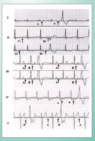
1 – Variation of the ST segment level – either elevated or depressed – in relation to preceding complexes (Illustrations 1-III, 1-IV, 2-II, 3-I, 3-II, 3-III and 3-IV).
2 – Variation of T wave amplitude – either higher or lower – whether previously positive, negative, or with minus-plus or plus-minus pattern (Illustrations 1-II, 1-III, 1-IV, 2-I, 2-III, 2-IV. 3-I and 3-III).
3 – Appearance of negative U wave (Illustrations 1-I, 1-V, 2-III and 2-IV).
4 – Appearance of upward or downward shift of diastolic baseline immediately before ventricular premature beat (Illustrations 1-II, 1-III, 2-I and 3-I).
Fig. 1 - Electrocardiographic Changes Preceding Ventricular Premature Beats.
I – Baseline downshifting during diastole triggered by a negative U wave.
II – Upshifting of diastolic baseline between A1 and B1 and small increase in T wave negativity between A2 and B2.
III – Downshifting of the ST segment and of diastolic baseline, between the A and B moments.
III – Increased downshifting of the ST segment from A to B.
V – Appearance of negative U wave (or downshifting of diastolic baseline), when A and B moments are compared.
Image
eder trezza eleCtrOCArdIOGrAphIC AbnOrMAlItIes preCedInG VentrICulAr preMAture beAts
Arq Bras Cardiol 2006; 87 : e219-e221
Comments
The electrogenesis of the premature beats is basically related to myocardial fibers automatism and dromotropism¹. During ventricular contraction – which in the EKG extends from the end of the QRS to the end of the T wave – myocardial fibers are in a refractory period. Refractoriness to new depolarization is absolute in early moments of contraction and relative at final moments. During diastole, fiber electric intracellular potentials are negative in relation to those on the membrane external surface (Phase 4 of action potential). The more negative the diastolic potential inside the fibers, the lower the possibility of spontaneous automatism manifestation. When the diastolic intracellular potential is less negative than it should be, myocyte electric charge distribution gets closer to the excitability threshold, above which depolarization is triggered. The abnormality most commonly related to the occurrence of ventricular premature
1. Moffa PJ. Introdução ao estudo das disritmias cardíacas. In: Moffa P e Sanches PCR. Eletrocardiograma Normal e Patológico. 1ª. Ed. Editado por Ramires JAF e Oliveira AS. São Paulo: Rocca Ltda; 2001: 207-22.
2. Rubart M, Zipes DP. Genesis of cardiac arrhythmias: electrophysiological considerations. In: Braunwald ER, Zipes DP and Libby P. Heart Disease. A Textbook of Cardiovascular Medicine. 6th ed. Philadelphia: W.B. Saunders
Company; 2001: 659-99.
3. Marchiliski FE, Schwartzman G, Gottlieb CD, González-Zuelgaray J, Callans DJ. Electrical events associated with arrhythmias initiation and stimulatrion techniques for arrhythmia prevention. In: Zipes DP and Jalife J. Cardiac Electrophysiology. From Cell to Bedside. 2nd ed. Philadelphia: W.B. Saunders Company; 1995: 863-77.
References
beats is heterogeneity of repolarization in different myocardial areas - a phenomenon known as dispersion, that sets condition for the occurrence of either the re-entry phenomenon or early depolarization of regional foci²,³.
Although time and spatial repolarization heterogeneity have been admitted to be commonly found in heart diseases – almost as often as ventricular premature beats themselves – the translation of such phenomena in conventional EKG remains unreported in the literature. The electrocardiographic patterns described herein demonstrate that the electric mechanisms underlying the triggering of ventricular premature beats can be manifested in surface tracings and deserve investigation in different patient groups, given that they are the same, irrespective of the underlying disease. Myocardial ischemia, inflammation, fibrosis, cardioactive drugs, electrolytic or acid-basic disorders, autonomic hyperactivity (particularly sympathetic), etc, usually present in heart or systemic diseases - may result in regional abnormalities of diastolic intracellular potentials, triggering premature beats.
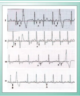
Fig. 2 - Electrocardiographic Abnormalities Preceding Ventricular Premature Beats.
I – Diastolic baseline downshifting from A to B.
II – Downshifting of the ST segment and modification of the T wave shape between the A and B moments.
III – Downshifting of the ST segment and appearance of negative U wave when A and B moments are compared.
IV – Increased ST segment downshifting, greater T wave negativity and appearance of negative U wave from A to B moments.
Fig. 3 - Electrocardiographic Abnormalities Preceding Ventricular Premature Beats.
I – Downshifting of ST segment and diastolic baseline, preceding premature beats, when moment A is compared to B and B1.
II – Reduction of the negative component of the T wave in the complex preceding premature beat in B in relation to A.
III – Greater downshifting of the ST segment and of the T negativity.
V – Downshifting of the ST segment in B when compared to A.