Clinicoradiological Session
Case 4/2006 – Thirteen-year-old Teenager with Single Left Ventricle,
Ventriculo-Arterial Discordance with Left Aorta, Pulmonary Stenosis
and Previous Pulmonary Systemic Shunt
Edmar Atik
Instituto do Coração do Hospital das Clínicas da FMUSP - São Paulo, SP, Brazil
Mailing Address: Edmar Atik •
InCor – Av. Dr. Enéas C. Aguiar, 44 - 05403-000 – São Paulo, SP, Brazil E-mail: [email protected]
Clinical findings
Thirteen-year-old white male teenager presented with cyanosis since birth, with early progression as of the 17th day of life that required the performance of a right systemic pulmonaryanastomosis, which was repeated in the left side at the age of 9 months. He has remained stable since then, with mild cyanosis and dyspnea, and delayed physical development. His physical examination revealed no respiratory distress, cyanosis +/++, and normal pulses. Blood pressure was 100/60 mmHg, heart rate was 71 bpm, oxygen saturation was 85%, weight was 27 kg, and height was 138 cm. The aorta was not palpable in the suprasternal notch. In the precordium, mild systolic impulses on the left sternal border could be observed, and a slightly forceful apex impulse was palpable at the 4th left intercostal space, limited to two fingertips. The first heart sound was accentuated, more intense in the mitral area than in the tricuspid area, and the second heart sound was accentuated and single in the pulmonic area, and was also more intense in the mitral area. A grade 3 protosystolic click was present in all the precordium. A grade 1-2/6 continuous heart murmur could be auscultated in the 2nd, 1st, and 3rd left intercostal spaces. The liver was not palpable.
The electrocardiogram showed a sinus rhythm and signs of right ventricular overload with a 10 mm R wave in V1, and a positive T wave in this lead. Q wave was absent in left precordial leads. These elements may suggest left ventricular overload, located at the right. SÂQRS: + 120 o, SÂT: -30 o.
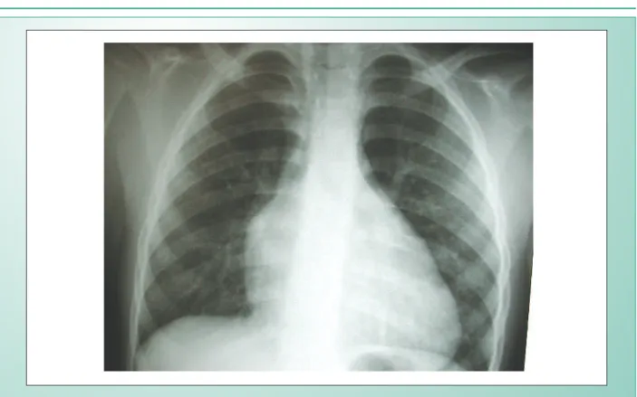
Radiograph imaging
The image shows a mild enlargement of the cardiac silhouette (CTI: 0.53) at the expense of a long left ventricular border. A bulging middle arch can be observed and the aortic arch right above is normal. The pulmonary vasculature is slightly increased, especially on the left side at the hilus and lower lobe. The right lower arch is not protuberant (Figure 1).
Diagnostic impression
This image suggests a heart disease like a ductus arteriosus/ ventricular septal defect among the non-cyanogenic heart diseases, because of the increased pulmonary blood flow with enlargement of the left ventricle and of the pulmonary
trunk. In cyanogenic heart diseases, the long middle arch can, however, correspond to the image originated by a rudimentary right ventricle located to the left and with ventriculo-arterial discordance as occurs in the single left ventricle, similar to the corrected transposition of the great arteries. Increased pulmonary blood flow would result from not severe pulmonary stenosis or from the flow through the systemic-to-pulmonary anastomosis.
Differential diagnosis
In the context of cyanogenic heart diseases with enlargement of the left ventricle a differential diagnosis could be made with anomalies such as tricuspid atresia, mitral atresia and pulmonary atresia with an intact ventricular septum. However, the bulging middle arch rules out these hypotheses.
Diagnostic confirmation
The clinical findings suggest cyanogenic heart diseases with right ventricle hypoplasia because of the accentuated heart sounds, more intense in the mitral area. Therefore, pulmonary and tricuspid atresia should be considered, in addition to the single ventricle. Among these conditions, the right ventricular overload suggests single left ventricle with ventriculo-arterial discordance, with the rudimentary right ventricle located in the left side, corroborating the radiograph imaging. The echocardiogram showed situs solitus, univentricular atrioventricular connection with the morphologically single left ventricle located to the right in relation to the left anterior rudimentary right ventricle. The ventriculo-arterial connection is discordant and a hypoplastic atresic pulmonary valve is observed. The pulmonary arteries are merging. The left pulmonary artery was 13 mm long and the right was 11.5 mm long. The left systemic-to-pulmonary anastomosis was patent and no dysfunction of the two atrioventricular valves was observed.
Management
Because of the high oxygen saturation and absence of heart failure we chose a conservative management. The Fontan operation will be duly considered according to the progression of the hypoxia.
Clinicoradiological Session
Edmar Atik
CASE 4/2006 – thiRtEEn-yEAR-old tEEnAgER With SinglE lEft vEntRiClE, vEntRiCulo-ARtERiAl diSCoRdAnCE With lEft AoRtA, pulMonARy StEnoSiS And pREviouS pulMonARy SyStEMiC Shunt
Arq Bras Cardiol 2006; 87 : 602-603
Fig. 1 - Chest radiograph shows the presence of a bulging middle arch corresponding to the rudimentary right chamber located to the left, with a long ventricular border originating in the single left ventricle to the right.