Letters to the Editor
Radiol Bras. 2016 Jul/Ago;49(4):267–276
273
Maurício Fabro1, Sara Raquel Fabro1, Rafael Santiago Oliveira de Sales1, Luiz Pedro de Souza Júnior1, Julian Catalan1
1. Hospital Santa Catarina de Blumenau, Blumenau, SC, Brazil. Mailing address: Dr. Maurício Fabro. Hospital Santa Catarina de Blumenau – Radiologia. Rua Ama-zonas, 301, Garcia. Blumenau, SC, Brazil, 89020-900. E-mail: mauriciofabro@ hotmail.com.
1971(5), the initial reports of the disease in the extraoral
gas-trointestinal tract not appearing until 2001(6).
Pulse granuloma is characterized by a chronic granuloma-tous reaction to a foreign body of vegetable origin(1), typically
in-digestible cellulose deposited under the mucosa(3). Most pulse
granuloma patients have a history of bowel disease, including di-verticulitis, fistula, perforation, ulcerative colitis, appendicitis, or anastomotic leakage(7), allowing the foreign body to reach the deep
layers of the intestinal wall. The oral cavity is the site most often affected, the occurrence of pulse granuloma at other sites being extremely rare(3). However, there have been reports of pulse
granu-loma in the stomach, small intestine, colon, peritoneum, mesen-tery, genitourinary tract, and skin(2,7).
Pulse granuloma predominantly affects males(7), of a broad
range of ages, cases having been described in patients from 13 to 85 years of age(7). The symptoms are vague and nonspecific(8),
occasionally including abdominal pain and discomfort(2).The
physi-cal examination is usually unremarkable, although a palpable mass can be identified(2,7).
The imaging evaluation of pulse granuloma is usually made either by ultrasound, the findings of which are often nonspecific, or by CT, which is more relevant because of its high sensitivity and specificity for the detection and characterization of foreign bodies in the gastrointestinal tract(8). Nevertheless, because
for-eign bodies of vegetable origin do not produce hyperintense im-ages, the diagnosis is not usually obtained by CT. Upper gas-trointestinal endoscopy is a useful tool in the study of gastric le-sions and allows the collection of material for histopathological evaluation. However, endoscopic biopsies are usually small and superficial, which can make it difficult to confirm the diagnosis of granuloma pulse(8). The diagnosis is made through exclusion on
http://dx.doi.org/10.1590/0100-3984.2015.0058
the basis of the histopathological findings(1). The possibility of
pulse granuloma should be considered in cases of expansive le-sions in the gastrointestinal tract(8), the main differential
diag-noses being adenocarcinoma, gastrointestinal stromal tumor, and leiomyoma(8). The definitive treatment is surgical intervention(1).
REFERENCES
1. Razavi A, Vlcek D, Kutten-Berger JJ. Oral pulse granuloma of the man-dible – a case report. Swiss Dent J. 2014;124:665–76.
2. Geramizadeh B, Mousavi SJ, Bananzadeh A. Omental mass caused by pericolic vegetable granuloma: a rare case report. Ann Colorectal Res. 2014;2:e20233.
3. Yeo NK, Eom DW, Lim HW, et al. Vegetable or pulse granuloma in the nasal cavity. Clin Exp Otorhinolaryngol. 2014;7:334–7.
4. Knoblich R. Pulmonary granulomatosis caused by vegetable particles. So-called lentil pulse pneumonia. Am Rev Respir Dis. 1969;99:380–9. 5. Lewars PH. Chronic periostitis in the mandible underneath artificial
den-tures. Br J Oral Surg. 1971;8:264–9.
6. Rhee DD, Wu ML. Pulse granulomas detected in gallbladder, fallopian tube, and skin. Arch Pathol Lab Med. 2006;130:1839–42.
7. Nowacki NB, Arnold MA, Frankel WL, et al. Gastrointestinal tract-de-rived pulse granulomata: clues to an underrecognized pseudotumor. Am J Surg Pathol. 2015;39:84–92.
8. Shan GD, Chen ZP, Xu YS, et al. Gastric foreign bogy granuloma caused by an embedded fishbone: a case report. World J Gastroenterol. 2014;20: 3388–90.
Caseous calcification of the mitral annulus: computed tomography features
Dear Editor,
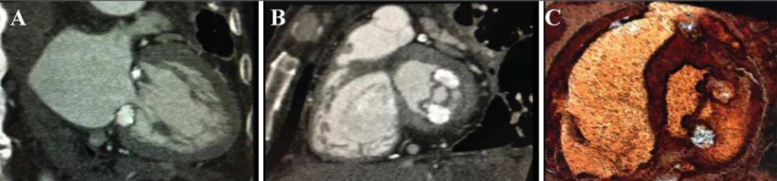
A 62-year-old patient with chronic kidney disease, who was undergoing treatment with intermittent dialysis, was admitted to the hospital for investigation of a complaint of progressively wors-ening dyspnea, despite the optimization of the dialysis. To eluci-date the case, ancillary tests were ordered, such tests including echocardiography. The echocardiography showed an expansive formation in the mitral valve, and cardiac computed tomography (CCT) was performed in order to better evaluate that finding (Fig-ure 1). The CCT identified a coarse caseous calcification between the anterior and posterior commissures, accompanied by a
sig-nificant reduction in the size of the mitral valve orifice, with a maximum aperture of 0.7 cm3
, as determined by planimetry. The CCT images allowed the diagnosis of degenerative caseous cal-cification of the mitral annulus.
Improving the use of imaging methods in the evaluation of cardiovascular diseases has been the objective of a number of recent studies in the radiology literature of Brazil(1–5). Caseous
calcification of the mitral annulus is a chronic degenerative pro-cess that usually involves the posterior mitral annulus(6). It is most
prevalent in elderly females(7) and in patients with chronic kidney
disease who are on hemodialysis(8–10). It is a rare disease,
account-ing for only 0.5–1.0% of all calcifications of the mitral annulus. Although rare, it is one of the major differential diagnoses of car-diac tumors, thrombi, vegetations, and abscesses(11).
Letters to the Editor
Radiol Bras. 2016 Jul/Ago;49(4):267–276
274
In most cases are asymptomatic patients, and the diagnosis is established by examination of cardiac imaging performed for other purposes. The symptoms, when present, correspond to pal-pitations, dyspnea, and syncope(11). The prognosis of caseous
de-generation of the mitral annulus is good, especially in patients who are asymptomatic, although some patients develop severe symptomatic valvular dysfunction; in the latter group of patients, the prognosis is poor and surgery should be considered(9,12).
On the CCT scans, we noted a hyperintense crescent-shaped mass or a well-defined oval-shaped mass with peripheral calcifica-tion, usually along the posterior mitral annulus, which was not enhanced after contrast administration(13). The heterogeneity of
the content of the mass was confirmed by the variation in its den-sity, which can range from negative Hounsfield units, suggest-ing fatty degeneration, to elevated Hounsfield units, suggestsuggest-ing a high protein content and structural calcification(14). The
cen-tral hypointensity was secondary to liquefaction of the calcium that fills the center of mass(11,13,15).
In this context of our findings in the case presented here, we can conclude that CCT helps confirm the diagnosis, allows the degree of mitral valve stenosis to be evaluated, and offers mea-sures to improve treatment strategies, especially those involving transcatheter or percutaneous transapical mitral valve implanta-tion. Therefore, CCT is considered an excellent tool for the diag-nosis of caseous degeneration of the mitral annulus.
REFERENCES
1. Neves PO, Andrade J, Monção H. Coronary anomalies: what the radiolo-gist should know. Radiol Bras. 2015;48:233–41.
2. Araújo Neto CA, Oliveira Andrade AC, Badaró R. Intima-media com-plex in the investigation of atherosclerosis in HIV-infected patients [Let-ter]. Radiol Bras 2014;47(1):x.
3. Brasileiro Junior VL, Luna AHB, Sales MAO, et al. Reliability of digital panoramic radiography in the diagnosis of carotid artery calcifications. Radiol Bras. 2014;47:28–32.
4. Ramos SMO, Glavam AP, Kubo TTA, et al. Optimization of a protocol for myocardial perfusion scintigraphy by using an anthropomorphic phantom. Radiol Bras. 2014;47:217–22.
Fernanda Boldrini Assunção1, Diogo Costa Leandro de Oliveira1, Alair Augusto Sarmet Moreira Damas dos Santos2, Marcelo Souto Nacif2
1. Complexo Hospitalar de Niterói (CHN), Niterói, RJ, Brazil. 2. Univer-sidade Federal Fluminense (UFF), Niterói, RJ, Brazil. Mailing address: Dra. Fernanda Boldrini Assunção. Hospital Universitário Antonio Pedro – Radiologia. Rua Marquês do Paraná, 303, 2º andar, Centro. Niterói, RJ, Brazil, 24030-900. E-mail: [email protected]. 5. Barranhas AD, Santos AASMD, Coelho-Filho OR, et al. Cardiac
mag-netic resonance imaging in clinical practice. Radiol Bras. 2014;47:1– 8.
6. Stamou SC, Braverman AC, Kouchoukos NT. Caseous calcification of the anterior mitral valve annulus presenting as intracardiac mass. J Thorac Cardiovasc Surg. 2010;140:e9–e10.
7. França LA, Rodrigues ACT, Vieira MLC, et al. Calcificação caseosa do anel mitral: relato de caso. Einstein. 2013;11:370–2.
8. Sequeira A, Morris L, Patel, B, et al. Calcific mitral stenosis in the he-modialysis patient. Hemodial Int. 2014;18:212–4.
9. Mozenska O, Sypula S, Celinska-Spoder M, et al. Mitral annulus caseous calcification mimicking cardiac mass in asymptomatic patient – multimodality imaging approach to incidental echocardiographic find-ing. Pol J Radiol. 2014;79:88–90.
10. Stone E, Cohn D, Deal C, et al. Calcific atrial mass in end-stage renal failure. Nephrol Dial Transplant. 1997;12:807–10.
11. Elgendy IY, Conti CR. Caseous calcification of the mitral annulus: a review. Clin Cardiol. 2013;36:E27–31.
12. Mallat N, Limeme M, Zaghouani H, et al. Caseous calcification of the mitral annulus on MDCT: a rare intracardiac mass. Acta Radiol Short Rep. 2013;2:2047981613502177.
13. Vanovermeire OM, Duerinckx AJ, Duncan DA, et al. Caseous calcifica-tion of the mitral annulus imaged with 64-slice multidetector CT and magnetic resonance imaging. Int J Cardiovasc Imaging. 2006;22:553– 9.
14. Harpaz D, Auerbach I, Vered Z, et al. Caseous calcification of the mitral annulus: a neglected, unrecognized diagnosis. J Am Soc Echocardiogr. 2001;14:825–31.
15. Ribeiro S, Salgado A, Salomé N, et al. Caseous calcification of the mitral annulus: a multi-modality imaging perspective. Rev Port Cardiol. 2012; 31:313–6.
http://dx.doi.org/10.1590/0100-3984.2015.0096
Aortic arch anomaly in an adult patient: a case of right aortic arch with aberrant left subclavian artery and Kommerell’s diverticulum
Dear Editor,
We report the case of a 54-year-old male presenting with vague symptoms of discomfort when swallowing. The patient un-derwent magnetic resonance imaging of the chest. The exami-nation showed right aortic arch with an aberrant left subclavian artery and Kommerell’s diverticulum (Figures 1 and 2).
Thoracic diseases of vascular origin have been the subject of a number of recent publications in the radiology literature of Bra-zil(1–5). First described by Fioratti et al., right aortic arch is an
un-common birth defect, of unknown cause, occurring in 0.05% of the general population. It is often asymptomatic but can be ac-companied by dysphagia and complications arising from the for-mation of an aneurysm. Such an aneurysm generally occurs at the origin of the left subclavian artery and is known as Kommerell’s aneurysm or Kommerell’s diverticulum, which can cause com-pression of mediastinal structures or can rupture spontaneously(6– 13). In children, the symptoms can also be associated with
exist-ing congenital cardiac abnormalities(7).
Various systems for classifying right aortic arch have been proposed. The most widely used classification system is that de-vised by Edwards, who described three main types of right aortic
arch: type I, with mirror-image branching of the major arteries; type II, with an aberrant subclavian artery; and type III, with an isolated subclavian artery (connected to the pulmonary artery via the ductus arteriosus)(8). In the case presented here, the variant
was classified as an Edwards type II right aortic arch, which ac-counts for approximately 40% of all cases(7).
In an autopsy study cited by Faucz et al.(7), 50% of cases of
right aortic arch were associated with an aberrant left subclavian artery, which can be located behind the esophagus (in 80%), be-tween the trachea and the esophagus (in 15%), or anterior to the trachea (in 5%). In some cases, right aortic arch is associated with a congenital heart defect(7,9,10).
The treatment of right aortic arch is generally surgical and is quite complex. Preoperative imaging tests are extremely impor-tant for the surgical planning, which relies heavily on knowledge of the anatomical distribution of the local structures, as well as of the size and extent of the aneurysm. Although outpatient treat-ment is an option, endovascular repair has been performed suc-cessfully(7,11).