678
Brazilian Journalof otorhinolaryngology 77 (5) SeptemBer/octoBer 2011
http://www.bjorl.org / e-mail: [email protected]
Billateral sudden deafness
Therezita M. Peixoto Patury Galvão Castro1, Laurisson Albuquerque da Costa2, Maria Eliza Alencar Nemezio3, Lucas José Sá da Fonseca4
1 PhD in Medicine - School of Medical Sciences of the Santa Casa de São Paulo (FCMSCSP). Adjunct Professor of Otorhinolaryngology - UNCISAL and UFAL. 2 6th year medical student - Federal University of Alagoas.
3 6th year medical student - Federal University of Alagoas. 4 6th year medical student - Federal University of Alagoas.
Universidade Federal de Alagoas (UFAL).
Send correspondence to: Av. Álvaro Otacílio, 3031 apto. 402. Ponta Verde, Maceió - AL. CEP: 57035-180.
Paper submited to the BJORL-SGP (Publishing Management System – Brazilian Journal of Otorhinolaryngology) on August 08, 2010; and accepted on October 12, 2010. cod. 7261
CASE REPORT
Braz J Otorhinolaryngol. 2011;77(5):678.
BJORL
Keywords: hearing loss, neurilemmoma, stroke, sudden.
.org
INTRODUCTION
Sudden hearing loss is described as a sensorineural hearing loss starting at 30 decibels (dB) in three of more contiguous frequencies, which can be severe and irreversible, which onset happens suddenly along 3 days. Au-ditory involvement is usually unilateral in 98%-99% of the cases1,2. It does not
have a well-defined etiological factor, and most are idiopathic; it can be due to vascular involvement, viral infection, autoimmune disorder or rupture of the intracochlear membrane; etiology confirmation is rather difficult, since many can be the causal agents. Mo-reover, these factors may manifest in a synergic way , giving the disorder a multifactorial etiology3,4. The acoustic
neuroma is considered a definitive cause of sudden hearing loss. The most frequent accompanying symptoms are tinnitus in 70% to 90% of the cases and dizziness in 20% to 40% of the cases4,5.
For the sudden hearing loss diagnosis, besides the detailed anam-neses and physical exam, a complete audiometric evaluation is paramount, brainstem evoked-response audiome-try (BERA), vestibular tests and trige-minal nerve assessment, as well as MRI with gadolinium and other tests in the pursue of the etiology5,6.
CASE PRESENTATION
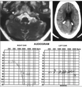
M.T.T.A., female, an 82 year-old retiree, coming from Maceió - AL, was seen in the ENT ward, where she complained of tinnitus and sudden hearing loss in her right ear for about 12 years, at the time she was diagno-sed with acoustic neuroma by means of an MRI (Figure 1). she also stressed that at about 18 months ago, after an emotional stress, she was affected by
a sudden hearing loss episode in her left ear, when a skull CT scan was or-dered for her, which showed a small hypodense area next to the right lateral ventricle horn (Figure 1), suggesting cerebral ischemic injury as the cause of the left-side sudden hearing loss. Audiology tests were carried out and upon tonal audiometry (Figure 1) we found a bilateral sensorineural hearing loss, and the patient still had a better hearing in her right ear. She was ad-vised to wear a hearing aid in both ears and go through multidisciplinary outpatient follow up by a geriatrician, cardiologist and a neurologist.
DISCUSSION
The case hereby reported is rare, it is a bilateral sudden hearing loss, of non-simultaneous occurrence, suggested by the tonal audiometry test6, as it shows bilateral
sensorineu-ral hearing loss above 30 dB in more
than three contiguous frequencies1,2.
Initially, she had right ear hearing loss together with tinnitus - a common symptom in 80% of the cases4,5, and
the hearing loss cause was established by the MRI, which showed an acoustic neuroma. Later, she had sudden hea-ring loss on the left side, of probable vascular involvement, which showed in the skull CT scan3,4.
FINAL REMARKS
The present report points to the likelihood of non-simultaneous sudden bilateral hearing loss, due to the partici-pation of numerous etiological factors which, added together, may cause se-vere loss to the bearer. Thus, we stress the importance of the early diagnosis of hearing involvement and the control of pathologies, especially vascular ones, which may cause sudden hearing loss, as in this clinical case and thus have a negative repercussion in the quality of life of the elderly.
REFERENCES
1. Wilson WR, Byl FM, Laird N. The efficacy of steroids in the treatment of idiopathic sudden hearing loss. A double-blind clinical study. Arch Otolaryngol. 1980;106(12):772-6. 2. Vasama JP, Linthicum FH Jr. Idiopathic
su-dden sensorioneural hearing loss: temporal bone histopathologic study. Ann Otol Rhinol Laryngol. 2000;109(6):527-32.
3. Guyot JP, Thielen K. Evolution of sudden deafness without treatment. Schweiz Med Wochenschr Suppl. 2000;116:93S-96S. 4. Maia RA, Cahali S. Surdez súbita. Rev Bras
Otorrinolaringol. 2004;70(2):238-48. 5. Hashisaki GT. Perda auditiva Sensorial
Súbi-ta. In: Bailey B & Johnson. Otorrinolaringolo-gia CirurOtorrinolaringolo-gia de Cabeça e Pescoço. 4ª edição: Rio de Janeiro. Ed. Revinter; 2010.p.1143-9. 6. Fitzgerald DC, Mark AS. Sudden hearing
loss: frequency of abnormal findings on contrast-enhanced MR studies. AJNR Am J Neuroradiol.1998;19(8):1433-6.