Periodontal Disease in Patients with Ischemic Coronary
Atherosclerosis at a University Hospital
Ana Lúcia Azevedo Barilli, Afonso Dinis Costa Passos, José Antônio Marin-Neto, Laércio Joel Franco
Faculdade de Medicina de Ribeirão Preto – USP, Ribeirão Preto, SP, Brazil, BrazilObjective: To verify the prevalence of periodontal disease (PD) in patients with ischemic heart disease. PD is a serious public dental health care problem with a differentiated distribution in regards to severity, age group, type of infection, co-morbidities and risk factors.
Methods: Four hundred and eighty patients were examined at the Ischemic Cardiopathy Outpatient Clinic of the Hospital das Clínicas of the Ribeirão Preto Medical School, São Paulo University, as well as 154 patients without heart disease from the same institution. Fifty-eight patients with heart disease and 62 patients without heart disease between the ages of 30 and 79 met the criteria to be included in the investigation. In accordance with recommendations of WHO (1999) the Community Periodontal Index (CPI) and the Periodontal Attachment Loss Index (PALI) were used.
Results: Houve predomínio de sextantes com DP moderada e grave nos pacientes com cardiopatia (76,3% versus 20,2%; p < 0,00001). Nesses pacientes, 1,1% dos sextantes exibiram saúde periodontal, contra 32,0% nos sem cardiopatia (p < 0,00001). No tocante à história pregressa das DP, 6,0% dos sextantes não exibiram perda de inserção entre os pacientes com cardiopatia, contra 68,0% nos sem cardiopatia (p < 0,00001). Observou-se biofilme dental em 100,0% dos pacientes com cardiopatia e em 82,3% dos sem cardiopatia (p < 0,001). Necessitavam de tratamento de bolsas periodontais > 6 mm, 79,3% dos pacientes com cardiopatia contra 9,7% dos sem cardiopatia (p < 0,0001).
Conclusion: PD was very prevalent in the groups studied with a higher degree of severity in those with ischemic heart diseases. The elevated prevalence of risk factors found, indicates that intervention strategies are required.
Key words: Periodontal disease, heart diseases, periodontal index, periodontal attachment loss.
Mailing Address: Ana lúcia Azevedo barilli •
Av. Bandeirantes, 3900 – 14049-900 – Ribeirão Preto, SP, Brazil
Periodontal disease is universal, representing a serious public dental health problem for both developing and industrialized countries1,2. In Brazil, PD is the second leading public oral health issue surpassed only by tooth decay3. Dental biofilm and calculus are important factors for the onset of PD, and are highly prevalent in adults and children worldwide. Nevertheless, the most serious manifestations present differentiated distributions that vary according to age group, type of infection, systemic problems and risk factors1,2,4.
While tooth decay results in demineralization of the teeth, PD is the result of periodontal tissue destruction caused by the action of toxic products released in the sub-gingival area by specific periodontal pathogens5. PD can also result from inflammatory and immunological responses caused by the presence of microorganisms and their toxic subproducts (lipopolysaccharides-LPS)5. It is important to recognize PD as an infectious disease caused by specific anaerobic bacteria, strongly suspected to be transmissible, multifactorial affecting populations and sites of risk6-10. PD is a chronic disease that
is usually assymptomatic5.
Recent studies6,11-21 demonstrate that the association between oral health and atherosclerosis is consistent in different population samples and that the oral conditions precede coronary events.
In PD there is a possibility of bacteremia due to the proximity of the infectious agents with the connective tissue and its vascular components. In moderate and advanced cases, the endotoxins (for example, LPS) of the microbial wall can stimulate the accumulation of plaque contributing to the formation of thrombi and atheroma plaque6,8,16. It is very possible that this physiopathological link is the conclusive explanation of the association between the two conditions.
probe along the cervical surface with the rounded tip inserted in the gingival sulcus and probing the entire extension. In the periodontal exam technique, the dentition is divided in six parts or sextants, and the sextant was only examined if there were two or more teeth present that were not indicated for extraction due to tooth decay.
The CPI codes are: 0 (healthy); 1 (bleeding when probed); 2 (calculus detected, but black band of the probe is visible); 3 (4-5mm periodontal pocket and gingival margin on black band on probe); 4 (periodontal pocket 6mm or more, black band on probe not visible); X (sextant excluded, less than two teeth present). Periodontal attachment loss (PAL) was observed in the same sextants in order to estimate the accumulated tissue destruction throughout the life of the periodontal attachment. The cementoenamel junction (CEJ) is exposed or visible when there is gingival recession and is used as a reference. This enables comparisons between the population groups without attempting to describe the extent of attachment loss in an individual. The PAL extent is recorded using the following codes: 0 (loss of attachment of 0-3 mm, CEJ is not visible and CPI value between 0-3); 1 (loss of attachment of 4-5 mm, CEJ within the black band); 2 (loss of attachment of 6-8 mm and CEJ between the upper limit of the black band and the 8.5 mm ring); 3 (loss of attachment of 9-11 mm and CEJ between the 8.5 mm and 11.5 mm rings); 4 (loss of attachment of 12 mm or more and CEJ beyond the 11.5 mm ring); X (sextant excluded and less than two teeth present). For both indexes the worst condition observed was registered; however this does not exclude the presence of less serious conditions at other sites of the sextants evaluated.
The information was entered into the Program EpiInfo 6.0 and analyzed with the Program Stata 6.0, using the chi-square test to verify associations. In the situations in which the reduced sample size inhibited the use of the chi-square test, the Fisher exact test was used. In all situations, the statistic significance limit adopted was equal to 0.05.
Results
In the initial stage 634 patients were examined between April 15 and September 19, 2002, of which 480 had heart disease and 154 did not.
Of the 480 patients with heart disease, 62.5% were males between the ages of 33 and 86 and 37.5% were females between the ages of 32 and 86. The average and median ages were 61.8 years and 62.5 years respectively, and 405 (84.4%) of the individuals were more than 50 years old. The results relative to the number of teeth revealed that 237 people (49.4%) were edentulous, 160 (33.3%) had between one and 19 teeth, 34 (7.1%) had between 20 and 24 teeth, 35 (7.3%) had between 25 and 28 teeth, and 14 (2.9%) had between 29 and 32 teeth. On the whole, 397 (82.7%) did not have enough healthy and/or restored natural teeth to perform normal oral functions and required major mouth rehabilitation interventions.
Of the 154 patients without heart disease, 66.2% were male and 33.8% were female. The ages of the males ranged from 21 to 61 years and for the females between 17 and 64. cultural classes, individuals that suffer from psychic stress and
with substantial genetic predisposition5,7,12,15,22,23.
Studies conducted in the past few decades11-21, demonstrate that people with advanced periodontal diseases present morbidity and mortality risks for cardiovascular disease (CVD) 25% higher than those with mild PD, even in the presence of other factors. It was also observed that people under 50 years of age with advanced PD have a 70% chance of developing CVD in the future13,19,21.
Since there are few studies regarding oral health in general and PD in particular, data relating to distribution and risk factors for these diseases are in short supply. The objective of the present investigation was to study oral conditions and requirements as well as the prevalence of PD in patients with ischemic heart disease with follow-up in a tertiary hospital, comparing the data with those obtained from a sample of patients without heart disease.
Methods
Patients from the Hospital das Clínicas (HCFMRP-USP) of the Ribeirão Preto Medical School, University of São Paulo with ischemic coronary atherosclerosis and a cardiac diagnosis confirmed of groups 124 and 125, CID 10, of the Ischemic Cardiopathy Outpatient Clinic were studied. As a comparison group, patients without ischemic heart disease from the Hepatitis Outpatient Clinic were used.
In the initial stage of the study, the patients were interviewed and examined in the medical office during their regularly scheduled appointment. During this first contact, data regarding oral conditions were collected and the potential participants of the periodontal investigation were identified. The interviews and oral exams for all candidates were performed by the same dental surgeon. Difficulties in recruiting patients with heart disease and a minimum number of 20 teeth as well as age matching with patients without heart disease altered the initial planning of the study sample size (100 people per group). At the Ischemic Cardiopathy Outpatient Clinic, 480 patients were initially evaluated and only 58 (12.1%) fulfilled the criteria to be included in the periodontal investigation. In the Hepatitis Outpatient Clinic, 154 patients were evaluated of which 62 (40.3%) were included in the second stage.
The patients between the ages of 30 and 79 and with a minimum of 20 functional teeth, were then placed in two new groups for periodontal evaluation. Even if the patients met the criteria for age and number of teeth, they were excluded if they had a previous history of bacterial endocarditis, congenital or rheumatic heart disease or HIV.
The average and median ages were 41 and 40 respectively and 117 (76.0%) were less than 50 years of age. The results of tooth counts were substantially different from those observed in the heart disease group. Ten people (6.5%) were edentulous, 48 (31.2%) had between one and 19 teeth, 19 (12.3%) had between 20 and 24 teeth, 45 (29.2%) had between 25 and 28 teeth and 32 (20.8%) had between 29 and 32 teeth. On the whole, 56 (37.7%) did not have a sufficient number of natural teeth to fulfill chewing, phonetic and aesthetic functions without major mouth rehabilitation.
In relation to dental treatment requirements, 282 (58.8%) of the heart disease patients required prosthetic rehabilitation, 200 (41.7%) required periodontal treatment, two (0.4%) required endodontic treatment, 51 (10.6%) required oral surgery (exclusively to remove damaged teeth), 69 (14.4%) required restoration procedures, one (0.2%) required other dental procedures and, one (0.2%) did not require any dental interventions. Among the patients without heart disease, 58 (37.7%) required prosthetic rehabilitation, 92 (59.7%) required periodontal treatment, none required endodontic treatment, 21 (13.6%) required oral surgery (exclusively to remove wisdom teeth); 23 (14.9%) required restoration procedures, five (3.2%) required other dental procedures and, 29 (18.8%) did not require any dental interventions.
In the periodontal investigation of the 58 patients with heart disease, 48 (82.8%) were men and ten (17.2%) were women. Ages ranged between 33 and 75 for the men and between 38 and 70 for the women. The median age for men was 53 years and for women 55. The average age for both genders was 53 years.
All required periodontal treatment, 70.7% required restoration procedures, 62.1% required counseling regarding routine exams for oral cancer prevention, 41.4% required prosthetic rehabilitation, 20.7% required oral surgery to remove condemned teeth, 3.4% required endodontic treatment and 3.4% required other procedures.
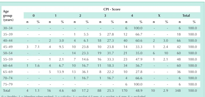
Based on the CPI, four (1.1%) sextants were healthy, 16 (4.6%) had bleeding during probing, 60 (17.2%) had calculus, 88 (25.3%) had periodontal pockets of 4-5 mm, 170 (48.9%) had periodontal pockets > 6 mm, and 10 (2.9%) sextants were excluded from the investigation as there were less than two teeth present (Tab.1). Eighty sextants (23.7%) were classified within the CPI scores indicating healthy gingiva or a minimal degree of PD (CPI = 0, 1 and 2), and 258 sextants (76.3%) were classified within the CPI scores indicating moderate to advanced degrees of PD (CPI = 3 and 4).
In reference to lack of periodontal treatment, 79.3% of the patients required treatment for periodontal pockets > 6 mm, 15.5% for treatment of periodontal pockets of 4-5 mm and 5.2% required other less complex periodontal treatments (Tab.2). Therefore, three (5.2%) patients required minimal interventions and 55 (94.8%) required interventions for moderate to maximum PD.
Based on the accumulated tissue destruction throughout the life of the periodontal attachment, measured by loss of attachment, 21 sextants (6.0%) presented no loss of attachment, 103 (29.6%) had loss of attachment between
4-5 mm, 149 (42.8%) loss of attachment between 6-8 mm, 57 (16.4%) loss of attachment between 9-11 mm, eight (2.3%) loss of attachment of 12 mm or more, and ten (2.9%) sextants were excluded from the exam. The categories “no loss of attachment” and “loss of attachment of 4-5 mm” were combined as these stages are not indicative of tooth loss and included 124 sextants (36.7%). All categories for loss of attachment of 6 mm and more were grouped together as these stages are indicative of tooth loss and included 214 sextants (63.3%).
Among the 62 patients without heart disease submitted to the periodontal investigation, 46 (74.2%) were male and 16 (25.8%) were female. Ages ranged from 30 to 61 for the males and from 30 to 60 for the females. The average and median ages for the men were 40 and 39 years and for the women 37 and 35 years, respectively.
It was observed that 51 (82.3%) patients in this group presented some type of problem of which 82.3% required periodontal treatment, 21.0% required restorations, 17.7% required prosthetic rehabilitation, 14.5% required surgery to remove condemned teeth, 9.7% required counseling regarding routine exams for the prevention of oral cancer, and 6.5% required other treatments.
Based on the CPI (Tab.3), healthy gingiva were observed in 119 sextants (32.0%), bleeding when probed in 55 (14.8%), calculus in 123 (33.1%), periodontal pockets of 4 to 5 mm in 58 (15.6%) and periodontal pockets > 6 mm in 17 (4.6%). The CPI scores for healthy gingiva and minimal degrees of PD (CPI = 0, 1 and 2) were combined and accounted for 297 sextants (79.8%), leaving 75 (20.2%) with moderate to maximum PD (CPI = 3 and 4).
Based on the number of people requiring periodontal treatment (Tab.4), it was observed that 41 patients (66.1%) required no or a minimal degree of treatment (CPI = 0, 1 and 2) and 21 (33.9%) required moderate to maximum degrees of treatment (CPI = 3 and 4).
Based on the accumulated tissue destruction throughout the life of the periodontal attachment, 253 sextants (68.0%) had no loss of attachment, 91 sextants (24.5%) had loss of attachment between 4 and 5 mm, and 28 sextants (7.5%) had loss of attachment of 6 to 8 mm. Loss of attachment greater than 8 mm was not observed in this group of patients. The categories “no loss of attachment” and “loss of attachment of 4-5 mm” were combined as these stages are not indicative of tooth loss and included 344 sextants (92.5%). All categories for loss of attachment of 6 mm or more were grouped together as these stages are indicative of tooth loss and included 28 sextants (7.5%).
Discussion
The elevated percentages of edentulous individuals and those with limited functions due to the low number of teeth are proof of the lack of adequate dental care at some point in their lives. This fact is also proven by the elevated number of individuals that required health care, particularly in regard to prostheses, periodontal treatment and oral cancer prevention. The worst conditions found among the individuals with heart disease are probably due to the advanced age of the members of this group (p < 0.0001) although the possible influence of PD in patients with ischemic heart problems cannot be disregarded. Thus, the greater severity of PD in patients with heart disease suggests that systemic factors could be simultaneously involved in the origin of the two diseases. However, age distribution discrepancies greatly limits result comparison when evaluating the two groups, particularly regarding the periodontal investigation, since PD is time dependent. Nevertheless, even when evaluated in the same age group, the two groups remained distinct regarding periodontal health. These results agree with the hypothesis that there could be a physiopathological relationship between CVD and PD in that PD is a risk factor for CVD.
The evaluation of general oral health conditions demonstrated that both groups required large percentages of dental treatments in all specialties. PD was very prevalent in the two study groups but much more severe in the patients with ischemic coronary atherosclerosis. Although not mentioned in the text, there was also a high prevalence of risk factors for PD, indicating the need to implement oral health care strategies that include preventative and educational measures for large population groups, early periodontal care and directed at groups with higher risks to develop PD (smokers, low socioeconomic and cultural classes, those with difficulty to modify inadequate habits of dental biofilm removal, people infected with HIV, diabetics, people with
psychomotor deficiencies and ischemic diseases).
In conclusion, the creation of methods to better orient patients regarding oral health care is essential. At the HCFMRP-USP clinic, diagnostic procedures and systematic treatment of periodontal diseases have been implemented, with emphasis placed on high risk groups, making quick and effective oral health care viable for those who need it.
The results of this study indicate the need to conduct new studies with larger samples and adequate age matching
Age group (years)
Periodontal Treatment Requirements
2 3 4 Total
n % n % n % n %
30–34 - - - - 1 100.0 1 100.0 35–39 - - - - 3 100.0 3 100.0
40–44 - - 1 9.1 10 90.9 11 100.0
45–49 1 14.3 1 14.3 5 71.4 7 100.0 50–54 - - 3 30.0 7 70.0 10 100.0 55–59 - - - - 8 100.0 8 100.0
60–64 - - 3 30.0 7 70.0 10 100.0
65–69 2 33.3 1 16.7 3 50.0 6 100.0 70–74 - - - - 1 100.0 1 100.0
75–79 - - - - 1 100.0 1 100.0
Total 3 5.2 9 15.5 46 79.3 58 100.0
2 = calculus removal + oral hygiene instruction (OHI); 3 = treatment of periodontal pocket 4-5 mm; 4 = treatment of periodontal pocket > 6 mm.
Table 2 - Periodontal treatment requirements for patients with heart disease according to age group. hCfMRP-USP, 2003 Age
group (years)
CPI - Score
0 1 2 3 4 X Total
n % n % n % n % n % n % n %
30–34 - - - 6 100.0 - - 6 100.0
35–39 - - - - 1 5.5 5 27.8 12 66.7 - - 18 100.0
40–44 - - 2 3.0 4 6.1 18 27.3 40 60.6 2 3.0 66 100.0
45–49 3 7.1 4 9.5 10 23.8 10 23.8 14 33.3 1 2.4 42 100.0 50–54 - - - - 14 23.3 19 31.7 21 35.0 6 10 60 100.0 55–59 - - 1 2.1 7 14.6 16 33.3 23 47.9 1 2.1 48 100.0 60–64 1 1.6 4 6.7 10 16.7 11 18.3 34 56.7 - - 60 100.0
65–69 - - 5 13.9 13 36.1 8 22.2 10 27.8 - - 36 100.0
70–74 - - - - 1 16.7 1 16.7 4 66.6 - - 6 100.0
75–79 - - - 6 100.0 - - 6 100.0
Total 4 1.1 16 4.6 60 17.2 88 25.3 170 48.9 10 2.9 348 100.0
0 = healthy; 1 = bleeding when probed; 2 = calculus; 3 = pocket 4-5 mm; 4 = pocket > 6 mm; X = excluded.
to accurately establish real oral health conditions in general population groups that receive care in clinics such as the one used in this study. They also indicate the requirement of Public Dental Care to be expanded to include preventive and rehabilitation care for age groups above 15 years of age.
Potential Conflict of Interest
No potential conflict of interest relevant to this article was reported.
Age group (years)
CPI - Score
0 1 2 3 4 Total
n % n % n % n % n % n %
30–34 25 20.8 21 17.5 59 49.2 15 12.5 - - 120 32.2
35–39 31 34.4 18 20.0 23 25.6 11 12.2 7 7.8 90 24.2 40–44 34 40.5 10 11.9 19 22.6 16 19.0 5 5.9 84 22.6
45–49 15 41.7 1 2.8 13 36.1 7 19.4 - - 36 9.7
50–54 8 44.4 2 11.1 6 33.3 2 11.1 - - 18 4.8
55–59 - - - - 2 33.3 4 66.7 - - 6 1.6
60–64 6 33.3 3 16.7 1 5.5 3 16.7 5 27.8 18 4.8
Total 119 32.0 55 14.8 123 33.1 58 15.6 17 4.6 372 100.0
70–74 - - - - 1 100.0 1 100.0
Total 3 5.2 9 15.5 46 79.3 58 100.0
0 = healthy; 1 = bleeding when probed; 2 = calculus; 3 = periodontal pocket 4-5 mm; 4 = periodontal pocket > 6 mm.
Table 3 - Community Periodontal Index (CPI) in patients without heart disease according to age group. hCfMRP-USP, 2003
Age group (years)
Periodontal Treatment Requirements
0 2 3 4 Total
n % n % n % n % n %
30–34 2 10.0 13 65.0 5 25.0 - - 20 100.0
35–39 3 20.0 7 46.7 3 20.0 2 13.3 15 100.0
40–44 4 28.6 5 35.7 2 14.3 3 21.4 14 100.0
45–49 1 16.7 3 50.0 2 33.3 - - 6 100.0
50–54 - - 2 66.7 1 33.3 - - 3 100.0
55–59 - - - - 1 100.0 - - 1 100.0
60–64 1 33.3 - - 1 3.33 1 3.33 3 100.0
Total 11 17.7 30 48.4 15 24.2 6 9.7 62 100.0
0 = no requirement; 1 = removal of biofilm + oral hygiene instruction (OHI); 2 = calculus removal; 3 = treatment of periodontal pocket 4-5 mm; 4 = treatment of periodontal pocket > 6 mm.
Table 4 - Periodontal treatment requirements for patients without heart disease according to age group. hCfMRP-USP, 2003
with heart disease
without heart disease
n % n %
Number of patients 40 69.0 59 95.1
CPI Score of 4 (presence
of periodontal pocket > 6
mm), by sextant
116 34.3 12 3.2
Treatment required for p o c k e t > 6 mm, by individual
34 58.6 5 8.1
Table 5 - Presence of periodontal pocket > 6 mm and complex periodontal treatment requirements in patients between the ages
References
1. Loe H, Anerud A, Boysen H, Morrison E. Natural history of periodontal
disease in man. Rapid, moderate and no loss of attachment in Sri-Lankan
tea laborers 14 to 46 years of age. J Clin Periodontol 1986; 13(5): 431-45. 2. Papapanou PN. Periodontal diseases: epidemiology. Ann Periodontol. 1996;
1(1): 1-36.
3. Brasil. Ministério da Saúde. Secretaria Nacional de Programas Especiais de Saúde. Divisão Nacional de Saúde Bucal. Levantamento epidemiológico em saúde bucal; Brasil, zona urbana, 1986. Brasília (DF), 1988. 137p. 4. Loe H, Theilade E, Jensen SB. Experimental gingivitis in man. J Periodontol
1965; 36: 177-87.
5. Listgarten MA. Nature of periodontal disease: pathogenic mechanisms. J Periodontal Res 1987; 22(3): 172-8.
6. Loesche WJ, Karapetow F, Pohl A, Kocher T. Plasma lipid and blood glucose in patients with destructive periodontal disease. J Clin Periodontol 2000; 27(8): 537-41.
7. Page RC. The pathobiology of periodontal disease may affect systemic disease: inversion of a paradigm. Ann Periodontol 1998; 3(1): 108-20. 8. Ross R. Atherosclerosis is an inflammatory disease. Am Heart J 1999; 138(5
Pt 2): S419-20.
9. Scannapieco FA. Position paper of The American Academy of Periodontology: periodontal disease as a potential risk factor for systemic disease. J Periodontol 1998l; 69(7): 841-50.
10. Williams RC, Offenbacher S. Periodontal medicine: the emergence of a new branch of periodontology. Periodontol 2000; 23: 9-12.
11. Beck J, Garcia R, Heiss G, Vokonas PS, Offenbacher S. Periodontal disease and cardiovascular disease. J Periodontol 1996; 67(10 Suppl): 1123-37. 12. Beck J. Periodontal implications: older adults. Ann Periodontol 1996; 1(1)
322-57.
13. DeStefano F, Anda RF, Kahn HS, Williamson DF, Russel CM. Dental disease and risk of coronary heart disease and mortality. Br Med J 1993; 306(6879): 688-91.
14. Glurich I, Grossi S, Albini B, et al. Systemic inflammation in cardiovascular and periodontal disease: comparative study. Clin Diagn Lab Immunol 2002; 9(2): 425-32.
15. Genco RJ. Current view of risk factors for periodontal diseases. J Periodontol 1996; 67(Suppl): 1041-9.
16. Herzberg MC, Meyer MW. Dental plaque, platelets, and cardiovascular disease. Ann Periodontol 1998; 3(1) 151-60.
17. Loesche WJ. Periodontal disease: link to cardiovascular disease. Compend Contin Educ Dent 2000; 21(6): 463-6, 468, 470.
18. Mattila KJ, Nieminen MS, Valtonen VV, et al. Association between dental health and acute myocardial infarction. Br Med J 1989; 298 (6676): 779-81.
19. Mattila KJ, Valtonen VV, Nieminen M, Huttunen JK. Dental infection and the
risk of new coronary events: prospective study of patients with documented
coronary artery disease. Clin Infect Dis 1995; 20(3): 588-92.
20. Mattila KJ, Valle MS, Nieminen MS, Valtonen VV, Hietaniemi KL. Dental infections and coronary atherosclerosis. Atherosclerosis 1993; 103(2): 205-11.
21. Morrison HI, Ellison LF, Taylor GW. Periodontal disease and risk of fatal coronary heart and cerebrovascular diseases. J Cardiovasc Risk 1999; 6(1): 7-11.
22. Haber J, Wattles J, Crowley M, Mandell R, Joshipura K, Kent RL. Evidence for cigarette smoking as a major risk factos for periodontitis. J Periodontol 1993; 64(1): 16-23.
23. Joshipura KJ, Rimm EB, Douglass CW, Trichopoulos D, Ascherio A, Willett WC. Poor oral health and coronary heart disease. J Dent Res 1996; 75(9): 1631-6.