Original Article
Predictive Value of Angina to Detect Coronary Artery Disease in
Patients With Severe Aortic Stenosis Aged 50 Years or Older
Aline Alves Vargas Gonçalves, Fabíola Lúcio Cardão, Maria Gabriela Gomes Soares, André Weksler, Clara Weksler,
Bernardo Rangel Tura, Paulo Roberto Dutra da Silva, Antônio Sérgio Cordeiro da Rocha
Instituto Nacional de Cardiologia Laranjeiras – Fundacor - Rio de Janeiro, RJ, Brazil
Objective: The objective of this study is to evaluate the value of angina pectoris as a predictor of CAD (coronary artery disease) in patients with AS (aortic stenosis) during and beyond the 5th decade of life.
Methods:The study population consisted of 186 consecutive patients with AS and ≥ 50 years of age, referred for surgical aortic valve replacement (AVR) between June 1989 and September 2004. Routine coronary angiography was performed for all patients. One hundred and one patients were males (54.3%) and 85 were females (45.7%), and the mean age was 66±8 years. One hundred and twenty-four patients (66.7%) had angina. The maximum transvalve gradient was 89.4±27.6 mmHg, and the aortic valve area measured 0.59±0.17 cm2. We calculated the sensitivity, specificity, positive and negative predictive values, as well as the likelihood ratio of a positive test result for angina in predicting the presence of CAD.
Results: Ninety-three patients (50%) had CAD. Of the 124 patients with angina, 68 (54.8%) had CAD, whereas of the 62 patients without angina, 25 had CAD (40.3%) (p=0.087). Therefore, the diagnostic sensitivity of angina to detect CAD was 73.1%, specificity was 39.7%, positive predictive value was 54.8%, negative predictive value was 59.6%, and the likelihood ratio of a positive test result was 1.6.
Conclusion: Angina pectoris is not a good predictor of CAD in patients with AS who are more than 50 years of age.
Key words:Angina pectoris, aortic valve stenosis, coronary diseases, heart valve diseases.
Mailing Address: Antônio Sérgio Cordeiro da Rocha •
Rua Roberto Dias Lopes, 220/201 22010-110 – Rio de Janeiro, RJ, Brazil
E-mail: [email protected]
Manuscript received April 4, 2005; Revised manuscript received January 8, 2006; Accepted March 30, 2006. Angina pectoris affects approximately two-thirds of patients
with severe aortic stenosis (AS)1. Angina is a symptom common to coronary artery disease (CAD) and severe aortic valvular stenosis. The prevalence of these two conditions increases with age, and the presence of aortic valve sclerosis alone increases the risk of cardiovascular death and acute myocardium infarction (AMI) by 50%2-4.
The surgical correction of both diseases within one single procedure has been shown to reduce the rates of intraoperative death and AMI, as well as late morbidity and mortality, compared to patients with significant CAD and severe AS who did not undergo MR (myocardial revascularization)at the time of aortic valve correction5.
Therefore, it would be of utmost importance to identify the presence of CAD based on clinical evidence. The symptom of angina could play a relevant role if its onset could truthfully indicate the presenceof associated CAD. The objective of this study is to assess the value of angina pectoris in predicting CAD in patients with AS who are over 50 years of age.
Methods
Between June 1989 and September 2004, patients referred for surgical correction of severe aortic valve stenosis, whether isolated or predominant, were screened at the Ministry of Health’s Instituto Nacional de Cardiologia Laranjeiras (Laranjeiras National Institute of Cardiology), in the city of Rio de Janeiro.
In order to participate in the study, patients had to meet the following criteria:
1. Inclusion criteria: a) age ≥ 50 years;
b) presence of severe AS, pure or predominant, with indication for surgery;
c) technically sound echocardiographic study to evaluate data.
2. Exclusion criteria: a) age under 50 years;
b) AS with no indication for surgery;
Original Article
gonçalves et al
PRedICTIVe VAlUe Of AngInA TO deTeCT COROnARy ARTeRy dISeASe In PATIenTS wITh SeVeRe AORTIC STenOSIS Aged 50 yeARS OR OldeR
Arq Bras Cardiol 2006; 87(6) : 641-644
lesions in the left coronary trunk (LCT), in 75% of patients with lesions in three vessels, in 81.3% of patients with lesions in two vessels, and in 62.5% of those with lesions in one vessel. Patients with lesions in two or more vessels or LCT were more affected by angina (81.1%) than those without CAD or with a lesion in one vessel (60.9%) (p=0.01).
Of the 124 patients with angina, approximately 68 (54.8%) had CAD, whereas of the 62 without angina, 25 (40.3%) had CAD (p=0.087). Therefore, sensitivity of angina to identify CAD was 73.1%; specificity was 39.7%; positive predictive value was 54.8%; negative predictive value was 59.6%, and thelikelihood ratio of a positive test resultwas 1.6.
Discussion
The incidence of CAD in patients with AS varies considerably depending on its prevalence in the population. Some studies show that the onset of degenerative calcific aortic stenosis bears many similarities with the process of CAD development, including the same risk factors: advanced age, male gender, systemic arterial hypertension, diabetes mellitus, and dyslipidemia3,4. The presence of CAD in patients with AS who need surgery has clinical significance, as this association translates into substantially increased surgical risks in cases where myocardial revascularization is not performed at the time of AVR5,8,9. Thus, it is of utmost importance to detect CAD before performing AVR.
With the improvement of noninvasive techniques for diagnosing and evaluating the severity of valve lesions, particularly echocardiography, cardiac catheterization has been reserved for diagnosing CAD. In this study, we tried to determine if angina pectoris, a very common symptom in both AS and CAD, could have a relevant role in the clinical detection of CAD and consequently dispense with cardiac catheterization. We observed that in a population of AS patients aged 50 years or older who needed AVR, approximately 67% experienced angina pectoris. Of these, approximately 55% had CAD (Tab. 1). These numbers are very similar to those reported in several studies in which about half of the patients with AS and angina also had CAD 8-10.
In our group of patients, however, CAD was also detected in about 40% of patients without angina. These data caused the sensitivity, specificity, positive and negative predictive values, and likelihood ratio to be relatively low within our population.
Comparable to the findings of our study, in a survey with 272 patients with AS and age > 18 years, Rapp et al. observed that diagnostic sensitivity, specificity, positive and negative predictive values of angina to identify CAD were low, and, therefore, of little clinical value for this assessment10.
Similar to what occurred in our study, other researchers reported an incidence of CAD ranging from 25% to 50% among AS patients without angina pectoris, which led them to recommend coronary angiography for all patients with AS regardless of the presence or absence of angina11-13. This opinion, however, is not shared by other researchers.
Based on a research about the relationship between angina pectoris and CAD in AS patientswho were of a more c) mild or severe aortic insufficiency;
d) moderate or severe mitral valvedamage; e) cardiomyopathy or myocarditis of any etiology; f) echocardiographic study not adequate for the analysis. AS was considered severe when the maximum gradient between the left ventricle (LV) and the aorta (AO) was greater than 50 mmHg, or the aortic valve area was smaller than 0.8 cm².
During that analysisperiod, 255 patients underwent surgery for the correction of severe AS.
Of the 255 patients, 186 met inclusion and exclusion criteria and were selected to participate in the study. One hundred and one (54.3%) were men and 85 (45.7%) were women, with ages ranging from 50 to 84 years (mean age was 66±8 years). All of them had symptoms consistent with severe aortic valve obstruction, i.e., 124 (66.7%) experienced angina, 130 (69.9%) experienced dyspnea, and 30 (16.1%) experienced syncope.
Angina pectoris was characterized as oppressive pain or discomfort in the anterior area of the chest triggered by physical exertion or emotions and relieved by rest or administration of sublingual nitrate. Dyspnea was detected when the patient had breathing difficulties associated with exertion or while lying down. Syncope was diagnosed in cases of loss of consciousness associated with physical exertion.
The maximum LV/AO gradient was 89.4±27.6 mmHg and the aortic valve area (AVA) measured 05.9±0.17 cm2. Aortic
stenosis etiology was determined based on echocardiographic, surgical, and anatomopathological findings.
All patients underwent continuous Doppler echocardiography b y c o n v e n t i o n a l t e c h n i q u e s . E c h o c a r d i o g r a p h i c measurements followed the “Guideline investigation norms for echocardiography equipment and techniques” 6. All of them were submitted to cardiac catheterization according to Sones’ or Judkins’ technique, which served mainly to assess the coronary arterial circulation7.
Statistical analysis - Numerical data are expressed as means and standard deviations. Non-paired two-tailed Student’s t test was used to compare continuous variables, and Fisher’s exact test was used to compare ratios. Bayes’ theorem was used to verify diagnostic sensitivity, specificity, positive and negative predictive values, and likelihood ratio of a positive test result for angina in identifying significant CAD. Significant CAD implied the existence of a ≥ 70%lumen obstruction in one or more main coronary arteries, or a ≥ 50% obstruction in the lumen of the left coronary artery trunk. The significance level was equal to or less than 5%.
Results
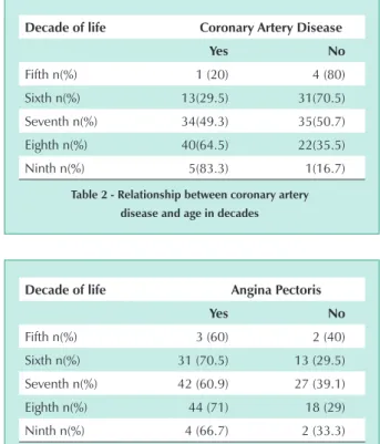
As shown on Table 1, 93 patients (50%) had CAD. The incidence of CAD gradually increased from the fifth (20%) to the ninth (83.3%) decades of life (p= 0.001) (Tab. 2). However, the occurrence of angina was not affected by age (Tab. 3).
Angina pectoris was present in68 of 93 patients with CAD (73.1%), compared to 56 of 93 patients without CAD (60.2%) (p=0.087). Angina was present in 100% of patients with
Original Article
gonçalves et al PRedICTIVe VAlUe Of AngInA TO deTeCT COROnARy ARTeRy dISeASe In PATIenTS wITh SeVeRe AORTIC STenOSIS Aged 50 yeARS OR OldeR
Arq Bras Cardiol 2006; 87(6) : 641-644
advanced age (> 70 years of age), Dangas et al., observed that angina in that population had a prognostic sensitivity of 78% and specificity of 82% in identifying CAD, and concluded that angina pectoris in that population strongly suggested the presence of CAD14.
Based on a study with 88 consecutive AS patients who needed AVR, Exadactylos et al, also suggested that coronary angiography would not be necessary in patients without angina15. Despite the fact that the authors of these two last studies suggested that coronary angiography could be unnecessary in cases of patients without angina, approximately 10.3% of the patients in the first study14 and 15% in the second study15 presented CAD in the absence of typical angina pectoris. In our study, in analyzing only the 76 patients over 70 years of age (data not expressed in the results),we verified that 68.6% of those with angina had CAD, whereas among those without angina 60% had CAD. Moreover, we demonstrated that, even though a significant association between aging and the presence of CAD (Tab. 2) was observed in our population, the same is not true with angina (Tab. 3).
Limitations of the study - This study evaluated patients with severe aortic valve stenosis who needed valve replacement;
therefore, our findings are not to be extended to the whole universe of patients with aortic stenosis.
We did not use noninvasive methods with physical or pharmacological stress which could help to discriminate
patients with and without CAD, as was very elegantly described by Avakian et al16 In that study, the researchers compared the value of single photon emission computed tomography (SPECT) with thallium during pharmacological stress with dipyridamole with that of coronary angiography in 110 patients with AS. They observed that SPECT was useful in excluding CAD in patients with typical angina, as well as in those asymptomatic or without typical angina. However, in a review article on the impact of the methods of perfusion imaging, Van Tosh concluded that, although these methods are accurate for AS patients, a regular study does not totally exclude the presence of CAD, and, therefore, these patients should undergo coronary angiography17.
Clinical implications - AS patients aged 50 years or older who need AVR should undergo coronary angiography before the surgery, regardless of the presence or absence of angina.
Conclusion
Angina pectoris is not a good predictor of the presence of CAD in patients with severe aortic valve stenosis beyond the fifth decade of life.
Age (years) 66±8
Men (%) 101 (54.3)
Women n (%) 85 (45.7)
Max. Grad. LV/AO (mmHg) 89.4±27.6
AVA (cm2) 0.59±0.17
Angina n(%) 124 (66.7)
Dyspnea n (%) 130 (69.9%)
Syncope n(%) 30 (16.1%)
CAD n(%) 93 (50)
CAD 1 vessel n(%) 40 (21.5)
CAD 2 vessels n(%) 32 (17.2)
CAD 3 vessels n(%) 16 (8.6)
LCTL n(%) 5 (2.7)
Max. grad. LF/AO = maximum gradient between the left ventricle and the aorta; CAD = coronary artery disease; LCTL = left coronary trunk lesion
Table 1 - Patient characteristics
decade of life Coronary Artery disease
yes no
Fifth n(%) 1 (20) 4 (80)
Sixth n(%) 13(29.5) 31(70.5)
Seventh n(%) 34(49.3) 35(50.7)
Eighth n(%) 40(64.5) 22(35.5)
Ninth n(%) 5(83.3) 1(16.7)
Table 2 - Relationship between coronary artery disease and age in decades
decade of life Angina Pectoris
yes no
Fifth n(%) 3 (60) 2 (40)
Sixth n(%) 31 (70.5) 13 (29.5)
Seventh n(%) 42 (60.9) 27 (39.1)
Eighth n(%) 44 (71) 18 (29)
Ninth n(%) 4 (66.7) 2 (33.3)
Table 3 - Relationship between angina and age in decades
References
1. Bonow RO, Braunwald E. Valvular heart disease. In: Zips DP, Libby P, Bonow RO, Braunwald E (eds). Braunwald’s heart disease: a textbook of cardiovascular medicine. 7th ed. Philadelphia: Elsevier Saunders; 2005. p. 1553-632.
2. Otto CM, Lind BK, Klitzman DW. Association of aortic-valve sclerosis with cardiovascular mortality in the elderly. N Engl J Med. 1999; 341: 142-7.
3. Ortlepp JR, Schmitz F, Bozoglu T, Hanrath P, Hoffmann R. Cardiovascular risk
factors in patientes with aortic stenosis predict prevalence of coronary artery disease but not of aortic stenosis: an angiographic pair matched case-control
study. Heart. 2003; 89: 1019-22.
4. Otto CM, O’Brien KD. Why is there discordance between calcific aortic stenosis and coronary artery disease? Heart. 2001; 85: 601-2.
Original Article
gonçalves et al
PRedICTIVe VAlUe Of AngInA TO deTeCT COROnARy ARTeRy dISeASe In PATIenTS wITh SeVeRe AORTIC STenOSIS Aged 50 yeARS OR OldeR
Arq Bras Cardiol 2006; 87(6) : 641-644
5. ACC/AHA guidelines for the management of patients with valvular heart
disease. A report of the American College of Cardiology/American Heart Association (Committee on management of patients with valvular heart
disease). J Am Coll Cardiol. 1998; 32: 1486-588.
6. Diretriz para investigação dos equipamentos e técnicas de exame para realização de exames ecocardiográficos. Arq Brasil Cardiol. 2004; 82 (supl 2): 1-10.
7. Diretriz de cirurgia nas valvopatias. Arq Brasil Cardiol. 2004; 82 (supl 5): 22-33.
8. Mandal AB, Gray IR. Significance of angina pectoris in aortic stenosis. Br Heart J. 1976; 38: 811-5.
9. Rahimtoola SH. Aortic valve disease. In: Fuster V, Alexander RW, O’Rourke RA, Roberts R, Kig III SO, Nash IS, Prystowsky EN (eds.). Hurst’s the heart, 11 ed. New York, McGraw Hill; 2005. p. 1643-67.
10. Rapp AH, Hillis LD, Cigarroa JE. Prevalence of coronary artery disease in patients with aortic stenosis with and without angina pectoris. Am J Cardiol. 2001; 87: 1216-7.
11. Green SJ, Pizzarello RA, Pastimanashan VT, Ong LY, Hall MH, Tertolani AJ.
Relation of angina pectoris to coronary artery disease in aortic valve stenosis.
Am J Cardiol. 1985; 55: 1063-5.
12. Hancock EN. Aortic stenosis, angina pectoris and coronary artery disease. Am Heart J. 1977; 93: 382-93.
13. Moraksi RE, Russel RO, Mantle SA, Rackley CE. Aortic stenosis, angina pectoris, coronary artery disease. Cathet Cardiovasc Diagn. 1976; 62: 157-64. 14. Dangas G, Khan S, Curry BH, Kini AS, Sharma SK. Angina pectoris in severe
aortic stenosis. Cardiology. 1999; 92: 1-3.
15. Exadactylos N, Sugrue DD, Oakley CM. Prevalence of coronary artery disease in patients with isolated aortic valve stenosis. Br Heart J. 1984; 51: 121-4.
16. Avakian SD, Grinberg M, Meneguetti JC, Ramires JA, Mansur AP. SPECT
dipyridamol scintigraphy for detecting coronary artery disease in patients
with isolated severe aortic stenosis. Int J Cardiol. 2001; 81: 21-7. 17. Van Tosh A. The value of myocardial perfusion imaging for diagnosing
coronary artery disease in patients with aortic valve stenosis. Adv Cardiol.
2002; 39: 61-9.