online | memorias.ioc.fiocruz.br
Trypanosoma cruzi: blood parasitism kinetics and their correlation with
heart parasitism intensity during long-term infection of Beagle dogs
Vanja M Veloso/+, Paulo MM Guedes, Isabel M Andrade, Ivo S Caldas, Helen R Martins,
Cláudia M Carneiro1, George LL Machado-Coelho2, Marta de Lana1, Lúcia MC Galvão3, Maria T Bahia, Egler Chiari3
Departamento de Ciências Biológicas, Instituto de Ciências Exatas e Biológicas 1Departamento de Análises Clínicas 2Departamentode
Ciências Médicas, Escola de Farmácia, Campus Universitário, Universidade Federal de Ouro Preto, 35400-000 Ouro Preto, MG, Brasil
3Departamento de Parasitologia, Instituto de Ciências Biológicas, Universidade Federal de Minas Gerais, Belo Horizonte, MG, Brasil
The goals of the present study were to evaluate the kinetics of blood parasitism by examination of fresh blood, blood culture (BC) and PCR assays and their correlation with heart parasitism during two years of infection in Beagle dogs inoculated with the Be-78, Y and ABC Trypanosoma cruzi strains. Our results showed that the parasite or its kDNA is easily detected during the acute phase in all infected animals. On the other hand, a reduced number of positive tests were verified during the chronic phase of the infection. The frequency of positive tests was correlated with T. cruzi strain. The percentage of positive BC and blood PCR performed in samples from animals inoculated with Be-78 and ABC strains were similar and significantly larger in relation to animals infected with the Y strain. Comparison of the positivity of PCR tests performed using blood and heart tissue samples obtained two years after infection showed two different patterns associated with the inoculated T. cruzi strain: (1) high PCR positivity for both blood and tissue was observed in animals infected with Be-78 or ABC strains; (2) lower and higher PCR posi-tivity for the blood and tissue, respectively, was detected in animals infected with Y strains. These data suggest that the sensitivity of BC and blood PCR was T. cruzi strain dependent and, in contrast, the heart tissue PCR revealed higher sensitivity regardless of the parasite stock.
Key words:Trypanosoma cruzi- Beagle dogs - parasitemia - blood culture - blood and tissue PCR
American trypanosomiasis is a protozoan infection caused by the flagellate Trypanosoma (Schizotrypanum) cruzi, and it is widespread throughout the American Continent. Approximately 13 million people living Cen-tral and South America are currently estimated to be in-fected and 14,000 annual deaths are associated with the infection (WHO 2005).
Different approaches have been used to diagnose Chagas disease. Serologic tests are regularly used to de-tect antibodies against T. cruzi. These tests have high sensitivity, but they lack specificity due to antigenic cross-reactivity with parasites such as Leishmania and
Trypanosoma rangeli (Marcon et al. 2002). In addition, positive results from blood culture (BC) and PCR have been found in patients previously demonstrating consis-tently negative serological tests (Ávila et al. 1993, Gomes et al. 1999, Castro et al. 2002), reinforcing the impor-tance and need for simultaneous use of parasitological and molecular tests in Chagas disease diagnosis.
Financial support: FAPEMIG, UFMG, UFOP, CNPq + Corresponding author: [email protected] Received 4 December 2007
Accepted 8 September 2008
The literature strongly suggests that the variable effi-cacies of parasitological methods, such as BC and xeno-diagnosis, in Chagas disease diagnosis (Portela-Lindoso & Shikanai-Yasuda 2003) are highly specific, but with low sensitivity (Chiari 1999).
Thus, molecular assays such as PCR have been pro-posed as good alternative tools for detection of T. cruzi
kDNA in human (Russomando et al. 1992, Wincker et al. 1994b, Gomes et al. 1999) and animal blood (Araújo et al. 2002, Guedes et al. 2004). According to Castro et al. (2002), PCR is more sensitive than BC in detecting circulating parasites in blood samples collected from chagasic patients during different time periods. Previous studies have shown PCR sensitivities of 45% to 100% for blood from chronic chagasic patients when compared to serological, xenodiagnosis or BC tests (Ávila et al. 1993, Wincker et al. 1994a, Gomes et al. 1998, Chiaramonte et al. 1999). These differences could be explained by var-iable levels of parasitemia in infected individuals living in different endemic areas.
These observations raise the question of whether the parasitemia detected in the peripheral blood could be re-lated to the tissue parasitism (TP) level and, consequently, to the pathogenesis of the chronic phase of the disease.
clini-cal manifestations identiclini-cal to those observed in human patients (Lana et al. 1988, 1992, Bahia et al. 2002), this model would be more suitable for long-term evaluations. Therefore, this investigation was undertaken to evaluate the blood parasitism level during long-term infection of Beagle dogs inoculated with three different strains of T. cruzi and its correlation with heart TP.
MATERIALS AND METHODS
T. cruzi strains - T. cruzi Berenice-78 (Be-78) strain (T. cruzi II) was isolated by xenodiagnosis in 1978 (Lana & Chiari 1986) from the first human case of Cha-gas disease; the Y strain (T. cruzi II) was isolated from an acute human case (Silva & Nussenzweig 1953); and the ABC strain (T. cruzi) was isolated from a chronic human case (Brener 1965).
Experimental animals - 24-month-old Beagle dogs representing both sexes from the kennel of the Univer-sidade Federal de Ouro Preto (UFOP), MG, Brazil, were used in this study. Animals were fed with commercial chow and water ad libitum.Before the study, they were treated with anti-helmintic drugs and vaccinated against several infectious diseases. Animals were inoculated in-traperitoneally with 4.0 x 10³ blood trypomastigotes/kg of
body weight of the T. cruzi Be-78, Y and ABC strains. This project was approved by the ethics committee for animal experimentation of UFOP. All procedures and experimental protocols were performed according to Colégio Brasileiro de Experimentação Animal instruc-tions for the use of animals in experimental condiinstruc-tions.
Fresh blood test (FB) - Parasitemia was determined microscopically by fresh blood collected from the margin-al ear vein daily from the 10th-30th day post-infection.
BC - Parasite detection was performed by culturing 10 mL blood samples and processing them according to Chiari et al. (1989) with some modifications. Blood sam-ples were collected 30 days after inoculation during the acute phase. During the chronic phase, BC was carried out at regular intervals of six months (four BC/dog). Im-mediately after collection, the blood was centrifuged at 1,000 g, 4°C for 10 min, in order to remove the plasma.
The packed blood cells were washed by centrifu-gation at 4°C in 5 mL of liver infusion tryptose (LIT) medium, resuspended in 5 mL of LIT medium and uni-formly distributed between two plastic tubes. Cultures were maintained at 28°C, homogenized weekly, and ex-amined monthly for 120 days.
Considering the number of positive BC tubes during the chronic phase, the animals were arbitrarily classified into one of three levels: “low parasitemia”: dogs with 0-1 positive tube; “medium parasitemia”: dogs with 2-4 positive tubes; and “high parasitemia”: dogs with 5-8 positive tubes.
Necropsy of the animals - All dogs were euthanized and 0.5cm3 fragments of the left and right atria, left and
right ventricles, interventricular septum and apex were obtained from each dog, cut and frozen at -70ºC for posterior parasite kDNA detection by PCR (T-PCR) or preserved in 10% formalin for examination of TP by im-munohistochemistry (IMH).
In blood PCR (B-PCR) - For PCR of blood, 10mL of blood was collected in parallel with BC. Blood samples were mixed with an equal volume of a 6 M guanidine hydrochloride-0.2 M EDTA solution and stored for two weeks at rt (Ávila et al. 1990), followed by boiling for 15 min before DNA extraction from 200 µL of the sample (Gomes et al. 1998).
In tissue PCR(T-PCR) - Tissue samples collected 24 months after infection were thawed, minced, subjected to alkaline lysis with 50 mM NaOH, heated in boiling water for 10 min and neutralized with 130 mM Tris-HCl (pH 7.0). The DNA was extracted according to Vago et al. (1996), using positive and negative controls.
PCR conditions - PCR amplification was performed in a total volume of 20 µL containing 0.1% Triton X-100, 10 mM Tris-HCl (pH 9.0), 75 mM KCl, 5 mM MgCl2, 0.2 mM (each) dATP, dTTP, dGTP and dCTP (Sigma), 1 µL of Taq DNA polymerase (Invitrogen), 20 pmol of S35 (5’ AAATAATGTACGGG(T/G)GAGATGCATGA3’) and S36 (5’ GGGTTCGATTGGGGTTGGTGT3’) prim-ers and 2 µL of DNA for each sample (Ávila et al. 1990). The reaction mixture was overlaid with 30 µL of mineral oil (Sigma) and subjected to 35 cycles of amplification in an automatic thermocycler (MJ Research programmable thermal controller PTC-100). The temperature profile was as follows: denaturation at 95°C for 1 min (with a longer initial time of 5 min at 95°C), 65°C for 1 min for primer annealing and 72°C for 1 min for extension, with a final incubation at 72°C for 10 min to extend the annealed primers. The PCR products were visualized by 6% poly-acrylamide gel electrophoresis followed by silver staining (Santos et al. 1993). All DNA extraction steps and reaction mixtures used for PCR were monitored and compared to positive and negative controls. The PCR was considered as negative after three DNA extractions of each sample and two amplifications of each extraction.
IMH - Tissue fragments were processed for embed-ding in paraffin and 5 µm-thick sections were examined. IMH staining was then performed according to the meth-odology of Barbosa (1985). Canine heart sections obtained during the acute phase of T. cruzi infection were used as positive control, and the absorbed antiserum samples or normal rabbit’s serum samples were used instead of rabbit anti-T. cruzi sera as negative controls. Examination was carried out using an optical microscope at 40X magnifica-tion and 30 fields were assessed per sample. The presence of T. cruzi antigens and amastigotes was assessed.
Statistical analyses - To compare the positivity among the different methods, the chi-square test was ap-plied. The results obtained by BC, B-PCR and T-PCR were evaluated by the McNemar chi-square test. Results were considered significant at p < 0.05.
RESULTS
Blood Parasitism
dif-ferent times depending on the T. cruzi strain inoculated (from the 19th-30th, 13th-21st and 13th-30th days post-inoculation in animals infected with Be-78, Y and ABC
T. cruzi strains, respectively). In addition, the number of tests necessary for parasite detection was variable among animals inoculated with different strains. The cumula-tive positivity of FB was similar among animals inocu-lated with Be-78 (18.7%) and Y (26.2%) and significantly higher in those inoculated with ABC (73.7%) (Table I). Thus, it was necessary to perform 5.3, 3.8 or 1.4 tests to detect the parasites in animals inoculated with strain Be-78, Y or ABC strain, respectively.
Similar results were observed for BC and PCR as-says performed during the acute phase. In general, the parasite was detected in the blood of infected animals by both tests, with the exception of a blood sample obtained from one dog inoculated with ABC strain that revealed negative results even after six repetitions.
Chronic phase(BC and B-PCR) - During the chron-ic phase, 100% of the animals inoculated with Be-78 and ABC strains presented at least one positive BC, whereas only 25% of the animals inoculated with Y strain revealed a positive BC. In addition, the positive test frequency for two years of evaluation was also simi-lar among animals inoculated with strains Be-78 (75%) and ABC strains (62.5%); however, a significantly lower (p < 0.05) number of positive BC were observed in ani-mals inoculated with Y strain (6.2%) (Table I).
Considering all positive BC tests performed during the chronic phase of infection, 61% (14/23) and 39% (9/23) were obtained in the first and second years, respectively, after inoculation. These results reveal a decrease in test positivity after the first year of evaluation. Interestingly, this decrease was only observed in animals inoculated with the ABC strain. In these dogs, 87.5% (7/8) of the positive BC were carried out during the first year of in-fection and 37.5% (p < 0.05)(3/8) were detected during the second year of infection. In contrast, the frequencies of positive BC tests performed using samples obtained from dogs inoculated with the Be-78 (75% and 75%) and Y (12.5% and 0%) strains were similar during the first and second year of examination, respectively.
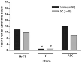
Parasitemia of animals was also evaluated by analy-sis of the frequency of positive tubes (eight tubes/dog) in relation to the number of positive BC per animal. Fig. 1 shows the frequency of positive tubes and BC in animals infected with the different T. cruzi strains. No signifi-cant differences were observed between the frequencies of positive tubes and the number of BC among the ani-mals inoculated with Be-78 and ABC strains, but these values were significantly higher (p < 0.05) than those observed for animals inoculated with Y strain.
Considering the number of positive tubes in BC, 33% of the animals were classified as presenting “low parasitemia”, 50% “median parasitemia” and 17% “high parasitemia”.
T. cruzi kDNA was detected in 40 (83.3%) out of the 48 blood samples obtained from dogs inoculated with different T. cruzi strains. In addition, B-PCR was posi-tive for at least one sample obtained from each evalu-ated animal. No significant difference was found in blood samples obtained from dogs inoculated with
Be-Fig. 1:frequency of positive tubes in blood culture (BC) and of positive BC performed during the chronic phase of Trypanosoma cruzi infec-tion of Beagle dogs inoculated with 4.0 x 10³ blood trypomastigotes of the Be-78, Y or ABC strains; *p < 0.05.
TABLE I
Fresh blood (FB) examination, blood culture (BC) and blood-PCR (B-PCR) results performed during the acute phase and chronic phase of the infection in Beagle dogs inoculated with 4.0 x 10³ blood trypomastigotes of the Be-78,
Y or ABC Trypanosoma cruzi strains
Time of infection
Acute phase Chronic phase
T. cruzi strains
Method/Total of tests Be-78 Y ABC Be-78 Y ABC
+FBb/Total of FB (%) 15/80 (18.7) 21/80 (26.2) 59/80 (73.7)a NP NP NP +BC/Total of BC (%) 4/4 (100) 4/4 (100) 4/4 (100) 12/16 (75) 1/16 (6.2)a 10/16 (62.5) +B-PCRc/Total of B-PCR (%) 4/4 (100) 4/4 (100) 3/4 (75) 16/16 (100) 9/16 (56.3)a 15/16 (93.7)
78 (100%) and ABC strains (93.7%). These indexes were significantly higher than those observed for animals in-oculated with strain Y (56.3%) (Table I). Considering all positive B-PCR reactions performed during the CP of in-fection, 52.5% (21/40) were obtained during the first and 47.5% (19/40) during the second year after inoculation of dogs, with similar positivity observed for both groups during the two years of observation. Representative B-PCR samples obtained from groups of dogs infected with different T. cruzi strains are shown in Fig. 2.
Tissue Parasitism
Acute phase (IMH) - In all animals necropsied during the acute phase of infection, amastigotes were detected in at least one of the six heart fragments examined. In a total of 24 fragments, the rate of positivity (amastigotes isolated/nests) according to the strain was 95.8% (23/24) for animals inoculated with Be-78 strain, 70.8% (17/24) for animals inoculated with ABC strain and only 29.2% (7/24) for dogs inoculated with Y strain.
The number of amastigotes varied among animals inoculated with different T. cruzi strains. Thus, 101 iso-lated amastigotes and 183 nests were observed in 720 microscopic fields of fragments obtained from the hearts of animals inoculated with Be-78 strain. Among animals inoculated with Y strain, an intense inflammatory pro-cesses and few amastigote nests (4/720 fields) were ob-served. However, a greater number of isolated amastigo-tes (52/720 fields) or small groups of amastigoamastigo-tes were observed to be associated with inflammatory process. Moreover, parasitism of the cardiac tissue in animals in-oculated with ABC strain showed intermediary intensity between animals inoculated with the Be-78 or Y strains, with 20 isolated amastigotes and 53 nests/720 fields ob-served. In these animals, TP was associated with the in-flammatory process (Fig. 3).
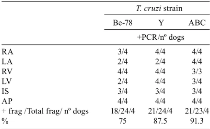
Chronic phase (IMH and T-PCR) - No parasites were found in any heart fragments obtained from animals during the chronic phase (24 months after inoculation) by IMH. However, DNA fragments of T. cruzi were de-tected in 84.5% of the tissue samples obtained from the left and right atria, left and right ventricles, interventric-ular septum and apex of dogs infected with Be-78, Y or ABC strains by T-PCR. Considering the different heart areas, T-PCR was positive for at least three samples from each animal evaluated. A total of 75%, 87.5% and 91.3% of the tissue samples were positive for animals infected
strain Be-78, Y or ABC. There was no significant differ-ence among animals infected with the different strains (Table II).
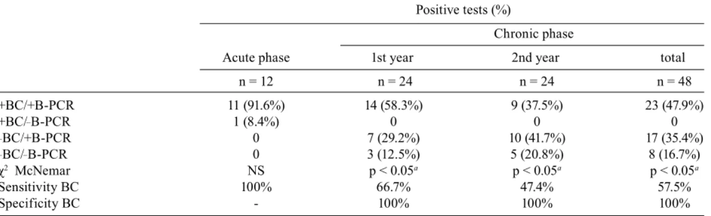
BC and B-PCR - During the acute phase of infection, 91.6% of the samples analyzed were positive for both BC and B-PCR, and 8.4% only for BC. These results indicate that BC and B-PCR showed similar sensitivity during this phase of the disease (Table III). During the chronic phase, 47.9% of the samples analyzed were positive for BC and B-PCR, 35.4% for B-PCR alone and 16.7% were negative for both tests (Table III). Positive B-PCR was significantly more frequent than positive BC. The sensi-tivities of BC in relation to B-PCR during the first and second years of infection for dogs inoculated with dif-ferent strains were 66.7% and 47.4%, respectively (Table III).
BC, B-PCR and T-PCR - The correlation between blood and TP was evaluated for BC, B-PCR and T-PCR performed in paired form 24 months after inoculation of
Fig. 2: representative gel showing the specific amplification of 330 bp fragments of kDNA minicircles of Trypanosoma cruzi in the blood of infected Beagle dogs (Bg 15, Bg 16, Bg 17 and Bg 18) with 4.0 x 10³ blood trypomastigotes of the Y strain during 24 months of evaluation. MW-DNA (100 pb ladder); Bg: Beagle dog; M: month; RC: reagents control; +C: infected/positive control dog; -C: negative control dog.
Fig 3: photomicrography of Beagle dogs myocardium infected with 4.0 x 10³ blood trypomastigotes of Be-78, Y or ABC Trypanosoma cruzi strains and euthanized at the acute phase. A: Be-78 strain: nests or isolated amastigotes (arrows) with absence of inflammatory process; B: Y strain: intense inflammatory process and absence of parasitism; C: ABC strain: moderate inflammatory process and isolated amasti- gotes; inserts: nests (upper) or isolated amastigotes (lower) observed in all strains. Imunohistochemistry Peroxidase Anti-Peroxidase. A, B and C: 160X; inserts: 1000X.
TABLE II
PCRs in different heart areas performed after necropsy of 12 Beagle dogs two years after inoculation with 4.0 x 10³ blood trypomastigotes of the Be-78, Y or ABC
strains of Trypanosoma cruzi
T. cruzi strain
Be-78 Y ABC
+PCR/nº dogs
RA 3/4 4/4 4/4
LA 2/4 2/4 4/4
RV 4/4 4/4 3/3
LV 2/4 4/4 3/4
IS 3/4 3/4 3/4
AP 4/4 4/4 4/4
+ frag /Total frag/ nº dogs 18/24/4 21/24/4 21/23/4
% 75 87.5 91.3
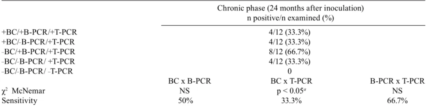
12 Beagle dogs with Be-78, Y or ABC T. cruzi strains (Fig. 4). The positivity was 33% (4/12) for BC, 66.7% (8/12) for B-PCR and 100% (12/12) for T-PCR. Consider-ing all dogs evaluated, a higher sensitivity was observed for T-PCR compared to that observed for the other tests performed. The sensitivity of BC in relation to B-PCR, BC in relation to T-PCR and B-PCR in relation to T-PCR were 50%, 33.3% and 66.7%, respectively (Table IV). On the other hand, the parasite was only detected in heart tissue by T-PCR in animals infected with Y strain. blood parasitism was not detected in any animal evaluated by BC or B-PCR (Fig. 4).
All tests performed using blood or tissue samples obtained from animals infected with Be-78 strain exhib-ited a similar frequency of positivity. A high correlation was observed between parasite levels in the peripheral blood and in heart tissue. In animals inoculated with ABC strain, the positivity of B-PCR and T-PCR was also similar, with the parasite detected with significant-ly lower frequency by BC. In animals infected with Y strain, the parasite was only detected in heart tissue by T-PCR. Blood parasitism was not detected in any animal evaluated by BC or B-PCR (Fig. 4).
DISCUSSION
Evaluation of the parasitism level of peripheral blood in chronic Chagas disease has been evaluated by several studies. In general, the sensitivity of the parasitological tests, xenodiagnosis and BC assays is related to the blood parasitism level, which could be related to genetic char-acteristics of the T. cruzi strains. These genetic charac-teristics vary according to geographic regions (Coura et al. 1984). However, due to ethical reasons, most of these studies were performed in humans during the acute or chronic phases of the infection and did not evaluate the relationship between the intensity of heart tissue and blood parasitism.
During the acute phase of the infection, the presence of the parasite was easily demonstrated in all animals by the three techniques used herein (FB, BC and B-PCR).
However, the variation in parasitemia levels among ani-mals infected with different T. cruzi strains was demon-strated by the frequency of positive FB tests. Considering the frequency of positive tests, we verified the necessity of performing a greater number of tests to detect para-sites in animals inoculated with the Be-78 strain com-pared to those inoculated with the Y and ABC strains. No correlation was observed between blood parasitism levels during the acute and chronic phases of infection because, during the chronic phase, animals inoculated with the Y strain showed significantly lower blood para-sitism levels than those inoculated with Be-78 and ABC, as evidenced by BC and B-PCR.
In the present work, a reduction in parasitemia levels throughout the chronic phase of the infection was dem-onstrated by the decrease in positive BC observed during the second year compared to the first year of evaluation.
TABLE III
Comparison between blood culture (BC) and blood-PCR (B-PCR) results obtained during the acute phase and chronic phase (two years) of the Trypanosoma cruzi infection of the 12 Beagle dogs inoculated with 4.0 x 10³ blood trypomastigotes of the
Be-78, Y or ABC strains
Positive tests (%)
Chronic phase
Acute phase 1st year 2nd year total
n = 12 n = 24 n = 24 n = 48
+BC/+B-PCR 11 (91.6%) 14 (58.3%) 9 (37.5%) 23 (47.9%)
+BC/_B-PCR 1 (8.4%) 0 0 0
_BC/+B-PCR 0 7 (29.2%) 10 (41.7%) 17 (35.4%)
_BC/_B-PCR 0 3 (12.5%) 5 (20.8%) 8 (16.7%)
χ2 McNemar NS p < 0.05a p < 0.05a p < 0.05a
Sensitivity BC 100% 66.7% 47.4% 57.5%
Specificity BC - 100% 100% 100%
a: p < 0.05, significant difference, when comparing positivity BC to B-PCR.
Interestingly, this decrease was detected in only animals infected with the ABC strain. On the other hand, the re-duction in parasite load throughout T. cruzi infection was not verified by B-PCR. In addition, PCR was more sen-sitive than BC for the detection of parasitemia or parasite kDNA in distinct blood samples collected from dogs in-fected with each parasite strain. The present results sug-gest that BC and B-PCR sensitivities are related to the parasite strain. An important difference was observed for BC, as well as PCR positivity performed using blood samples from animals infected with the three different T. cruzi strains studied. BC and/or PCR detected the para-site or parapara-site kDNA in approximately 97% of the tests carried out for animals infected with Be-78 and ABC strains. On the other hand, only 56.3% of the animals infected with Y strain were positive. The difference in positivity for BC could be explained by morphological differences in the strains considered, as previously has demonstrated that T. cruzi strains with a predominance of slender blood trypomastigotes are more sensitive to host immune mechanisms than strains with a predomi-nance of large or stout trypomastigotes (Brener 1969). This hypothesis was suggested by other authors who verified different courses of parasitemia in dogs inocu-lated with Be-62 or Be-78 strains, with a predominance of slender and large trypomastigotes (Lana et al. 1992, Araújo et al. 2002) associated with lower and higher BC, xenodiagnosis and PCR, respectively. According to Coura et al. (1984), the sensitivity of parasitological tests seems to reflect the different genetic constitution of circulating T. cruzi strains, which vary according to geo-graphical regions and naturally with the polymorphism of the blood trypomastigote.
Arbitrary classification of the animals according to the parasitemic level based on BC was also associated with the T. cruzi strain assessed. Thus, animals with “high parasitemia”, presented a higher frequency of pos-itive B-PCR and IMH, while the opposite was observed in animals classified as “low parasitemia”. In this sce-nario, the low positivity of BC and PCR performed us-ing peripheral blood samples obtained from animals in-fected with strain Y reflect the low parasitemia of these
animals. Additionally, negative BC and PCR results for patients and animals with positive serology have been reported. This fact has been explained by the occurrence of patients with low parasitemia, for whom parasites could rarely or never be detected (Castro et al. 2002).
A high frequency of T. cruzi kDNA in representative portions of the heart was observed for all animals evalu-ated 24 months after infection. These results showed, as previously reported in human chronic Chagas disease (Lages-Silva et al. 2001, Elias et al. 2003), that fragments of T. cruzi kDNA remain in the heart tissue after long-term infection of dogs. On the other hand, Machado et al. (2001) did not detect T. cruzi DNA in the heart tissue of dogs inoculated with different T. cruzi strains using primers that amplify a 195 bp repetitive sequence of the genomic DNA, in contrast to the PCR condition used herein. However, the differences in the primers and PCR methodologies used require consideration.
With regard to the IMH results, the parasitism and inflammatory process pattern was again different, sug-gesting a large influence of the parasite strain on the results obtained for injuries incurred during the acute phase of infection. During the chronic phase, the IMH reaction was negative for all heart fragments. The low sensitivity of the IMH at this stage of infection has al-ready been demonstrated for dog tissues by other authors (Caliari et al. 1994, Machado et al. 2001, Cruz et al. 2006). Lages-Silva et al. (2001) also verified that PCR exhibits greater sensitivity than IMH for detecting the presence of T. cruzi in the esophagus of chagasic patients during the chronic phase of the infection.
Taken together, these results corroborate the impor-tance of T. cruzi strains in the sensitivity of parasitological techniques (FB, BC, B-PCR and IMH) used for diagno-sis of infection. Furthermore, our data provide definitive evidence of long-term persistence of T. cruzi in the heart tissue of dogs infected with different T. cruzi strains.
ACKNOWLEDGMENTS
To Lívia de Figueiredo Diniz and Geovam Crepalde, for the care of Beagle dogs, and the Rede Mineira de Bioterismo da FAPEMIG.
TABLE IV
Comparison between blood culture (BC), blood-PCR (B-PCR) and tissue-PCR (T-PCR) results obtained after 24 months of
Trypanosoma cruzi infection of the 12 Beagle dogs inoculated with 4.0 x 10³ blood trypomastigotes of the Be-78, Y or ABC strains
Chronic phase (24 months after inoculation) n positive/n examined (%)
+BC/+B-PCR/+T-PCR 4/12 (33.3%)
+BC/_B-PCR/+T-PCR 4/12 (33.3%)
_BC/+B-PCR/+T-PCR 8/12 (66.7%)
_BC/_B-PCR/ +T-PCR 4/12 (33.3%)
_BC/_B-PCR/ _T-PCR 0
BC x B-PCR BC x T-PCR B-PCR x T-PCR
χ2 McNemar NS p < 0.05a NS
Sensitivity 50% 33.3% 66.7%
REFERENCES
Araújo FM, Bahia MT, Magalhães NM, Martins-Filho OA, Veloso VM, Carneiro CM, Tafuri WL, Lana M 2002. Follow-up of ex-perimental chronic Chagas’ disease in dogs: use of polymerase chain reaction (PCR) compared with parasitological and serologi-cal methods. Acta Trop 81: 21-31.
Ávila HA, Gonçalves AM, Nehme NC, Morel CM, Simpson L 1990. Schizodeme analysis of Trypanosoma cruzi stocks from South and Central America by analysis of PCR-amplifiable minicircle variable region sequences. Mol Biochem Parasitol42: 175-188.
Ávila HA, Pereira JB, Thiemann O, De Paiva E, DeGrave W, Morel CM, Simpson L 1993. Detection of Trypanosoma cruzi in blood specimens of chronic patients by polymerase chain reaction am-plification of kinetoplast minicircle DNA: comparison with se-rology and xenodiagnosis. J Clin Microbiol31: 2421-2426.
Bahia MT, Tafuri WL, Caliari MV, Veloso VM, Carneiro CM, Coelho GL, Lana M 2002. Comparison of Trypanosoma cruzi infection in dogs inoculated with blood or metacyclic trypomastigotes of Berenice-62 and Berenice-78 strains via intraperitoneal and con-junctival routes. Rev Soc Bras Med Trop35: 339-345.
Barbosa AJA 1985. Método imunocitoquímico para a identificação de amastigotas do Trypanosoma cruzi em cortes histológicos de rotina. Rev Inst Med Trop Sao Paulo 27: 293-297.
Brener Z 1965. Comparative studies of different strains of Trypano-soma cruzi. Ann Trop Med Parasitol 59: 19-26.
Brener Z 1969. The behavior of slender and stout forms of Trypano-soma cruzi in the blood-stream of normal and immune mice. Ann Trop Med Parasitol 63: 215-220.
Caliari MV, Lana M, Oliveira ER, Barbosa AJ, Tafuri WL 1994. Immunocytochemical study of tissue parasitism of dog adrenal glands in experimental Chagas’ disease. Parasite 1: 397-400.
Castro AM, Luquetti AO, Rassi A, Rassi GG, Chiari E, Galvão LM 2002. Blood culture and Polymerase Chain Reaction for the diag-nosis of the chronic phase of human infection with Trypanosoma cruzi. Parasitol Res88: 894-900.
Chiaramonte MG, Frank FM, Furer GM, Taranto NJ, Margni RA, Malchiodi EL 1999. Polymerase chain reaction reveals Trypano-soma cruzi infection suspected by serology in cutaneous and mu-cocutaneous leishmaniasis patients. Acta Trop 72: 295-308.
Chiari E 1999. Chagas disease diagnosis using polymerase chain re-action, hemoculture and serologic methods. Mem Inst Oswaldo Cruz 94 (Suppl. I): 299-300.
Chiari E, Dias JCP, Lana M, Chiari CA 1989. Hemocultures for the parasitological diagnosis of human chronic Chagas’ disease. Rev Soc Bras Med Trop 22: 19-23.
Coura JR, Abreu LL, Dubois LEG, Correia-Lima F, Arruda JRE, Wellcox HPF, Anunzioto N, Pestana W 1984. Morbidade da do-ença de Chagas. II - Estudos seccionais em quatro áreas de campo no Brasil. Mem Inst Oswaldo Cruz 79: 101-124.
Cruz RE, Macedo AM, Barnabe C, Freitas JM, Chiari E, Veloso VM, Carneiro CM, Bahia MT, Tafuri WL, Lana M 2006. Further ge-netic characterization of the two Trypanosoma cruzi Berenice strains (Be-62 and Be-78) isolated from the first human case of Chagas disease (Chagas, 1909). Acta Trop 97: 239-246.
Elias FE, Vigliano CA, Laguens RP, Levin MJ, Berek C 2003. Analy-sis of the presence of Trypanosoma cruzi in the heart tissue of three patients with chronic Chagas’ heart disease. Am J Trop Med Hyg 68: 242-247.
Gomes ML, Galvão LM, Macedo AM, Pena SD, Chiari E 1999.
Cha-gas’ disease diagnosis: comparative analysis of parasitologic, mo-lecular, and serologic methods. Am J Trop Med Hyg 60: 205-210.
Gomes ML, Macedo AM, Vago AR, Pena SDJ, Galvão LMC, Chiari E 1998. Trypanosoma cruzi: Optimization of Polymerase Chain Reaction for detection in human blood. Exp Parasitol88: 28-33.
Guedes PM, Urbina JA, de Lana M, Afonso LC, Veloso VM, Tafu-ri WL, Machado-Coelho GL, ChiaTafu-ri E, Bahia MT 2004. Activ-ity of the new triazole derivative albaconazole against Trypano-soma (Schizotrypanum) cruzi in dog hosts. Antimicrob Agents Chemother 48: 4286-4292.
Lages-Silva E, Crema E, Ramirez LE, Macedo AM, Pena SD, Chia-ri E 2001. Relationship between Trypanosoma cruzi and human chagasic megaesophagus: blood and tissue parasitism. Am J Trop Med Hyg 65: 435-441.
Lana M, Chiari CA 1986. Caracterização biológica comparativa das ce-pas Berenice-78 de Trypanosoma cruzi, isoladas da mesma pacien-te em diferenpacien-tes períodos. Mem Inst Oswaldo Cruz 81: 247-253.
Lana M, Chiari E, Tafuri WL 1992. Experimental Chagas’ disease in dogs. MemInstOswaldoCruz 87: 59-71.
Lana M, Tafuri WL, Caliari MV, Bambirra EA, Chiari CA, Leite VHR, Barbosa AJA, Toledo MJO, Chiari E 1988. Fase crônica cardíaca fibrosante da tripanosomíase cruzi experimental no cão.
Rev Inst Med Trop Sao Paulo 21: 113-121.
Machado EM, Fernandes AJ, Murta SM, Vitor RW, Camilo DJJ, Pi-nheiro SW, Lopes ER, Adad SJ, Romanha AJ, Dias JCP 2001. A study of experimental reinfection by Trypanosoma cruzi in dogs.
Am J Trop Med Hyg 65: 958-965.
Marcon GE, Andrade PD, de Albuquerque DM, Wanderley JS, de Almei-da EA, Guariento ME, Costa SC 2002. Use of a nested Polymerase Chain Reaction (N-PCR) to detect Trypanosoma cruzi in blood sam-ples from chronic. Diagn Microbiol Infect Dis 43: 39-43.
Portela-Lindoso AA, Shikanai-Yasuda MA 2003. Chronic Chagas’ disease: from xenodiagnosis and hemoculture to polymerase chain reaction. Rev Saude Publica 37: 107-115.
Russomando G, Figueredo A, Almiron M, Sakamoto M, Morita K 1992. Polymerase chain reaction-based detectionof Trypanoso-ma cruzi DNA in serum. J Clin Microbiol302: 864-868.
Santos FR, Pena SDJ, Epplen JT 1993. Genetic and population study of a Y-linked tetranucleotide repeat DNA polymorphism with a simple non-isotopic technique. Hum Genet 90: 655-656.
Silva LHP, Nussenzweig V 1953. Sobre uma cepa de Trypanosoma cruzi altamente virulenta para o camundongo branco. Folia Clin Biol 20: 191-203.
Vago AR, Macedo AM, Adad SJ, Reis DA, Corrêa-Oliveira R 1996. PCR detection of Trypanosoma cruzi DNA in esophageal tissues of patients with chronic digestive Chagas’ disease. Lancet 348: 891-892.
WHO - World Health Organization 2005. Tropical Disease Research: progress 2003-2004. Special Programme for Research & Train-ing in Tropical Diseases. Programme Report 17, Geneva, 31-33.
Wincker P, Bosseno MF, Britto C, Yaksic N, Cardoso MA, Morel CM, Breniere SF 1994a. High correlation between Chagas’ disease se-rology and PCR-based detection of Trypanosoma cruzi kineto-plast DNA in Bolivian children living in an endemic area. FEMS Microbiol Lett124: 419-423.