Trypanosoma cruzi
: Immunoglobulin isotype profiles during the acute phase
of canine experimental infection with metacyclic or blood trypomastigotes
W. Coura-Vital
a, C.M. Carneiro
a,b, H.R. Martins
c, M. de Lana
c, V.M. Veloso
c, A. Teixeira-Carvalho
d,
M.T. Bahia
c, R. Corrêa-Oliveira
e, O.A. Martins-Filho
d, W.L. Tafuri
a, A.B. Reis
a,b,e,*aLaboratório de Imunopatologia, Núcleo de Pesquisas em Ciências Biológicas/NUPEB, Instituto de Ciências Exatas e Biológicas, Universidade Federal de Ouro Preto,
Ouro Preto, MG, CEP 35400-000, Brazil
bDepartamento de Análises Clínicas, Escola de Farmácia, Universidade Federal de Ouro Preto, Ouro Preto, MG, Brazil
cLaboratório de Doença de Chagas, Núcleo de Pesquisas em Ciências Biológicas, Universidade Federal de Ouro Preto, Ouro Preto, MG, Brazil dLaboratório de Biomarcadores de Diagnóstico e Monitoração, Centro de Pesquisas René Rachou, Fundação Oswaldo Cruz, Belo Horizonte, MG, Brazil eLaboratório de Imunologia Celular e Molecular, Centro de Pesquisas René Rachou, Fundação Oswaldo Cruz, Belo Horizonte, MG, Brazil
a r t i c l e
i n f o
Article history:
Received 13 December 2007
Received in revised form 1 August 2008 Accepted 18 August 2008
Available online 26 August 2008
Keywords: Trypanosoma cruzi
Chagas disease Canine model
Class and subclass of immunoglobulins Parasitemia
a b s t r a c t
A detailed investigation has been carried out about the serological profiles of groups of dogs experimen-tally infected with metacyclic (MT) or blood (BT) trypomastigotes of Berenice-78Trypanosoma cruzi strain. Peripheral blood was collected from infected dogs and uninfected controls, weekly during 35 days following the acute phase of infection, and immunoglobulin profiles were determined by ELISA. Dogs infected with BT exhibited unaltered levels of IgG2, increases in IgM, IgE, IgA, IgG and IgG1. In contrast, dogs infected with MT presented unaltered levels of IgE and IgG1 and an increase in IgM, IgA, IgG and IgG2 levels. Compared with the MT group, animals infected with BT showed significant increases in IgM on days 7, 14 and 28, in IgA on days 7, 14 and 21, in IgE on days 7 and 14, in IgG on days 14 and 28, and in IgG1 on days 7, 14 and 21. Parasitemia levels of the infected animals were measured over the same time period. No correlations were found between the immunoglobulin profiles and the parasi-temia levels. The results demonstrated that the inoculum source (BT or MT) influence the immunoglob-ulin isotype profile that may drive distinct outcome of acute canine Chagas disease.
Ó2008 Elsevier Inc. All rights reserved.
1. Introduction
The etiological agent of Chagas disease is the flagellate proto-zoanTrypanosoma cruzi. In regions that are endemic for the dis-ease, the main mechanism of infection is by contamination with metacyclic trypomastigotes (MT) eliminated by triatomine vectors (Dias, 2006). Blood transfusion represents the second most impor-tant infection mechanism, especially in urban areas where vectors are not present (Sanchez-Guillen et al., 2002). Whilst MT and epimastigotes are commonly found in triatomine vectors, amastig-otes and blood trypomastigamastig-otes (BT) represent, respectively, the main proliferative intracellular and bloodstream forms found in the vertebrate hosts (Andrade and Andrews, 2005). Although both MT and BT forms ofT. cruziare fully functional with respect to par-asite–host interaction and/or target cell invasion (Meirelles et al.,
1982; Ramirez et al., 1993), the two forms differ regarding their surface molecules (Yoshida, 2006).
Trypanosoma cruziinfections are associated with immunological and immunopathological reactions that may result from non-spe-cific polyclonal activation (Montes et al., 2006) or suppressive ef-fects (Abrahamsohn and Coffman, 1995). In addition, immune control of the infection involves the participation of CD4+ and
CD8+T cells (Sher and Coffman, 1992; Tarleton et al., 1992).
To-gether with cellular immune responses, anti-parasite humoral re-sponses also play a major role in the control of parasitemia and in resistance toT. cruziinfection (Krettli and Brener, 1976).
Anti-Trypanosoma cruzi IgG antibodies are produced at the beginning of the acute phase of the disease (Lana et al., 1991; Carneiro et al., 2007) and react mainly with surface molecules of the infective trypomastigote forms. However, most epitopes shared by trypomastigote and epimastigote forms belong to inter-nal antigens (Umezawa et al., 1996), a fact that may explain why epimastigote antigens do not result in high IgG reactivity with acute phase sera. The major antibody isotypes involved in the elim-ination of blood forms of the parasite and in decreasing mortality rates are IgG1 and IgG2 (Brodskyn et al., 1989; Cordeiro et al., 2001).
0014-4894/$ - see front matterÓ2008 Elsevier Inc. All rights reserved. doi:10.1016/j.exppara.2008.08.001
* Corresponding author. Address: Laboratório de Imunopatologia, Núcleo de Pesquisas em Ciências Biológicas/NUPEB, Instituto de Ciências Exatas e Biológicas, Universidade Federal de Ouro Preto, Ouro Preto, MG, CEP 35400-000, Brazil. Fax: +55 21 31 3559 1680.
E-mail address:alexreis@nupeb.ufop.br(A.B. Reis).
Contents lists available atScienceDirect
Experimental Parasitology
Dogs may be employed as standardized experimental models, reflecting the natural history of evolution of human Chagas disease, since these animals develop acute and chronic phases of the infec-tion with morphological and clinical characteristics that are very similar to those observed in man (Andrade and Andrade, 1980; Lana et al., 1992).Bahia et al. (2002)studied parasitemia and histopa-thological aspects and demonstrated that the source of the inocu-lum remarkably influence the evolution of the infection in dogs.
In order to gain an insight into the effect of the inoculum source on the humoral immune response in infected animals, we have performed a detailed comparative analysis of the anti-T. cruzi immu-noglobulin profiles during the acute phase of infection in experi-mental dogs that had been inoculated with BT or MT forms ofT. cruzi.
2. Materials and methods
Details of the study were presented to and approved by the Eth-ical Committee of the Universidade Federal de Ouro Preto, Minas Gerais, Brazil. All procedures described were carried out in compli-ance with current Brazilian criteria relating to Experimental Biology and Medicine as described in the guidelines issued by theColégio Brasileiro de Experimentação Animal (2006). All of the animals em-ployed in this study were maintained in the central animal facility of the Universidade Federal de Ouro Preto, Minas Gerais, Brazil.
2.1. Parasites, dogs and study design
MT and BT forms of the Berenice-78 (Be-78)1T. cruzistrain (Lana
and Chiari, 1986) were isolated, respectively, from nymphs of Tria-toma infestansand female Swiss Webster mice that had been previ-ously infected in our laboratory.
Before the beginning of the study, 12 dogs born in the kennel of the Universidade Federal de Ouro Preto, MG, Brazil, belonging to two different litters, were treated with anti-helminthics and immunized against more common canine infectious pathogens. Animals were maintained in quarantine for 16 weeks receiving drinking water and a balanced commercial feedad libitum.
When the animals completed 120 days of age, groups of 4 dogs were inoculated intraperitoneally with either MT or BT forms of Be-78T. cruzistrain (2000/kg body weight), while the remaining 4 dogs were maintained uninfected as control group. Each group of 4 dogs was maintained in isolated place until the beginning of the experiment.
2.2. Parasitemia evaluation
Parasitemia in the infected animals was evaluated since the 10th day after infection until day 35. The number of parasites in fresh blood collected by vein-puncture of the ear veins was determined under the optical microscope according toBrener (1962). Briefly this methodology consists in examination of 5
l
L of infected blood on the microscope slide covered by 2222 mm slide. Parasites present in 50 microscope fields were counted and multiplied by the micro-scope factor (calculated for each objective). The parasitemia curves represent the mean value of blood trypomastigotes/0.1 mL of blood from all infected dogs of each group at each time point.2.3. Parasite antigen
Soluble antigen from epimastigotes ofT. cruziY strain were ob-tained from an axenic culture of the protozoan at exponential
growth phase in liver infusion tryptose (LIT) medium. Parasites were washed three times by centrifugation (800g; 10 min) in phosphate buffer saline (pH 7.2), followed by three ultrasound cycles (each of 1 min at 40 W) with a Sonifier Cell DisruptorÒ
(Branson Sonic Power Co., Danbury, CT, USA) cooled in an ice bath. The sonicated material was centrifuged (18,500 rpm; 1.5 h; 4°C) and the supernatant
trans-ferred to dialysis tubes and dialyzed against phosphate buffer saline for 36 h, with four phosphate buffer saline changes every 6 h. Finally, the remaining material was filtered under aseptic conditions through a 0.22
l
m filter and an aliquot was taken to determine pro-tein concentration by the method ofLowry et al. (1951). The bulk of the sample was adjusted to a protein concentration of 1000l
g/mL and stored in small aliquots at 70°C prior to use.2.4. Determination of immunoglobulin profiles by enzyme linked immunosorbent assay
Peripheral blood (5 mL) was collected from the brachial cepha-lic veins of the experimental dogs prior to inoculation (on day 0) and weekly thereafter (on days 7, 14, 21, 28 and 35), and placed in vials in the absence of anticoagulant. Serum samples were stored at 70°C and enzyme linked immunosorbent assays (ELISA)
were carried out using a modified version of theVoller et al. (1976)
method. Polystyrene microtiter plates (MaxiSorpTM surface, Nalge Nunc Int., Rochester, NY, USA) were coated with 200
l
L of a solu-tion antigenic (Final concentrasolu-tion = 0.5l
g/well) from epimastig-otes of T. cruzi in carbonate buffer (pH 9.6) and incubated overnight at 4°C. After incubation, the plates were washed fourtimes with phosphate buffered saline (PBS) containing 0.05% Tween 20, and blocked for 45 min at 37°C with 100
l
L of foetalbo-vine serum (5%) in PBS per well. Plates were then incubated at 37°C for 45 min with 100
l
L of serum sample diluted 1:80, thewells were washed three times and incubated at 37°C for 45 min
with specific monoclonal peroxidase-conjugated antibodies (Beth-yl Laboratories Inc., Montgomery, TX, USA) previously diluted as follows: goat anti-dog IgG1(anti-heavy chain specific), 1:16,000; IgM (anti-
l
chain specific), 1:500; IgA (anti-a
chain specific), 1:500; IgE (anti-e
chain specific), 1:1000, or sheep anti-dog IgG and IgG2 (both anti-heavy chain specific), 1:16,000. Following four further washes (as above), reaction was initiated by the addition of 100l
L of 0.1 M citrate buffer (pH 5.0) containing 0.03%o -phenyl-enediamine and 0.012% H2O2, followed by incubation at 37°C for10 min. The reaction was stopped by the addition of 32
l
L of 2.5 M H2SO4and the absorbance value (OD492 nm) measured withan ELISA plate reader (Molecular Devives, E Max, USA).
2.5. Statistical analysis
Statistical analyses were performed with the aid of the Prism 4.0 software package (Prism Software, Irvine, CA, USA). Data col-lected for the BT and MT groups at identical times after infection were compared using the Mann–Whitney test. Repeated measure-ment analyses for the longitudinal study were performed using the Friedman test followed by Dunn’s multiple comparison test. Spear-man’s rank tests were employed in order to investigate correla-tions between immunoglobulin levels and parasitemia curves. In all cases, differences were considered statistically significant when the probabilities of equality (p-values) were60.05.
3. Results
3.1. Immunological parameters
The kinetic profiles of the anti-T. cruziisotypes IgM, IgA, IgE, IgG, IgG1 and IgG2 were determined by ELISA assay over a period of 35 1 Abbreviations used:Be-78, Berenice-78; BT, blood trypomastigotes; C, uninfected
days following inoculation of experimental dogs with BT or MT, and were compared with those determined in the uninfected con-trol group (C) of animals. Correlations between parasitemia and immunoglobulin profiles in the BT and MT groups were also inves-tigated during the acute phase of infection.
3.1.1. Kinetic profiles of IgM, IgA and IgE
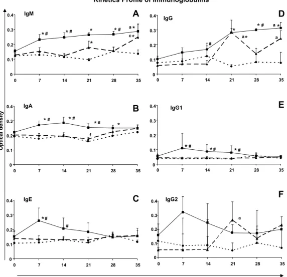
The longitudinal analysis of the BT group revealed that, although levels of IgM were already elevated on day 7 in compar-ison with the pre-inoculation (day 0) level, and increased continu-ously thereafter, the increase only became statistically significant (p60.05) on day 35 (Fig. 1A). IgM reactivities in the BT group were significantly higher than those of the MT group on days 7, 14 and 28, and on all test days in comparison with the control group. Within the MT group, there was a significant increase in IgM level on day 35 compared with day 14, whilst in comparison with the control group, IgM levels were significantly higher on days 21 and 35.
IgA reactivities in the BT group were significantly higher than those of the MT group on days 7, 14 and 21, and on days 7, 14, 21 and 28 days in comparison with the control group. The MT
group showed no significant differences in relation to IgA levels compared with the control group throughout the experimental period (Fig. 1B).
No significant alterations in the levels of IgE were detected in the longitudinal analysis of the BT and MT groups (Fig. 1C). How-ever, increased levels of IgE were observed in the BT group com-pared with the MT group on days 7 and 14, and on day 7 in comparison with the control group. No significant differences were observed in IgE levels between the MT and the control groups throughout the experimental period
3.1.2. Kinetic profiles of IgG, IgG1 and IgG2
The longitudinal analysis of the BT group showed that IgG reac-tivity was significantly higher on day 35 compared with the pre-inoculation (day 0) level (Fig. 1D). IgG levels in the BT group were significantly higher than those of the MT group on days 14 and 28, and on days 21, 28 and 35 in comparison with the control group. Within the MT group, there were significant increases in IgG levels on days 21 and 35 compared with day 0, whilst in comparison with the control group, the IgG level was significantly higher only on day 21.
0 0.1 0.2 0.3 0.4
0 7 14 21 28 35
IgG
a a
a
*
*
*
*
#
# IgM
c a
*
*
*
#*
#*
*
#*
IgA
f
*
*
*
*
# #
#
IgG1
*
#*
*
# #
Optical density
Days after infection
Kinetics Profile of Immunoglobulins
IgE
*
# #IgG2
a
A
B
C
D
E
F
00.1 0.2 0.3 0.4
0 7 14 21 28 35
0 0.1 0.2 0.3 0.4
0 7 14 21 28 35
0 0.1 0.2 0.3 0.4
0 7 14 21 28 35
0 0.1 0.2 0.3 0.4
0 7 14 21 28 35
0 0.1 0.2 0.3 0.4
0.5
0 7 14 21 28 35
Fig. 1.Kinetic profiles of immunoglobulins in dogs experimentally infected with blood trypomastigotes ( BT) or metacyclic trypomastigotes ( MT) ofT. cruzi
strain Be-78, and in uninfected dogs (control group C ). They-axis represents mean ELISA absorbance values (determined at 492 nm). The symbols # and*indicate,
No significant differences in the levels of IgG1 were observed within the BT and MT groups, or between the MT and the control groups, during the whole of the experimental period (Fig. 1E). However, increased IgG1 reactivities were detected in the BT group compared with both the MT and the control groups on days 7, 14 and 21.
No significant differences in the IgG2 levels were observed within the BT group, or between the BT, MT and control groups, during the experimental period (Fig. 1F). In contrast, the MT group showed a significant increase n IgG2 reactivity on day 21 compared with day 0.
3.2. Parasitological parameters and correlation with immunoglobulin profile
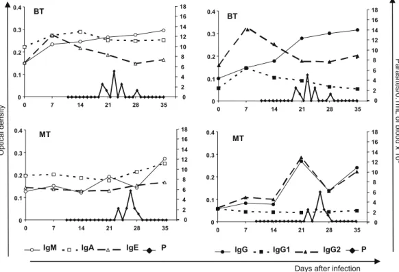
Infection was confirmed by parasitemia in all 8 dogs that had been inoculated with BT or MT forms of Be-78T. cruzistrain, how-ever, no mortality, signs or symptoms of acute phase were ob-served during the period of evaluation. The pre-patent period was significantly higher in MT group, but no differences in patent periods were observed, between BT and MT infected dogs (Fig. 2). The parasitemia peak of 6250 trypomastigotes per 5
l
L of blood observed on the 26th day after inoculation of dogs with MT, was similar to that found in BT animals (i.e. 5000 trypomastigotes per 5l
L of blood) on the 22nd day after infection.During the experimental period, the BT group showed increased levels of IgM and IgG, which remained high even after negative parasitemia. On the other hand the levels of IgA, IgE, IgG1 and IgG2 remained stable in this group throughout the whole experi-mental period, irrespective of the presence or absence of parasite-mia. In this context other IgG isotypes (IgG3 and/or IgG4) can be inducing the increase of IgG total levels after 21 days.
In the MT group, constant levels of IgA, IgE and IgG1 were main-tained during the whole period of infection. In contrast, elevated
levels of IgM, IgG and IgG2 were observed throughout the experi-mental period and, although the reactivities decreased somewhat at the parasitemia peak, they increased again even after parasite-mia was no longer detectable. No correlations were observed
be-tween parasitemia and the levels of the different
immunoglobulins or IgG isotypes evaluated (Fig. 2).
4. Discussion
In the present study, 4-month-old dogs were used since it has been shown that young animals reproduce with greater fidelity the parasitological, histopathological and clinic characteristics that are observed in the acute form of human Chagas disease (Andrade and Andrade, 1980; Lana et al., 1992). Dogs were infected with MT and BT forms ofT. cruzion the basis that different sources of inoc-ulum may generate distinct infections involving different humoral responses (Brener and Gazzinelli, 1997; Bahia et al., 2002).
Carneiro et al. (2007)recently studied parasitemia in dogs that had been experimentally infected with MT and BT forms ofT. cruzi, and demonstrated that the pre-patent period was 23 days in the MT group and 19 days in the BT group, findings that are in agree-ment with those of the present study. A longer pre-patent period in the MT group may indicate that the trypomastigote–host cell inter-action occurs more slowly, since these animals presented a longer sub-patent period compared with those in the BT group. Therefore, others factors can be influenced the pre-patent period, such as nat-ural antibodies or acute phase lectins, elements from the natnat-ural immune response (Meirelles et al., 1982; Araújo-Jorge et al., 1989). Similar parasitemia profile has been observed for Be-78 as well as otherT. cruzistrains in canine model for Chagas disease, despite the inoculum source (Lana et al., 1992; Bahia et al., 2002).
Previous studies have demonstrated that variations in the membrane proteins of the MT and BT forms ofT. cruzican give rise to differences in the characteristics of cell invasion (Yoshida, 2006).
Days after infection P 0
0.1 0.2 0.3 0.4
0 7 14 21 28 35 42
BT
0 2 4 6 8 10 12 14 16 18
MT
IgG
IgM IgA IgE P IgG1 IgG2
0 0.1 0.2 0.3 0.4
0 7 14 21 28 35 4242
BT
Optical density
0 0.1 0.2 0.3 0.4
0 7 14 21 28 35 4242
MT
0 2 4 6 8 10 12 14 16 18 0 2 4 6 8 10 12 14 16 18
Paras
ite
s/0.
1mL
of
blo
od
x
10
3
0 0.1 0.2 0.3 0.4
0 7 14 21 28 35 42
0 2 4 6 8 10 12 14 16 18
So, the kinetics of different classes of antibodies were investigated in animals infected with MT and BT. It was verified that the level of IgM increased from the day of inoculation of animals of the BT group, and became significantly higher on day 35. Moreover, the level of IgM in this group was higher than that of the MT group in three of the five weekly assays performed.Lana et al. (1991)also observed increasing levels of IgM from the 2nd week following infection with MT until the 2nd and 3rd month when assayed by ELISA and indirect immunofluorescence (IFI).
In the present study, elevated levels of IgA were detected in the BT group compared with the MT and control groups, and the in-crease was statistically significant from the 1st week after inocula-tion. However, the levels of IgA did not alter during the experimental period confirming the report of Guedes et al. (2007), who observed low levels of IgA during the acute phase of Chagas disease in dogs. In humans, increased IgA may be associ-ated with the digestive form of Chagas disease (Sá Ferreira et al., 1983). On the other hand,Matsumoto et al. (1993)did not observe a correlation between IgA and the digestive form.
With respect to IgE, the present investigation revealed elevated reactivities in the BT group on the 7th day after infection, followed by a decline. Earlier studies of Chagasic patients did not identify in-creased levels of IgE in comparison with non-infected individuals (Geller et al., 1978), suggesting that this class of immunoglobulins is not important in human forms of the disease.
In the present work, progressive increases in the levels of IgG were observed in the BT group, with oscillating levels in the MT group, from the 2nd week after inoculation. Similar results were observed byGuedes et al. (2007)in studies of Beagles infected with BT fromT. cruzistrain Be-78. It is clear that total IgG may be de-tected in the serum of experimentally infected dogs during the early stages of the disease, thus corroborating the findings ofLana et al. (1991).
In the BT group of infected animals, the levels of IgG1 were higher than in the MT and control groups at different periods of infection. In general, however, the IgG1 levels were low in the pres-ent study, but this may have been due to the short experimpres-ental period employed since infected Beagles showed levels of IgG1 that were low at the beginning of the infection but subsequently in-creased (Guedes et al., 2007). The profile observed for IgG2 in the present study was similar to that of total IgG in the MT group, but opposite to that of total IgG in the BT group. However, statisti-cal analysis did not reveal significant differences in the levels of IgG2 in the BT group during the experimental period.
Analysis of IgG isotypes by flow cytometry (Cordeiro et al., 2001) demonstrated that IgG1 and IgG3 are the main lytic antibod-ies present in humans, and that patients with undetermined forms of the disease showed high levels of IgG1 suggesting an immuno-protective role for this immunoglobulin. Similar results have been obtained using murine models (Lima-Martins et al., 1985; Pyrrho et al., 1998). It is not known, however, if canine IgG1 has the same biological function as human IgG1. More recently, conjugates able of detecting canine IgG3 and IgG4 specifically have been produced (Quinnell et al., 2003), and these will allow further insights to be gained in understanding the function of the immunoglobulins in experimental canine Chagas disease.
When the Ig kinetic curves obtained in the present study were superimposed upon the parasitemia curves, it could be seen that increases in the levels of IgG, IgG2, IgM, IgA and IgE occurred prior to the peak of parasitemia in both MT and BT groups (with the exception of IgA and IgE levels in the BT group). Such increases may result from the activation of B lymphocytes before parasite-mia attained a high level. However, during the acute phase of the infection no correlations were observed between immunoglobulin profiles and parasitemia, a finding that is in agreement with that of
Lana et al. (1991).
Our results suggest that the inoculum source (BT or MT) influ-ence the immunoglobulin isotype profile that may drive distinct outcome of acute canine Chagas disease. The knowledge of the immunoglobulin profiles may contribute to the overall under-standing of immunopathogenesis in experimental canine Chagas disease.
Acknowledgments
The authors express their appreciation to the kennel staff of the Universidade Federal de Ouro Preto for their hard work and special dedication during the execution of this project. The authors are also grateful for the use of facilities at CEBIO, Universidade Federal de Minas Gerais and Rede Mineira de Bioterismo. We also thank FAPEMIG for financial support.
References
Abrahamsohn, I.A., Coffman, R.L., 1995. Cytokine and nitric oxide regulation of the immunosuppression inTrypanosoma cruziinfection. Journal of Immunology 155, 3955–3963.
Andrade, Z.A., Andrade, S.G., 1980. Pathology of experimental Chagas disease in dogs. Memórias do Instituto Oswaldo Cruz 75, 77–95.
Andrade, L.O., Andrews, N.W., 2005. TheTrypanosoma cruzihost-cell interplay: location, invasion, retention. Nature Reviews. Microbiology 3, 819–823. Araújo-Jorge, T.C., Sampaio, E.P., De Souza, W., Meirelles Mde, N., 1989.
Trypanosoma cruzi: the effect of variations in experimental conditions on the levels of macrophage infection in vitro. Parasitology Research 75, 257–263. Bahia, M.T., Tafuri, W.L., Caliari, M.V., Veloso, V.M., Carneiro, C.M., Coelho, G.L., Lana,
M., 2002. Comparison ofTrypanosoma cruziinfection in dogs inoculated with blood or metacyclic trypomastigotes of Berenice-62 and Berenice-78 strains via intraperitoneal and conjunctival routes. Revista da Sociedade Brasileira de Medicina Tropical 35, 339–345.
Brener, Z., 1962. Therapeutic activity and criterion of cure on mice experimentally infected withTrypanosoma cruzi. Revista do Instituto de Medicina Tropical de São Paulo 4, 389–396.
Brener, Z., Gazzinelli, R.T., 1997. Immunological control of Trypanosoma cruzi
infection and pathogenesis of Chagas’ disease. International Archives of Allergy and Immunology 114, 103–110.
Brodskyn, C.I., Silva, A.M., Takehara, H.A., Mota, I., 1989. IgG subclasses responsible for immune clearance in mice infected withTrypanosoma cruzi. Immunology and Cell Biology 67 (6), 343–348.
Carneiro, C.M., Martins-Filho, O.A., Reis, A.B., Veloso, V.M., Araujo, F.M.G., Bahia, M.T., de Lana, M., Machado-Coelho, G.L.L., Gazzinelli, G., Correa-Oliveira, R., Tafuri, W.L., 2007. Differential impact of metacyclic and blood trypomastigotes on parasitological, serological and phenotypic features triggered during acute
Trypanosoma cruziinfection in dogs. Acta Tropica 101, 120–129.
Colégio Brasileiro de Experimentação Animal, 2006. Legislação e ética. Available from:<http://www.cobea.org.br/>accessed January 2006.
Cordeiro, F.D., Martins-Filho, O.A., da Costa Rocha, M.O., Adad, S.J., Correa-Oliveira, R., Romanha, A.J., 2001. Anti-Trypanosoma cruziimmunoglobulin G1 can be a useful tool for diagnosis and prognosis of human Chagas’ disease. Clinical and Diagnostic Laboratory Immunology 8, 112–118.
Dias, J.C., 2006. Notes aboutTrypanosoma cruziand its bio-ecology characteristics with agents of the transmission by meals. Revista da Sociedade Brasileira de Medicina Tropical 39, 370–375.
Geller, M., Geller, M., Flaherty, D.K., Black, P., Capanema-Souza, A.P., 1978. Serum IgE levels in Chagas’ disease. Clinical Allergy 8, 383–385.
Guedes, P.M., Veloso, V.M., Caliari, M.V., Carneiro, C.M., Souza, S.M., de Lana, M., Chiari, E., Bahia, M.T., Galvao, L.M., 2007.Trypanosoma cruzihigh infectivity
in vitrois related to cardiac lesions during long-term infection in Beagle dogs. Memórias do Instituto Oswaldo Cruz 102, 141–147.
Krettli, A.U., Brener, Z., 1976. Protective effects of specific antibodies inTrypanosoma cruziinfections. Journal of Immunology 116, 755–760.
Lana, M., Chiari, C.A., 1986. Caracterização biológica comparativa das cepas Berenice-78 deTrypanosoma cruzi, isoladas da mesma paciente em diferentes períodos. Memórias do Instituto Oswaldo Cruz 81, 247–253.
Lana, M., Vieira, L.M., Machado-Coelho, G.L., Chiari, E., Veloso, V.M., Tafuri, W.L., 1991. Humoral immune response in dogs experimentally infected with
Trypanosoma cruzi. Memórias do Instituto Oswaldo Cruz 86, 471–473. Lana, M., Chiari, E., Tafuri, W.L., 1992. Experimental Chagas’ disease in dogs.
Memórias do Instituto Oswaldo Cruz 87, 59–71.
Lima-Martins, M.V., Sanchez, G.A., Krettli, A.U., Brener, Z., 1985. Antibody-dependent cell cytotoxicity against Trypanosoma cruziis only mediated by protective antibodies. Parasite Immunology 7, 367–376.
Lowry, O.H., Rosebrough, N.J., Farr, A.L., Randall, R.J., 1951. Protein measurement with the Folin phenol reagent. The Journal of Biological Chemistry 193, 265– 275.
serodiagnosis of different clinical forms of Chagas’ disease. Journal of Clinical Microbiology 31, 1486–1492.
Meirelles, M.N., Chiari, E., de Souza, W., 1982. Interaction of bloodstream, tissue culture-derived and axenic culture-derived trypomastigotes ofTrypanosoma cruziwith macrophages. Acta Tropica 39, 195–203.
Montes, C.L., Costa-Rodriguez, E.V., Mucci, J., Zuniga, E.I., Campetella, O., Gruppi, A., 2006. ATrypanosoma cruziantigen signals CD11b+ cells to secrete cytokines that promote polyclonal B cell proliferation and differentiation into antibody-secreting cells. European Journal of Immunology 36, 1474–1485.
Pyrrho, A.S., Moraes, J.L., Pecanha, L.M., Gattass, C.R., 1998.Trypanosoma cruzi: IgG1 and IgG2b are the main immunoglobulins produced by vaccinated mice. Parasitology Research 84, 333–337.
Quinnell, R.J., Courtenay, O., Garcez, L.M., Kaye, P.M., Shaw, M.A., Dye, C., Day, M.J., 2003. IgG subclass responses in a longitudinal study of canine visceral leishmaniasis. Veterinary Immunology and Immunopathology 91, 161–168. Ramirez, L.E., Lages-Silva, E., Soares Junior, J.M., Chapadeiro, E., 1993. Experimental
hamster infection by Trypanosoma cruzi: the chronic phase. Revista da Sociedade Brasileira de Medicina Tropical 26, 253–254.
Sá Ferreira, J.A., Galvao-Castro, B., Macedo, W., Castro, C., 1983. Immunoglobulins and other serological parameters in Chagas’ disease: evidence for increased IgA
levels in the chronic digestive form. Clinical and Experimental Immunology 52, 266–270.
Sanchez-Guillen, M.C., Barnabe, C., Guegan, J.F., Tibayrenc, M., Velasquez-Rojas, M., Martinez-Munguia, J., Salgado-Rosas, H., Torres-Rasgado, E., Rosas-Ramirez, M.I., Perez-Fuentes, R., 2002. High prevalence anti-Trypanosoma cruzi
antibodies, among blood donors in the State of Puebla, a non-endemic area of Mexico. Memórias do Instituto Oswaldo Cruz 97, 947–952.
Sher, A., Coffman, R.L., 1992. Regulation of immunity to parasites by T cells and T cell-derived cytokines. Annual Review of Immunology 10, 385–409. Tarleton, R.L., Koller, B.H., Latour, A., Postan, M., 1992. Susceptibility of beta
2-microglobulin-deficient mice toTrypanosoma cruziinfection. Nature 356, 338– 340.
Umezawa, E.S., Shikanai-Yasuda, M.A., Stolf, A.M., 1996. Changes in isotype composition and antigen recognition of anti-Trypanosoma cruzi antibodies from acute to chronic Chagas disease. Journal of Clinical Laboratory Analysis 10, 407–413.
Voller, A., Bidwell, D.E., Bartlett, A., 1976. Enzyme immunoassays in diagnostic medicine. Theory and Practice. Bull. WHO 53, 55–65.