Acute respiratory failure caused by organizing
pneumonia secondary to antineoplastic therapy
for non-Hodgkin’s lymphoma
Insuficiência respiratória aguda causada por pneumonia em
organização secundária à terapia antineoplásica para linfoma
não Hodgkin
INTRODUCTION
Acute respiratory failure secondary to interstitial lung diseases is a serious condition with high mortality, which requires an early etiological diagnosis and speciic treatment. In this respect, this is a signiicant challenge in clinical practice because there are multiple diagnostic hypotheses, including infectious diseases, pulmonary embolism, organizing pneumonia (OP), acute interstitial pneumonia, alveolar hemorrhage, eosinophilic pneumonia and radiation pneumonitis among others.(1)
Infectious diseases are usually the irst cause to come to mind because they represent the most common etiology. However, noninfectious causes are also common and should be evaluated, especially in cases with atypical presentation and progression.(2)
he current article reports a case of acute respiratory failure secondary to OP related to chemotherapy treatment for non-Hodgkin’s lymphoma.
CLINICAL CASE
he male patient, who was 37 years old from Brasilia (DF), was admitted to the intensive care unit (ICU) with a condition of dry cough for 4 days with a fever Adriell Ramalho Santana1, Fábio Ferreira
Amorim1,2, Paulo Henrique Alves Soares3,
Edmilson Bastos de Moura2, Marcelo de Oliveira
Maia2
1. Escola Superior de Ciências da Saúde - ESCS - Brasília (DF), Brazil.
2. Adult Intensive Care Unit, Hospital Santa Luzia - Brasília (DF), Brazil.
3. Grupo Acreditar - Brasília (DF), Brazil.
ABSTRACT
Interstitial lung diseases belong to a group of diseases that typically exhibit a subacute or chronic progression
but that may cause acute respiratory
failure. he male patient, who was 37 years of age and undergoing therapy for non-Hodgkin’s lymphoma, was admitted with cough, fever, dyspnea and acute hypoxemic respiratory failure. Mechanical ventilation and antibiotic therapy were initiated but were associated with unfavorable progression. horacic computed tomography showed bilateral pulmonary “ground glass” opacities. Methylprednisolone pulse therapy was initiated with satisfactory response because the patient had used three drugs related to organizing pneumonia (cyclophosphamide,
doxorubicin and rituximab), and the clinical and radiological symptoms were suggestive. Organizing pneumonia may be idiopathic or linked to collagen diseases, drugs and cancer and usually responds to corticosteroid therapy. he diagnosis was anatomopathological, but the patient’s clinical condition precluded performing a lung biopsy. Organizing pneumonia should be a diferential diagnosis in patients with apparent pneumonia and a progression that is unfavorable to antimicrobial treatment.
Keywords: Cryptogenic organizing pneumonia; Respiratory insuiciency; Drug toxicity; Lung diseases, interstitial; Lymphoma, non-Hodgkin/drug therapy; Tomography, X-ray computed; Case reports
This study was conducted at the Adult Intensive Care Unit, Hospital Santa Luzia - Brasília (DF), Brazil.
Conflicts of interest: None.
Submitted on November 22, 2012 Accepted on December 19, 2012
Corresponding author:
Fábio Ferreira Amorim
Coordenação de Pesquisa e Comunicação Científica
SMHN Quadra 03, conjunto A, Bloco 1, Edifício FEPECS
he patient had no history of tobacco smoking, prior lung disease or occupational or environmental exposure. Upon a physical examination, the patient was tachycardic (115 beats per minute), normotensive (115/68 mmHg) and tachypneic (28 breathing cycles per minute) and had difuse crackles on pulmonary auscultation. Laboratory tests showed leukopenia (3,100 leucocytes/mm³ with 18% rods and 48% segmented), 273 U/L lactate dehydrogenase and 2.33 mg/dL ultra-sensitive C-reactive protein. he arterial blood gasometry showed 44
mmHg oxygen pressure (PaO2) and 82% oxygen saturation
(SaO2). he patient showed 62 mmHg PaO2 and 90% SaO2
following administration of supplementary oxygen via a Venturi mask with 50% fraction of inspired oxygen (FiO2). horacic computed tomography (CT) indicated extensive pulmonary opacities with a predominantly “ground glass” appearance and bilateral difuse involvement (Figure 1).
he introduction of noninvasive mechanical ventilation and empiric broad-spectrum antibiotic therapy (piperacillin/ tazobactam, clarithromycin, trimethoprim/ sulfamethoxazole and linezolid) was initially chosen.
On the 2nd day of ICU admission, the patient’s respiratory mechanics and gas exchange worsened with a drop in SaO2, and an orotracheal intubation (OTI) and introduction of invasive mechanical ventilation (IMV) with a protective ventilation
strategy were chosen. On the 6th day of ICU admission,
the patient showed an improvement of gas exchange and respiratory mechanics, and he was extubated after performing the spontaneous breathing test.
he white blood cell count was in the normal range, and the PaO2/FiO2 ratio was 69. he blood, urine and bronchoalveolar lavage cultures were negative, as was the analysis of acid-alcohol-resistant bacilli (AARB).
On the 19th day following ICU admission, the patient
had an IMV PaO2/FiO2 ratio of 74 and 88 mmHg PaCO2
with 15 cmH2O positive end-expiratory pressure (PEEP)
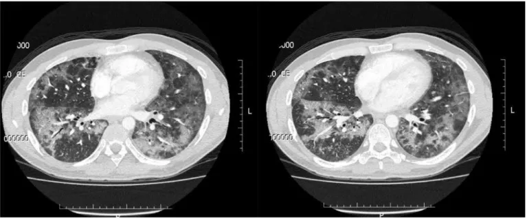
and 6 L/min tracheal gas insulation. At that time, the OP diagnostic hypothesis was suggested. Pulse therapy with methylprednisolone was initiated (1 g per day for 5 days), and the use of clarithromycin was maintained (500 mg twice daily). Upon ending the pulse therapy (23rd day), the patient showed a 116 PaO2/FiO2 ratio. A thoracic CT showed a large reduction of “ground glass” pulmonary opacities on the 25th hospital day (Figure 2).
A tracheostomy was performed on the 27th day of ICU
admission, and on the 31st day, the patient was removed
from mechanical ventilation, showing a 402 PaO2/FiO2
ratio. he patient was discharged from the ICU on the 42nd day of ICU admission and was discharged from the hospital 8 days later. he patient still showed consolidated “ground glass” pulmonary opacities bilaterally based on the therapeutic monitoring thoracic CT at hospital discharge, which were more pronounced in the posterior and lateral basal segments of the inferior lobes, albeit with signiicant decrease in the extent of pulmonary opacities (Figure 3). he patient maintained outpatient follow-up and used corticosteroid therapy with 60 mg of prednisone per day for another 6 months.
DISCUSSION
OP is a clinical entity that may be cryptogenic (formerly known as bronchiolitis obliterans with OP or BOOP (bronchiolitis obliterans organizing pneumonia)) or secondary to connective tissue diseases, infections, radiation therapy, aspiration, drugs and cancer among other causes.(3-7) hat classiication is essential because treatment of the underlying disease or discontinuation of
contact with the aggressive factor is critical for treating secondary OP.
OP is considered a rare disease with an incidence of 1.96/100 thousand, but the number of cases has increased in the last 20 years, primarily afecting adults in the fourth and ifth decades of life with no gender predominance. he mortality ranges from 5 to 27% in studies and is apparently higher in secondary OP.(5-8)
he typical histopathological pattern is identiied
Figure 2 - Thoracic computed tomography following completion of pulse therapy. Bilateral pulmonary “ground glass” opacities predominantly affecting the lingula of the left lung with considerable reduction compared to the previous exam. Consolidated pulmonary opacities in the posterior and lateral basal segments of the lower lobes with pneumomediastinum and bilateral small pleural effusion.
granulation tissue (Masson bodies) and a predominance of alveolar inlammation over the small airways disease (bronchiolitis).(4,6)
he symptoms are nonspeciic and usually progress subacutely or chronically with clinical symptoms similar to those of community-acquired pneumonia (CAP), which often delays the diagnosis and treatment. he use of antibiotic therapy for the empirical
treatment of infection is inefective,(3-6) although
macrolides are used in the treatment of patients with mild symptoms or as an adjuvant therapy, given their
immunomodulatory properties.(9,10) he disease usually
develops after a prodrome, wherein the patient shows lu-like symptoms, including fever, fatigue, dry cough and dyspnea. Typically, the diagnosis is not suspected in the irst 4 to 10 weeks. he physical examination is nonspeciic, and inspiratory crackles occur in two thirds
of cases and more commonly in secondary OP.(3-6)
Radiological indings are also nonspeciic, thereby leading to an indication for lung biopsy to conirm the diagnosis. In a recent study performed by
Drakopanagiotakis et al.,(6) consolidation was noted in
82% of patients on thoracic radiography, which was bilateral in 68.8% of cases with a possible migratory pattern. he density of alveolar iniltrate may vary from “ground glass” to consolidation, which may comprise a few centimeters to a full impairment of a pulmonary lobe or show the appearance of nodules or masses with an air bronchogram in certain instances on high-resolution thoracic CT. his alveolar iniltrate is predominantly peripheral but may have a peribronchovascular location (bronchocentric pattern) less frequently.(7)
Cases of spontaneous regression are rare, and occasionally, OP develops rapidly and progressively, as in the case described in this report, thereby leading to acute respiratory failure. Depending on the extent of injury, certain patients meet the clinical criteria for a diagnosis of acute respiratory distress syndrome (ARDS), albeit with a diferent histological pattern and without the difuse alveolar damage typical of ARDS. In those cases, a diagnostic suspicion is crucial because the patients usually respond well to treatment with methylprednisolone pulse therapy (125 to 250 mg every 6 hours), which is administered for 3 to 5 days and is followed by oral corticosteroid therapy with prednisone (1 to 1.5 mg/kg of weight per day)
secondary OP has a worse prognosis. he combination of immunosuppressants can deliver satisfactory results in certain patients who fail to improve with the use of corticosteroids.(6,7,10) Usually, corticosteroid therapy is suspended within 3 to 6 months, but relapses are common (13-58%) and are more frequent in patients with hypoxemia, difuse alveolar iniltrate, tobacco
smoking and secondary OP.(5,6)
Regarding the case reported in this study, it is noteworthy that the lung is an organ that is often afected through injuries resulting from adverse reactions to antineoplastic drugs. hese lesions may manifest through a wide variety of clinical syndromes, and OP is one of those entities. Cyclophosphamide, doxorubicin and rituximab have already been reported to cause OP among the drugs used in the antineoplastic treatment of this patient.(11-13)
A limitation of the current report was the failure to perform a histopathological examination to conirm the diagnosis. he severity of clinical symptoms, including unfavorable outcomes following the initial treatment and the high risk of performing surgical procedures, in combination with the pattern of lesions on the thoracic CT and history of exposure to drugs known to be related to the development of secondary OP led to the decision to treat the OP with methylprednisolone pulse therapy. However, that therapy has a high risk of complications that must be stressed. Moreover, a surgical (thoracoscopic or open) lung biopsy remains the gold standard for an OP diagnosis,(3,6) and this procedure is indicated prior to introducing corticosteroid therapy in the absence of contraindications.
OP is a disease that has increased in incidence over recent years and may occasionally progress into acute respiratory failure. he diagnosis of OP is diicult because it shows clinical symptoms similar to CAP, but a thorough medical history evaluation, especially regarding exposure to drugs causing secondary OP, can be helpful when considering this diagnosis.
CONCLUSIONS
RESUMO
Doenças difusas do parênquima pulmonar pertencem
a um grupo de doenças de evoluçãogeralmente subaguda
ou crônica, mas que podem determinar insuficiência
res-piratória aguda. Paciente masculino, 37 anos, em terapia para linfoma não Hodgkin, admitido com tosse seca, fe-bre, dispneia e insuficiência respiratória aguda hipoxêmica.
Iniciadas ventilação mecânica e antibioticoterapia, porém
houve evolução desfavorável. Tomografia computadorizada de tórax mostrava opacidades pulmonares em “vidro fosco” bilaterais. Devido ao paciente ter feito uso de três drogas relacionadas à pneumonia em organização (ciclofosfamida, doxorrubicina e rituximabe) e quadros clínico e radiológico
serem sugestivos, iniciou-se pulsoterapia com metilpredni-solona com boa resposta. Pneumonia em organização pode ser idiopática ou associada a colagenoses, drogas e neopla-sias, e geralmente responde bem a corticoterapia. O diag-nóstico é anatomopatológico, mas condições clínicas do paciente não permitiam a realização de biópsia pulmonar. Pneumonia em organização deve ser diagnóstico diferencial em pacientes com aparente pneumonia de evolução desfa-vorável ao tratamento antimicrobiano.
Descritores: Pneumonia em organização criptogênica; Insuiciência respiratória; Toxicidade de drogas; Doenças pulmonares intersticiais; Linfoma não Hodgkin/quimioterapia; Tomograia computadorizada por raios X; Relatos de casos
REFERENCES
1. Pinheiro BV. Infiltrado pulmonar no paciente crítico: a importância da biópsia pulmonar. J Bras Pneumol. 2006;32(5):xxiii-xxiv.
2. Monteiro AS, Addor G, Nigri DH, Franco CA. Biópsia pulmonar a céu aberto em pacientes sob ventilação mecânica e com infiltrado pulmonar difuso. J Bras Pneumol. 2005;31(3):212-8.
3. Cottin V, Cordier JF. Cryptogenic organizing pneumonia. Semin Respir Crit Care Med. 2012;33(5):462-75.
4. Epler GR. Bronchiolitis obliterans organizing pneumonia. Arch Intern Med. 2001;161(2):158-64. Review.
5. Drakopanagiotakis F, Polychronopoulos V, Judson MA. Organizing pneumonia. Am J Med Sci. 2008;335(1):34-9. Review.
6. Drakopanagiotakis F, Paschalaki K, Abu-Hijleh M, Aswad B, Karagianidis N, Kastanakis E, et al. Cryptogenic and secondary organizing pneumonia: clinical presentation, radiographic findings, treatment response, and prognosis. Chest. 2011;139(4):893-900.
7. Cordier JF. Cryptogenic organising pneumonia. Eur Respir J. 2006;28(2):422-46. Review.
8. Gudmundsson G, Sveinsson O, Isaksson HJ, Jonsson S, Frodadottir H, Aspelund T. Epidemiology of organising pneumonia in Iceland. Thorax. 2006;61(9):805-8.
9. Stover DE, Mangino D. Macrolides: a treatment alternative for bronchiolitis obliterans organizing pneumonia? Chest. 2005;128(5):3611-7
10. Epler GR. Bronchiolitis obliterans organizing pneumonia, 25 years: a variety of causes, but what are the treatment options? Expert Rev Respir Med. 2011;5(3):353-61.
11. Maldonato F, Limper AH, Jett JR. Pulmonary toxicity associated with systemic antineoplastic therapy: Clinical presentation, diagnosis, and treatment. Uptodate.com [Internet]. 2012 [cited 2012 Nov 16; last updated: Mar 29, 2012]. Available from: http://www.uptodate.com/ contents/pulmonary-toxicity-associated-with-systemic-antineoplastic-therapy-clinical-presentation-diagnosis-and-treatment
12. Maldonato F, Limper AH, Jett JR. Pulmonary toxicity associated with antineoplastic therapy: molecularly targeted agents. Uptodate.com [Internet]. 2012 [cited 2012 Nov 16; last updated: Mar 29, 2012]. Available from: http://www.uptodate.com/contents/pulmonary- toxicity-associated-with-antineoplastic-therapy-molecularly-targeted-agents?source=see_link