Is a safety guidewire needed for retrograde ureteroscopy?
WILSON RICA MOLINA JUNIOR1, RODRIGO R. PESSOA2, RODRIGO DONALÍSIODA SILVA3, DIEDRA GUSTAFSON4, LETICIA NOGUEIRA4, ALEX MELLER5*
1Associate Professor, University of Colorado, Chief of Endourology Division, Denver Health Medical Center, Denver, CO, USA 2Urology Resident, University of Colorado, Denver, CO, USA
3Assistant Professor of Surgery, Division of Urology, University of Colorado, and Department of Urology, Denver Health Medical Center, Denver, CO, USA 4Reseach Coordinator, University of Colorado, Denver, CO, USA
5Assistant Doctor, Endourology Division, Universidade Federal de São Paulo, São Paulo, SP, Brazil
S
UMMARYStudy conducted at Universidade Federal de São Paulo (Unifesp),
São Paulo, SP, Brazil, and at Denver Health Medical Center, University of
Colorado, Denver, CO, USA
Article received: 7/3/2017 Accepted for publication: 7/21/2017
*Correspondence:
Departamento de Urologia, Unifesp Address: Rua Napoleão de Barros, 715,
Vila Clementino São Paulo, SP – Brazil
Postal code: 04024-002 [email protected]
http://dx.doi.org/10.1590/1806-9282.63.08.717
Introduction: It is generally advised to have a safety guidewire (SGW) present during ureteroscopy (URS) to manage possible complications. However, it increases the strenght needed to insert and retract the endoscope during the procedure, and, currently, there is a lack of solid data supporting the need for SGW in all procedures. We reviewed the literature about SGW utilization during URS.
Method: A review of the literature was conducted through April 2017 using PubMed, Ovid, and The Cochrane Library databases to identify relevant studies. The primary outcome was to report stone-free rates, feasibility, contraindications to and complications of performing intrarenal retrograde flexible and semi-rigid URS without the use of a SGW.
Results: Six studies were identified and selected for this review, and overall they included 1,886 patients where either semi-rigid or flexible URS was performed without the use of a SGW for the treatment of urinary calculi disease. Only one study reported stone-free rates with or without SGW at 77.1 and 85.9%, respectively (p=0.001). None of the studies showed increased rates of complications in the absence of SGW and one of them showed more post-endoscopic ureteral stenosis whenever SGW was routinely used. All studies recommended utilization of SGW in complicated cases, such as ureteral stones associated with significant edema, ureteral stricture, abnormal anatomy or difficult visualization.
Conclusion: Our review showed a lack of relevant data supporting the use of SGW during retrograde URS. A well-designed prospective randomized trial is in order.
Keywords: safety guidewire, ureteroscopy, retrograde intrarenal surgery, meta-analysis, kidney stone, ureteral calculi.
I
NTRODUCTIONUreteroscopy (URS) has become the standard of care for treating urolithiasis less than 2 cm, mainly due to the development of small flexible ureteroscopes, the improve-ment of laser lithotripsy and the quality of disposable materials.1 It is generally advised to have a safety guidewire
(SGW) present during URS to allow placement of a
ure-teral stent in order to manage possible complications.2,3
However, there is a lack of solid data to support this long-standing principle in endourology.
The forces needed to insert and retract the endoscope during URS with an SGW in place are considerably
high-er when compared with procedures that not involve SGW.4
Although not completely proved, this fact raises the ques-tion that placement of an SGW could eventually increase the risk of harming the ureter in some patients.
Moreover, some data advocate that working without an SGW often facilitates access, scope manipulation and stone basketing. There is less friction passing the uretero-scope over than alongside a guidewire and increased torque to rotate the scope.5
On the other hand, as patient safety should continue to be the highest priority, having an SGW during the entire procedure may be advised because of the risk of ureteral
injury requiring prompt placement of ureteral stent.6
The following publication aimed to look at SGW utilization during URS, reviewing the current literature available for both semi-rigid and flexible URS.
M
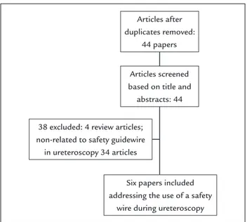
ETHODdatabases to identify relevant studies. Six separate search-es were done by applying the following free-text search terms: “Safety guidewire ureteroscopy,” “Safety guidewire flexible ureteroscopy,” “Safety wire ureteroscopy,” “Safe-ty wire retrograde intrarenal surgery” and “Safe“Safe-ty wire upper ureter.” Article selection was done based on Pre-ferred Reporting Items for Systematic Reviews and Meta--Analyses (PRISMA) criteria7 (Figure 1). Titles of articles
were first reviewed to determine whether they might fit the inclusion criteria. After assessing the abstract, a more detailed subsequent assessment was performed by look-ing at the full text. References of included studies were also reviewed to identify additional studies of interest.
Two reviewers (R.P and W.M) independently screened all the titles and abstracts to minimize selection bias. The quality of the evidence was evaluated based on compre-hensiveness of the data and precision of the reporting according to the criteria provided by the Centre for Evi-dence-Based Medicine in Oxford, UK (website, same 18 as Cryometa). Only studies where an SGW was both used and omitted in the same cohort of patients were includ-ed. The initial literature search identified 72 potentially relevant studies. Their titles and abstracts were screened for relevance, resulting in 44 potential articles after ex-cluding duplicate results. Four reports were excluded because they were review URS articles and 35 were ex-cluded because they didn’t specifically addressed the use or not of an SGW. Therefore, five articles were included and one additional record was added after reference list survey (Figure 1). The primary outcome was to report feasibility, contraindications to and complications of performing intrarenal retrograde flexible and semi-rigid URS without the use of an SGW. Secondary outcomes were to compare stone-free rates and complications be-tween cases where an SGW was used or omitted for the treatment of ureteral and kidney stone disease. Patients were considered stone-free if they had remnant fragments of up to 2 mm in follow-up tomography or intravenous urography six weeks to three months after the main pro-cedure. The Clavien-Dindo classification was used to
report complication.8
R
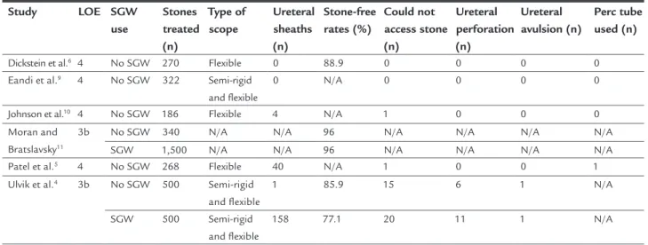
ESULTSSix studies (Table 1) were identified and selected for this review. Overall, they included 1,886 patients, and either semi-rigid or flexible URS was performed without the use of an SGW for the treatment of urinary calculi disease. Four of them were retrospective observational
non-com-parative studies (level of evidence 4)6,9-11 and two were
retrospective observational non-consecutive comparative studies (level of evidence 3b).4,11
Johnson et al.10 studied retrospectively a
single-sur-geon prospective database of flexible URS. A total of 186 patients were submitted to wireless flexible URS for the treatment of intrarenal stones. They reported a stone-free rate of 90, 89 and 75% after primary therapy of intra-renal calculi of < 1.0 cm, 1.0 to 2.0 cm, and > 2.0 cm, respec-tively. Stone-free rates after primary treatment of ure-teral calculi were 93, 96 and 100% for proximal, medial and distal third location, respectively. Inability to access the lower pole was reported in six cases and inability to reach the kidney, in one. There were no false passages or ureteral perforations secondary to endoscope placement. Minor complications were limited to postoperative py-elonephritis in five individuals and gross hematuria in three, both treated successfully with antibiotics and with
conservative measures, respectively.10
Dickstein et al.6 reported their experience with
flex-ible URS for the treatment of ureteropelvic junction (54) and renal calyces (216) stones in 270 consecutive patients. In all cases, lithotripsy was performed with a Holmium:YAG laser until calculi pulverization, without the use of a basket for extraction of fragments. The average stone size was 9.1±3.5 mm, and stone-free rate was 88.9% (240 of 270). There were no intraoperative complications, no cases of lost access, ureteral perforation, avulsion, or the need for a percutaneous nephrostomy tube placement (PCNT). However, the authors still recommended the use of an SGW in cases of complicated cases, such as encrusted ureteral stents, ureteral stricture requiring
Articles after duplicates removed:
44 papers
38 excluded: 4 review articles; non-related to safety guidewire
in ureteroscopy 34 articles
Six papers included addressing the use of a safety
wire during ureteroscopy Articles screened based on title and
abstracts: 44
dilation and concomitant longstanding obstructive ure-teral stones.6
Two other groups reported independently their results of semi-rigid and flexible URS for the treatment of stone disease without an SGW.5,9 Eandi et al.9 reported no
intra-operative complications related to lack of a safety wire over 322 semi-rigid and flexible URS performed for the treatment of urolithiasis. Patel et al.5 described their
ex-perience with flexible URS for the treatment of calyceal and pelvic stones on 268 patients with the use of a work-ing wire alone. In all, 20% of the patients needed ureteral dilation, and 15% had a ureteral access sheath placed intra operatively. The overall complication rate was 2.6%. There were no intraoperative complications (no ureteral avulsions or ureteral perforations). Overall, six patients had urinary tract infection (Clavien grade II), two of whom needed post procedure hospital admission and treatment with intravenous antibiotics. One patient had a urinary reten-tion (Clavien grade I). Access into the renal pelvis was obtained in all patients except for one who had multiple ureteral strictures necessitating a nephrostomy tube
place-ment with subsequent percutaneous nephrolithotomy.5
However, the authors acknowledge that their study in-cluded only patients with kidney stones and that, for the treatment of concomitant ureteral stones associated with significant edema, ureteral strictures, abnormal anatomy or difficult visualization, a safety wire should be placed.5
The only two available comparative studies in the literature that studied the role of an SGW for semi-rigid and flexible URS are depicted in Table 1. Moran and
Brat-slavsky11 compared a single urologist’s experience with
flexible ureteroscopic laser lithotripsy without the use of an SGW to a contemporary, large single-center’s experi-ence with 11 treating urologists. A total of 340 flexible ureteroscopies were performed over a single working wire placed prior to laser lithotripsy, whereas 1,500 laser lith-otripsies were done at a single center with an SGW in place. Targeted stone destruction occurred in 98% of these cases and the stone-free rates were lower in 96% (326/340) for those that did not use an SGW. Failures in this cohort were infrequent and occurred in seven patients with high grade obstruction and/or impacted calculi. On the other side, in the entire series of 1,500 patients the targeted stone destruction occurred in 98% and stone-free rate was 96%, results identical to the technique without the safety wire. There were no complications in the group without
a safety wire secondary to loss of upper tract access.11
Ulvik et al.12 compared the results of URS for the
treat-ment of ureteral stones at two different hospitals where the SGW was either routinely used or omitted. Both groups
had 500 patients each. Pretreatment stone status differed in many aspects between groups. The hospital where an SGW was routinely used treated more proximal stones, more cases with obstruction and more urgent cases. As a result, flexible endoscopes were employed in 39.8 and 4.4% of the procedures in the group with an SGW and without it, respectively (p<0.0005). Similarly, access sheaths were used in 31.6% of the cases in the group with SGW compared to only one case in the group without it (p<0.0005).12
The reported success rates of passing the ureteroscope through the ureteral orifice, the ability to access the ure-teral stone and the ability to place a ureure-teral stent when needed after the endoscopy were not significantly
differ-ent between the two groups of patidiffer-ents.12 There was no
significant difference in the overall intraoperative com-plication rates at the two hospitals. The overall stone-free rates were 77.1% and 85.9% with and without the SGW, respectively (p=0.001). However, according to the stone location, the stone-free rates were 61.2 and 70.2% for up-per (p=0.135), 72.6 and 81.1% for mid (p=0.305), and 89.8 and 93.9% for lower ureteral stones (p=0.102) with and without SGW, respectively. A significant increase in the number of patients (14 patients, 3.4%) was found to have post endoscopic ureteral stenosis at the hospital where the SGW was routinely used than at the hospital where
an SGW was omitted (six patients, 1.2%), p=0.039.12
D
ISCUSSIONThe advantage of using an SGW is to ensure a prompt stent placement in an event of a major ureteral
perfora-tion or bleeding precluding continuing URS.3,13 However,
what we found on the literature is that the cumulative evidence that endorse the routine use of an SGW during URS is very weak (level of evidence grade C). It seems that there is a belief that the routine use of an SGW may not be necessary and may even be deleterious, mainly due to the fact that working without a safety wire often facilitates access to the kidney (less friction passing the ureteroscope), scope manipulation (less torque to rotate the scope), and makes it easier to laser and basket fragments.5,9,12 Moreover,
many publications have described their successful experi-ence with both semi-rigid and flexible URS for the treat-ment of both ureteral and renal stones without the use of an SGW.5,6,9-12
guide-wires and nitinol baskets have raised the safety and precision of the procedure to a new level. Despite technological prog-ress, endoscopic intervention can still result in unpredictable and difficult to solve situations. Therefore, we concur with the recommendations to use an SGW whenever a more dif-ficult procedure is anticipated such as in cases with ureteral edema, ureteral strictures, abnormal anatomy, sub-optimal visualization, encrusted ureteral stents and concomitant longstanding obstructive ureteral stones.5,6
The main limitation of our study is the low level of evi-dence of the articles available. Most of them are retrospec-tive analysis of series of cases without a compararetrospec-tive group. Moreover, the best comparative available study has a lot of limitations itself, as described previously. However, it should be noted that this major drawback is also present in the literature supporting the use of ureteral stents after URS. In conclusion, our review showed a lack of relevant data supporting the use of SGW during retrograde URS. A well-designed prospective randomized trial is necessary.
R
ESUMOFio guia de segurança é necessário na ureteroscopia?
Introdução: O uso de fio guia de segurança (FGS) costu-ma ser recomendado para a realização de ureteroscopia para prevenir e solucionar complicações durante o proce-dimento. Seu uso, porém, aumenta a força necessária para manipular o aparelho endoscópico dentro da luz ureteral e, atualmente, existe uma carência de dados consistentes que indiquem o uso do FGS em todos os procedimentos.
Método: Uma revisão da literatura foi realizada em abril de 2017 utilizando as ferramentas PubMed, Ovid e The
Cochrane Library para identificar estudos relevantes. O desfecho primário da análise foi reportar taxas de reso-lução dos cálculos, viabilidade, contraindicações e com-plicações relacionadas ao não uso do FGS.
Resultados: Seis estudos foram incluídos na análise, to-talizando 1.886 pacientes, nos quais foi realizada urete-roscopia semirrígida ou flexível sem o uso do FGS no tratamento de cálculos renais ou ureterais. Somente um estudo relatou taxa livre de cálculos com ou sem FGS, sendo 77,1 e 85,9%, respectivamente (p=0.001). Todos os estudos mostraram não haver aumento da taxa de com-plicação na ausência do FGS e um deles relatou aumento de estenose ureteral pós-endoscopia no grupo que utilizou o FGS. Todos os estudos recomendam o uso do FGS em casos complicados, como cálculos ureterais associados a edema de mucosa, estenose ureteral, anomalias anatômi-cas ou dificuldade de visualização do cálculo.
Conclusão: Nossa revisão mostrou que faltam dados rele-vantes para justificar o uso do FGS durante a ureteroscopia.
Palavras-chave: fio guia, ureteroscopia, cirurgia intrarrenal retrógrada, metanálise, litíase renal, cálculos ureterais.
R
EFERENCES1. de la Rosette J, Denstedt J, Geavlete P, Keeley F, Matsuda T, Pearle M, et al.; CROES URS Study Group. The clinical research office of the endourological society ureteroscopy global study: indications, complications, and outcomes in 11,885 patients. J Endourol. 2014; 28(2):131-9.
2. Sprunger JK, Herrell SD 3rd. Techniques of ureteroscopy. Urol Clin North
Am. 2004; 31(1):61-9.
3. Bagley DH, Kuo RL, Zeltser IS. An update on ureteroscopic instrumentation for the treatment of urolithiasis. Curr Opin Urol. 2004; 14(2):99-106. 4. Ulvik Ø, Wentzel-Larsen T, Ulvik NM. A safety guidewire influences the
pushing and pulling forces needed to move the ureteroscope in the ureter: a clinical randomized, crossover study. J Endourol. 2013; 27(7):850-5.
TABLE 1 Summary outcomes of selected publications.
Study LOE SGW
use
Stones treated (n)
Type of scope
Ureteral sheaths (n)
Stone-free rates (%)
Could not access stone (n)
Ureteral perforation (n)
Ureteral avulsion (n)
Perc tube used (n)
Dickstein et al.6 4 No SGW 270 Flexible 0 88.9 0 0 0 0
Eandi et al.9 4 No SGW 322 Semi-rigid
and lexible
0 N/A 0 0 0 0
Johnson et al.10 4 No SGW 186 Flexible 4 N/A 1 0 0 0
Moran and Bratslavsky11
3b No SGW 340 N/A N/A 96 N/A N/A N/A N/A
SGW 1,500 N/A N/A 96 N/A N/A N/A N/A
Patel et al.5 4 No SGW 268 Flexible 40 N/A 1 0 0 1
Ulvik et al.4 3b No SGW 500 Semi-rigid
and lexible
1 85.9 15 6 1 N/A
SGW 500 Semi-rigid
and lexible
158 77.1 20 11 1 N/A
5. Patel SR, McLaren ID, Nakada SY. The ureteroscope as a safety wire for ureteronephroscopy. J Endourol. 2012; 26(4):351-4.
6. Dickstein RJ, Kreshover JE, Babayan RK, Wang DS. Is a safety wire necessary during routine flexible ureteroscopy? J Endourol. 2010; 24(10):1589-92. 7. Moher D, Liberati A, Tetzlaff J, Altman DG; PRISMA Group. Preferred
reporting items for systematic reviews and meta-analyses: the PRISMA statement. PLoS Med. 2009; 6(7):e1000097.
8. Dindo D, Demartines N, Clavien PA. Classification of surgical complications: a new proposal with evaluation in a cohort of 6336 patients and results of a survey. Ann Surg. 2004; 240(2):205-13.
9. Eandi JA, Hu B, Low RK. Evaluation of the impact and need for use of a safety guidewire during ureteroscopy. J Endourol. 2008; 22(8):1653-8.
10. Johnson GB, Portela D, Grasso M. Advanced ureteroscopy: wireless and sheathless. J Endourol. 2006; 20(8):552-5.
11. Moran ME, Bratslavsky G. Changing paradigm during routine flexible ureteroscopy and Holmium:YAG laser lithotripsy: need for safety wires? J Endourol 2003; 17:A225.
12. Ulvik Ø, Rennesund K, Gjengstø P, Wentzel-Larsen T, Ulvik NM. Ureteroscopy with and without safety guide wire: should the safety wire still be mandatory? J Endourol. 2013; 27(10):1197-202.