FLEXIBLEURETERORENOSCOPYINPOSITIONORFUSIONANOMALY: ISITFEASIBLE?
REV ASSOC MED BRAS 2017; 63(8):685-688 685
ORIGINAL ARTICLE
Flexible ureterorenoscopy in position or fusion anomaly:
Is it feasible?
RAFAEL HADDAD ASTOLFI1, GUSTAVO FRESCHI2, FERNANDO FIGUEIREDO BERTI2, NELSON GATTAS3, WILSON RICA MOLINA JUNIOR4,
ALEX MELLER5*
1MD, Urology Resident at Universidade Federal de São Paulo (Unifesp), São Paulo, SP, Brazil 2MD, Urology Division, Unifesp, São Paulo, SP, Brazil
3MD, Lecturer of Urology, Unifesp, São Paulo, SP, Brazil
4Associate Professor, University of Colorado, and Head of the Endourology Department, Denver Health Medical Center, University of Colorado, Denver, CO, USA 5MD, Assistant Physician, Urology Division, Unifesp, São Paulo, SP, Brazil
S
UMMARYStudy conducted at Universidade
Federal de São Paulo (Unifesp), São Paulo, SP, Brazil, and at Denver
Health Medical Center, University of Colorado, Denver, CO, USA
Article received: 7/3/2017
Accepted for publication: 7/21/2017
*Correspondence: Disciplina Urologia
Address: Rua Napoleão de Barros, 715 São Paulo, SP – Brazil
Postal code: 04024-002 [email protected]
http://dx.doi.org/10.1590/1806-9282.63.08.685
Objective: To analyze the results of lexible ureterorenoscopy (F-URS) with holmium laser in the treatment of kidney stones with ectopic and fusion anomalies (horseshoe kidney and rotation anomalies).
Method: We reviewed data from 13 patients with fusion and ectopic renal anomalies that underwent F-URS from April 2011 to April 2017. We analyzed demographic and clinical data (age, gender, BMI, anatomical abnormality, location and dimension of the renal calculi) and perioperative data (method of treatment, stone-free rate, number of days with DJ catheter and perioperative complications). Results: The mean stone size was 12.23 +/- 5.43 mm (range 6-22mm), located in the inferior (58.33%) and middle (16.76%) calyceal units, renal pelvis (16.67%) and multiple locations (8.33%). All 13 patients were treated with Ho-Yag laser, using dusting technique (25%), fragmentation and extraction of the calculi (58.33%) and mixed technique (16.67%). We did not have any severe perioperative complication. After 90 days, nine patients (75%) were considered stone free. Conclusion: Our data suggest that F-URS is a safe and feasible choice for the treatment of kidney stones in patients with renal ectopic and fusion anomalies.
Keywords: urolithiasis, kidney calculi, kidney diseases, fused kidney.
INTRODUCTION
Nephrolithiasis is an increasingly common condition, af-fecting 5-15% of the world’s population and mainly indi-viduals at a productive age between the second and sixth decade of life.1 In recent years in Brazil, according to Da-tasus, the number of hospital admissions and costs for the treatment of this condition has increased, with a total expenditure of BRL 29.2 million/year with hospital admis-sions alone, causing a high impact on public health.2
Renal anomalies are relatively rare. Horseshoe kidney (HK) represents the most common fusion anomaly, with an incidence of 0.25%, while the incidence of pelvic kidney varies from 1/2,100 to 1/3,000 and the variance of crossed renal ectopia is 1/1,000.3 These conditions make it even more challenging to treat urinary lithiasis, with lower success rates in endourologic procedures and increased
intraoperative risks due to anatomical differences in renal structure, rotation, and vasculature.4,5
Extracorporeal lithotripsy (ESWL) and percutaneous nephrolithotripsy (PCNL) are currently the most common treatment methods for kidneys with fusion or position abnormalities.6-8 The choice of lexible ureterorenoscopy with holmium laser – Yag (Ho-Yag) as the irst line of treatment for stones < 20 mm has been increasing due to important technological advances, but only a few studies have reported their results on anomalous kidneys.
OBJECTIVE
ASTOLFI RH ETAL.
686 REV ASSOC MED BRAS 2017; 63(8):685-688
rates, operative time, dificulty accessing the calyces and complications.
METHOD
Data collection
We prospectively collected data from 13 patients with fu-sion or position abnormalities submitted to the F-URS between April 2011 and April 2017 at the Hospital São Paulo (Federal University of São Paulo – Unifesp, SP, Bra-zil) and at the Denver Health Medical Center (University of Colorado, CO, USA). Demographic and clinical data (age, gender, BMI, anatomical abnormalities, size and location of the stone), as well as perioperative data (stone treatment method, stone-free index, DJ catheter time and perioperative complications) were collected from the medical records. All patients underwent a control exam within 90 days, either by non contrast-enhanced com-puted tomography for lithiasis investigation or simple abdominal X-ray. The tomography protocol used the low--dose radioactive modulation technique, with the exception of patients with BMI > 30.9 The abdominal X-ray, in turn, was used for monitoring patients with radiopaque stones and viewed in this examination prior to surgery.
Surgical technique
The surgical procedures were performed by two endou-rologists with extensive experience in F-URS (AM, WRM), all under general anesthesia and in a lithotomy position. After performing asepsis and placing sterile ields, cystos-copy was performed with identiication of the ureteral meatus looking for abnormalities (duplicity). In all cases, after positioning the guidewire, a semi-rigid retrograde ureteroscopy was performed followed by an attempt to pass an 11/13 Fr or 12/14 ureteral sheath (Boston Scien-tiic). After access to the renal pelvis with the lexible ure-teroscope (Storz Flex X2, Oympus URFP5) through the ureteral sheath, a 200 or 273 μm laser iber was used for the treatment of the stone, adjusted according to the stone’s location and composition (pulverization, frag-mentation and removal or mixed technique). To perform the mobilization or the removal of stones, we used a 1.9 Fr Zero Tip nitinol stone retrieval basket or 1.9 Fr Escape model (Boston Scientiic). In all cases, a double J catheter was used postoperatively. Patients in whom residual frag-ments < 2 mm were found in the control exams after 90 days were considered as stone free.
RESULTS
A total of 13 patients (six male and seven female) with anomalous kidney stones (ive with rotational defects
and eight with horseshoe kidneys) were submitted to the F-URS between 2011 and 2017. A non contrast-enhanced abdominal CT was used to determine the dimensions of the stones, with a mean value of 12.23 mm +/- 5.43 mm (ranging from 6 to 22 mm), mostly distributed in only one calycinal group (58.33% in upper calyx, 16.67% in medium calyx, 16.67% in pelvis and 8.33% in multiple calyces). All patients were treated with Ho-Yag laser, with fragmentation and removal of stones in seven cases (58.33%), pulverization in three cases (25%) and mixed technique in two cases (16.67%).
In relation to perioperative complications, there were no intraoperative complications and only one patient with a rotational defect had a mild complication in the irst 24 hours after the procedure (hematuria). There were no patients with Clavien III or IV complications during postoperative monitoring. The DJ catheter was maintained for an average of nine days +/- 3.46 (ranging from 6 to 14 days). Ninety (90) days after the procedure, nine patients were stone free (75%), while residual stones were identiied in only three cases (25%) (Tables 1 and 2).
DISCUSSION
Renal fusion and positional anomalies are related to an increase in the frequency of kidney stones.10-12 Anatomic factors associated with concomitant metabolic disorders contribute to this condition, and make endoscopic treat-ment dificult.13-15
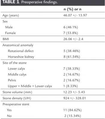
TABLE 1 Preoperative indings.
n (%) or n
Age (years) 46.07 +/- 13.97
Sex Male Female
6 (46.1%) 7 (53.8%)
BMI 26.06 +/- 2.4
Anatomical anomaly Rotational defect Horseshoe kidney
5 (38.46%) 8 (61.54%)
Site of the stone Lower calyx Middle calyx Pelvis
Upper + Middle + Lower calyx
7 (58.33%) 2 (16.67%) 2 (16.67%) 1 (8.33%)
Stone volume (mm) 12.23 +/- 5.43
Stone density (UH) 924 +/- 328.01
Preoperative stent Yes
No
FLEXIBLEURETERORENOSCOPYINPOSITIONORFUSIONANOMALY: ISITFEASIBLE?
REV ASSOC MED BRAS 2017; 63(8):685-688 687
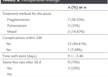
TABLE 2 Perioperative indings.
n (%) or n
Treatment method for the stone Fragmentation
Pulverization Mixed
7 (58.33%) 3 (25%) 2 (16.67%)
Complications within 24h No
Yes
12 (84.61%) 1 (7.69%)
Time with stent (days) 9 +/- 3.46
Stone-free rate after 30 d Yes
No
9 (75%) 3 (25%)
PCNL is the chosen option for the treatment of anomalous kidney stones, especially for stones larger than 20 mm, with stone-free rates between 80 and 90%.16-20 The success of the procedure is impaired by features such as renal pelvis and anteriorly positioned calyces, vascular abnormalities and different anatomical relationships with adjacent organs, which increases the risk of perioperative compli-cations and the dificulty of the procedure.6-8 A routine preoperative abdominal CT scan can reduce the risk of visceral injury in PCNL, especially in pelvic and horseshoe kidneys.20,21 Auxiliary methods to aid puncture, such as laparoscopy or ultrasonography, have been described, and present good results.16,17,22 However, the potential sever-ity of these lesions, in addition to increasing the inherent cost of these auxiliary procedures, favors the search for more conservative treatments.
ESWL remains an interesting option for anomalous kidneys due to its non-invasive nature, although ana-tomical variations (high ureter implantation, JUP steno-sis, etc.) make it dificult to pass stones in a signiicant number of patients, and complementary procedures are usually required.1,2 The stone-free rates in anomalous kidneys vary in the literature and depend on the dimen-sions of the stones. Sheir et al.23reported a general success rate of ESWL in anomalous kidneys of 72.2%, with only 46.1% for stones > 15 mm.1 Tunc et al.,24 in turn, reported a rate of 92% for stones < 10 mm, but 34% for those great-er than 30 mm.3 Coupled with lower eficiency of ESWL in eliminating larger stones, Ray et al.13 has pointed out that 51% of their patients needed an additional procedure, but that little improvement occurred after the second session, revealing a limitation in the number of attempts that could be made.
The technological advances in lexible ureteroscopy have allowed its use to be expanded, and it is
increas-ingly used in cases of renal anomalies, especially horseshoe kidneys. Its greater delection capacity (up to 270°), cou-pled with progressively thinner laser ibers and the devel-opment of nitinol stone extractors have allowed the access and treatment of stones located in lower calyces or errat-ically-positioned calyces, leading to stone-free rates rang-ing from 70 to 88.2% in up to 1.5 sessions for stones < 30 mm in diameter.25-28 Techniques such as reallocation of stones from the lower calyx to the middle or upper calyx aid in the success of the procedure by facilitating frag-mentation, as well as increasing the useful life of the ap-paratus by avoiding excessive use of delection. For cases with residual calculi, ESWL, PCNL or another F-URS session can be performed, but conservative treatment should not be ruled out when possible. In our series of cases, we obtained a stone-free rate of 75% for stones with a diameter of 12.22 mm (+/- 5.43 mm), with minimal complication rates (one case of transient hematuria), re-inforcing data in the current literature that F-URS is currently a safe and effective procedure for the treatment of stones < 30 mm in anomalous kidneys.
CONCLUSION
Patients with renal position and fusion anomalies are predisposed to the formation of stones and lower success rates in interventional procedures. Although tradition-ally ESWL and PCNL are the treatments of choice for these patients, advances in F-URS technology have now allowed them to be treated less invasively and with excel-lent results.
RESUMO
Ureterorrenolitotripsia lexível no tratamento de cálculos em rins anômalos: Qual a viabilidade?
Objetivo: Analisar os resultados da ureterorrenolitotripsia lexível (ULT-F) no tratamento de cálculos em rins com anomalia de posição e de fusão (rins em ferradura e rins com vício de rotação).
Método: Realizamos a coleta prospectiva dos dados de 13 pacientes com anomalias de fusão e de posição sub-metidos a ULT-F entre abril de 2011 e abril de 2017. Ana-lisaram-se dados clínicos (idade, gênero, IMC, anorma-lidades anatômicas, dimensão e localização dos cálculos) e perioperatórios (método de tratamento do cálculo, índice de stone free, tempo de cateter DJ e complicações perioperatórias).
ASTOLFI RH ETAL.
688 REV ASSOC MED BRAS 2017; 63(8):685-688
em sua maioria distribuídos em apenas um grupo calici-nal (58.33% em grupo calicial inferior, 16.67% em grupo calicial médio, 16,67% em pelve e 8,33% em múltiplos cálices). Todos os pacientes foram tratados com utilização de laser Ho-Yag, com fragmentação e retirada de cálculos em sete casos (58,33%), pulverização em três casos (25%) e técnica mista em dois casos (16,67%). Não houve com-plicações intraoperatórias ou pós-operatórias graves. Após 90 dias, nove pacientes tornaram-se stone free (75%). Conclusão: A ULT-F apresenta-se como método seguro e eicaz no tratamento de litíase em rins com anomalia de posição e de fusão.
Palavras-chave: urolitíase, cálculos renais, rim fundi-do, nefropatias.
REFERENCES
1. Lotan Y. Economics and cost of care of stone disease. Adv Chronic Kidney Dis. 2009; 16(1):5-10.
2. Korkes F, Silva JLS, Heilberg IP. Custo do tratamento hospitalar da litíase uriná-ria para o Sistema Único de Saúde brasileiro. Einstein. 2011; 9(4 Pt 1):518-22. 3. Shapiro E, Bauer SB, Chow JS. Anomalies of the upper urinary tract. In: Wein AJ,
ed. Campbell’s urology. 10th ed. Philadelphia: W.B. Saunders; 2012. p.3106-43. 4. Bozkurt OF, Tepeler A, Sninsky B, Ozyuvali E, Ziypak T, Atis G, et al. Flexible
ureterorenoscopy for the treatment of kidney stone within pelvic ectopic kidney. Urology. 2014; 84(6):1285-9.
5. Grasso M, Beaghler M, Loisides P. The case for primary endoscopic management of upper urinary tract calculi: II. Cost and outcome assessment of 112 primary ureteral calculi. Urology. 1995; 45(3):372-6.
6. Bhatia V, Biyani CS. Calculus disease in duplex system: role of extracorporeal shockwave lithotripsy. Urol Int. 1993; 50(3):164-9.
7. Serrate R, Regué R, Prats J, Rius G. ESWL as the treatment for lithiasis in horseshoe kidney. Eur Urol. 1991; 20(2):122-5.
8. Etemadian M, Maghsoudi R, Abdollahpour V, Amjadi M. Percutaneous ne-phrolithotomy in horseshoe kidney: our 5-year experience. Urol J. 2013; 10(2):856-60.
9. Fulgham PF, Assimos DG, Pearle MS, Preminger GM. Clinical effectiveness protocols for imaging in the management of ureteral calculous disease: AUA technology assessment. J Urol. 2013; 189(4):1203-13.
10. Gutierrez R. Role of anomalies of kidneys and ureter in causation of surgical conditions. JAMA. 1936; 106(3):183-9.
11. Gupta NP, Mishra S, Seth A, Anand A. Percutaneous nephrolithotomy in abnormal kidneys: single-center experience. Urology. 2009; 73(4):710-4.
12. Yohannes P, Smith AD. The endourological management of complications associated with horseshoe kidney. J Urol. 2002; 168(1):5-8.
13. Ray AA, Ghiculete D, D’A Honey RJ, Pace KT. Shockwave lithotripsy in patients with horseshoe kidney: determinants of success. J Endourol. 2011; 25(3):487-93.
14. Esuvaranathan K, Tan EC, Tung KH, Foo KT. Stones in horseshoe kidneys: result of treatment by extracorporeal shock wave lithotripsy and endourology. J Urol. 1991; 146(5):1213-5.
15. Kirkali Z, Esen AA, Mungan MU. Effectiveness of extracorporeal shockwave lithotripsy in the management of stone-bearing horseshoe kidneys. J Endourol. 1996; 10(1):13-5.
16. Mosavi-Bahar SH, Amirzargar MA, Rahnavardi M, Moghaddam SM, Babbolhavaeji H, Amirhasani S. Percutaneous nephrolithotomy in patients with kidney malformations. J Endourol. 2007; 21(5):520-4.
17. Osther PJ, Razvi H, Liatsikos E, Averch T, Crisci A, Garcia JL, et al; Croes PCNL Study Group. Percutaneous nephrolithotomy among patients with renal anomalies: patient characteristics and outcomes; a subgroup analysis of the clinical research office of the endourological society global percutaneous nephrolithotomy study. J Endourol. 2011; 25(10):1627-32. 18. Rana AM, Bhojwani JP. Percutaneous nephrolithotomyin in renal anomalies
of fusion, ectopia, rotation, hypoplasia, and pelvicalyceal aberration: uniformity in heterogeneity. J Endourol. 2009; 23(4):609-14.
19. Matlaga BR, Kim SC, Watkins SL, Kuo RL, Munch LC, Lingeman JE. Percutaneous nephrolithotomy for ectopic kidneys: over, around, or through. Urology. 2006; 67(3):513-7.
20. Binbay M, Istanbulluoglu O, Soikerim M, Beytur A, Skolarikos A, Akman T, et al. Effect of simple malrotation on percutaneous nephrolithotomy: a matched pair multicenter analysis. J Urol. 2011; 185(5):1737-41. 21. Skoog SJ, Reed MD, Gaudier FA Jr, Dunn NP. The posterolateral and the
retrorenal colon: implication in percutaneous stone extraction. J Urol. 1985; 134(1):110-2.
22. Holman E, Tóth C. Laparoscopically assisted percutaneous transperitoneal nephrolithotomy in pelvic dystopic kidneys: experience in 15 successful cases. J Laparoendosc Adv Surg Tech A. 1998; 8(6):431-5.
23. Sheir KZ, Madbouly K, Elsobky E, Abdelkhalek M. Extracorporeal shock wave lithotripsy in anomalous kidneys: 11-year experience with two second-generation lithotripters. Urology. 2003; 62(1):10-5.
24. Tunc L, Tokgoz H, Tan MO, Kupeli B, Karaoglan U, Bozkirli I. Stones in anomalous kidneys: results of treatment by shock wave lithotripsy in 150 patients. Int J Urol. 2004; 11(10):831-6.
25. Symons SJ, Ramachandran A, Kurien A, Baiysha R, Desai MR. Urolithiasis in the horseshoe kidney: a single-centre experience. BJU Int. 2008; 102(11):1676-80.
26. Weizer AZ, Springhart WP, Ekeruo WO, Matlaga BR, Tan YH, Assimos DG, et al. Ureteroscopic management of renal calculi in anomalous kidneys. Urology. 2005; 65(2):265-9.
27. Molimard B, Al-Qahtani S, Lakmichi A, Sejiny M, Gil-Diez de Medina S, Carpentier X, et al. Flexible ureterorenoscopy with holmium laser in horseshoe kidneys. Urology. 2010; 76(6):1334-7.