311
Bras J Rheumatol 2009;49(3):308-14 CASE REPORT
Received on 22/07/08. Approved on 18/01/2009. We declare no conflict of interest.
Study carried out in Physical Medicine and Rehabilitation Service at Hospital de Matosinhos, Portugal.
1. Resident Doctor at Internato Complementar de Reumatologia at the Rheumatology Service at Centro Hospitalar do Alto Minho, Ponte de Lima, Portugal 2. Doctorate Student in Forensic Sciences, Department of Biology and Forensic Genetics, Delegation from Centro do Instituto Nacional de Medicina Legal, Universidade de Coimbra, Portugal
3. Physiatrist Doctor at the Physical Medicine and Rehabilitation Service at Hospital de Matosinhos, Portugal
Correspondence to: Centro Hospitalar do Alto Minho, 04990-041 – Ponte de Lima. Phone 258909500. Fax: 259909501. E-mail: [email protected]
INTRODUCTION
Camurati-Engelmann disease or progressive diaphyseal dysplasia is a rare autosomal dominant genetic disease of the bone metabolism, characterized by limb pain and muscular weakness, and cortical thickening of the diaphyses of long bones. Myopathic waddling gait and pain in the affected limb are the most common clinical manifestations. Due to its development and rarity, diagnosis is frequently established with delay and
dificulty. This article describes the development of a patient, the dificulty in diagnosing Camurati-Engelmann disease and
the relevance of considering diagnosis of bone dysplasia in
non-speciic pain of limbs and other diseases that occur with
osteosclerosis and/or hyperostosis in its differential diagnosis.
CASE REPORT
A 46-year-old man, who worked at an airport and whose parents were not kindred, from Mozambique. Since his childhood, he had predominantly proximal pains in lower limbs, described as of bone and muscular origin, and progressive reduction of
Camurati-Engelmann disease: typical
manifestations of a rare disease
Mónica Bogas1, Vanessa Bogas2, Frederico Pinto3
ABSTRACT
Camurati-Engelmann Disease or progressive diaphyseal dysplasia is a rare disease, characterized by limb pain and muscular weakness, and cortical thickening of the diaphyses of long bones. The authors report a case of a male patient with manifestations since his childhood, whose diagnosis was established later on, when he was an adult, with the dise-ase already progressed, and when the same manifestations began in one of his sons. The importance of the differential diagnosis regarding other diseases concurrent with osteosclerotic and/or hyperostotic changes is emphasized here. Description of its evolution along the years is rarely found in the literature.
Keywords: Camurati-Engelmann disease, progressive diaphyseal dysplasia.
muscular strength, acquiring waddling gait with myopathic characteristics. Pains had a mixed pace and an important nocturnal component. As time went on, algic complaints became less intense and physical and psychological adaptation to the case has allowed him to have a life within society normal parameters; he had a normal profession, got married and had children. He learned techniques to wake up and to mask waddling, denying limitations in daily and professional tasks. In the beginning of his case history and for some years, he looked for medical orientation, but the investigation was inconclusive, and there was not any data in the study carried out that would support the neuromuscular disease diagnosis or signs of malignity. Four decades after the beginning of symptoms, evidence of radiographic changes in the femoral diaphyses, not detected previously, again raised attention to the elucidation of his case history. To objective examination, relevant changes were described such as reduction in proximal leg strength with positive Gowers’ maneuver and leg muscle
atrophy. Deep tendon relexes were normal and there was no
Bogas et al.
312 Bras J Rheumatol 2009;49(3):308-14
In the analytical study, there were no changes in hemogram, sedimentation velocity, renal function, phosphocalcic metabolism and alkaline phosphatasis. At that time, his older 5-year-old son began to present a similar case history, with hereditary etiology as part of the medical thought. Diagnosis of Camurati-Engelmann Disease was then considered and confirmed in both of them by detecting the C.652C > T mutation (p.Arg218Cys) in heterozygosity in the exon 4 of
the TGFβ1 gene. When asked, patient denied similar family
antecedents. His other daugher was assymptomatic; no genetic change compatible with this disease was found.
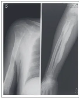
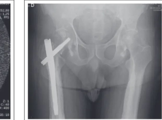
More recently, the patient suffered a fracture of the right femoral neck and trochanter after a traffic accident, and underwent surgery for a proximal femur nailing and functional rehabilitation treatments. Cortical sclerosis and thickening, typical radiologic changes of disease, were found in long bones of his arms and legs in bilateral and symmetric ways, without any change in his hands and feet (Figures 1, 2, 3, 4, and 5). A computed axial tomography study showed narrow marrow canal of the bones involved (Figure 6). Cranial radiography was normal.
The patient did not present postoperative complications and recovered autonomous gait with physical therapy treatments. Surveillance was maintained in external physiatrics appointments for 5 months.
DISCUSSION
Camurati-Engelmann disease or progressive diaphyseal dysplasia is a rare autosomal dominant genetic disease of the bone
metabolism, characterized by limb pain and muscular weakness, and cortical thickening of the diaphyses of long bones.1
A TGFβ1 gene mutation in the chromosome 19q13, which
translates increasing activity of that protein, seems to be in the origin of this clinical entity.20 Case history consists of limb pain, muscular weakness, abnormal gait similar to myopatic and easy fatigue.3 The disease starts in childhood and it is usually manifested before 30 years old.1,3 The beginning age is not foreseeable, but it seems that it tends to be earlier and/or with a more severe phenotype in successive generations.4 However, the disease development seems to vary, and case history may present
an indolent course or even seem to be quiescent, which dificults
diagnosis, even when there are developed radiographic changes.5 Generally, bone damage is bilateral and starts in femoral and
tibial diaphyses, slowly advancing to the ibula, humerus, radius,
and cubitus, with progressive bone deformity.1,6,7,8 Although less frequently, cranium and pelvis may also be involved.1,6,9 Increasing osteoblastic activity in the affected region may be early detected by skeletal scintigraphy.1,6 Epiphyses and joint spaces are generally preserved, although during development they may be secondarily involved.7 Case history may occasionally be followed up by systemic manifestations, like anemia, leucopenia and hepatosplenomegalia, stenosis, and marrow endangerment.10,1,11 Increasing globular sedimentation velocity or alkaline phosphatasis may occur.6,10 Although neuromuscular endangerment may be suggested, it is not part of the case history.12,3
Induced changes in bone remodelation are thought to affect the osteoclastic reabsorption process or osteoblast formation. It would
be in accordance with the TGFβ1 protein role in stimulating and
Figure 1. Stressed thickening and cortical bilateral sclerosis of femur.
Figure 2. Cortical sclerosis and thickening of leg bones, more evident in the right tibia.
313
Bras J Rheumatol 2009;49(3):308-14
Camurati-Engelmann disease: typical manifestations of a rare disease
suppressing bone.2 Despite that, changes in the classic markers of bone reabsorption or formation are not consistent.13
In Camurati-Engelmann disease, unlike other bone metabolism diseases, low impact fracture are rare.7 Microscope bone changes are uncertain, thus biopsy is useful only to exclude other causes.7 Orthopedic surgery, associated with perioperative complications in other diseases of the bone metabolism (e.g.: Paget’s disease), has rarely been described in patients with Camurati-Engelmann, and little is known about its development and prognosis. In the case described, there was not complication during and postoperatively for a 4 month period of surveillance in external appointments of physiatrics and orthopedics.
In the differential diagnosis of the disease, dysplasia of cranial hyperostosis like Van Buchem disease, osteosclerosis and sclerostosis, and entities like craniodiaphyseal dysplasia and familial hyperphosphatasemia should be considered.7,14 Additionally, due to the possibility of any temporal latency between the occurrence of symptoms in a limb and its collateral, other situations with pain, swelling of long bones and similar radiographic changes should be considered, such as osteosarcoma, osteomyelitis and Paget’s disease.7,14 Due to
the severity of some of these diseases and the need for speciic
treatment, diagnostic study should be quickly initiated. Secondary effects of corticosteroids, considered adverse in most situations, may be used to treat Camurati-Engelmann Disease.1,15 Its capacity to induct apoptosis and interfere in proliferation and differentiation of osteoblasts and osteocytes is responsible for reducing mineral bone density, which is useful in this situation. Dexamethasone, prednisolone and
deflazacort, in doses corresponding to 0.5-1 mg/kg/day of prednisolone, were successfully used to treat pain and constitutional symptoms.1,15 However, these drugs do not change the disease progress.
The sole objective of the therapy with non-steroidal
anti-inlammatory drugs is analgesia, and there is no effect on the
bone metabolism.1 The use of bisphosphonate is controversial, and there are different descriptions of improvement and symptom aggravation with its administration.1,16 Physical therapy treatment may be established with the main goal to increase muscular strength and to avoid tendinous retractions.1
Surgery should be assigned in case of enlargement of spinal canal and neurologic decompression.17,18,19
REFERÊNCIAS REFERENCES
1. Janssens K, Vanhoenacker F, Bonduelle M, Verbruggen L, Van Maldergem L, Ralston S et al. Camurati-Engelmann disease:
review of the clinical, radiological, and molecular data of 24 families and implications for diagnosis and treatment. J Med Genet 2006;43(1):1-11.
2. Janssens K, ten Dijke P, Ralston SH, Bergmann C, Van Hul W. Transforming growth factor-beta 1 mutations in Camurati-Engelmann disease lead to increased signaling by altering either activation or secretion of the mutant protein. J Biol Chem 2003;278(9):7718-24.
3. Bondestam J, Pihko H, Vanhanen SL, Brander A, Toiviainen-Salo S, Marttinen E et al. Skeletal dysplasia presenting as a
neuromuscular disorder - report of three children. Neuromuscul Disord 2007;17(3):231-4.
4. Saraiva JM. Anticipation in progressive diaphyseal dysplasia. J Med Genet 2000;37(5):394-5.
Figure 4. Cortical sclerosis and thickening of the left cubitus, radius and umerus.
Figure 5. Left femur TAC Cortical thickening with narrow marrow canal.
Bogas et al.
314 Bras J Rheumatol 2009;49(3):308-14
5. Naveh Y, Kaftori JK, Alon U, Ben-David J, Berant M. Progressive diaphyseal dysplasia: genetics and clinical and radiologic manifestations. Pediatrics 1984;74(3):399-405.
6. Kumar B, Murphy WA, Whyte MP. Progressive diaphyseal dysplasia (Engelmann disease): scintigraphic-radiographic-clinical correlations. Radiology 1981;140(1):87-92.
7. Kaftori JK, Kleinhaus U, Naveh Y. Progressive diaphyseal dysplasia
(Camurati-Engelmann): radiographic follow-up and CT indings.
Radiology 1987;164(3):777-82.
8. Vanhoenacker FM, Janssens K, Van Hul W, Gershoni-Baruch R, Brik R, De Schepper AM. Camurati-Engelmann disease. Review of radioclinical features. Acta Radiol 2003;44(4):430-4.
9. Moumoulidis I, De R, Ramsden R, Moffat D. Unusual otological manifestations in Camurati-Engelmann’s disease. J Laryngol Otol 2006;120(10):892-5.
10. Crisp AJ, Brenton DP. Engelmann’s disease of bone--a systemic disorder? Ann Rheum Dis 1982;41(2):183-8.
11. Mondal RK, Karmakar B, Chandra PK, Mukherjee K. Ghosal type hemato-diaphyseal dysplasia: a rare variety of Engelmann’s disease. Indian J Pediatr 2007;74(3):291-3.
12. Yoshioka H, Mino M, Kiyosawa N, Hirasawa Y, Morikawa Y, Kasubuchi Y et al. Muscular changes in Engelmann’s disease. Arch
Dis Child 1980;55(9):716-9.
13. Hernández MV, Peris P, Guañabens N, Alvarez L, Monegal A, Pons F
et al. Biochemical markers of bone turnover in Camurati-Engelmann
disease: a report on four cases in one family. Calcif Tissue Int 1997;61(1):48-51.
14. Seeger LL, Hewel KC, Yao L, Gold RH, Mirra JM, Chandnani VP
et al. Ribbing disease (multiple diaphyseal sclerosis): imaging and
differential diagnosis. Am J Roentgenol 1996;167(3):689-94.
15. Minford AMB, Hardy GJ, Forsythe WI, Fitton JM, Rowe VL. Engelmann’s disease and the effect of corticosteroids. J Bone Joint Surg Br 1981;63(4):597-600.
16. Inaoka T, Shuke N, Sato J, Ishikawa Y, Takahashi K, Aburano T
et al. Scintigraphic evaluation of pamidronate and corticosteroid
therapy in a patient with progressive diaphyseal dysplasia (Camurati-Engelmann disease). Clin Nucl Med 2001;26(8):680-2.
17. Lazzarone C, Cartesegna M, Crova M, Calorio D. Progressive diaphyseal dysplasia: Camurati-Engelmann’s disease. Ital J Orthop Traumatol 1983;9(1):109-14.
18. Simpson RK Jr, Fisher DK, Gall GK, Rose JE. Fatal cerebellar herniation secondary to Camurati-Englemann’s disease. J Neurol Neurosurg Psychiatry 1988;51(10):1349-52.