61
C
ASER
EPORTPtose miogênica na distrofia
muscular oculofaríngea
Miogenic ptosis in oculopharyngeal
muscular dystrophy
Hellen Cristina Paraguassu Macedo
1, José Ricardo Mouta Araújo
2, Roberto Freitas de Castro Leão
3, Gabriel Ângelo
Ribeiro da Silva
3, Caroline Galvão Leite
31,3 Residence Program in Ophthalmology, Hospital Universitário Bettina Ferro de Souza, Belém, PA, Brazil. 2 Department of Oculoplastics, Hospital Universitário Bettina Ferro de Souza, Belém, PA, Brazil.
R
ESUMORelato de caso de distrofia muscular oculofaríngea, doença genética de herança autossômica dominante e uma das causas de ptose miogênica adquirida. A paciente apresentou quadro de ptose palpebral bilateral e disfagia, achados clínicos típicos da doença, foi submetida a tratamento cirúrgico da ptose, com bom resultado estético e funcional.
Descritores: Blefaroptose/cirurgia; Distrofia muscular oculofaríngea; Disfagia; Músculos oculomotores/fisiopatologia; Relato de casos
A
BSTRACTThe authors report a case of oculopharyngeal muscular dystrophy, an autosomal dominant genetic disease, which leads to miogenic ptosis. This patient presented bilateral palpebral ptosis and dysphagia and underwent ptosis surgical treatment, with a good functional and aesthetic result. Keywords: Blepharoptosis/surgery; Muscular dystrophy, oculopharyngeal; Dysphagia; Oculomotor muscles/physiopathology; Case reports
Rev Bras Oftalmol.2016; 75 (1): 61-3
The authors declare não conflicts de interests.
Received for publication 31/10/2012 - Accepted for publication 12/02/2015
Study conducted at the Hospital Universitário Bettina Ferro de Souza, Belém, PA, Brazil.
62
I
NTRODUCTIONP
tosis is an anatomical disorder characterized by the positioning of the upper eyelid (UE) below its normal position, usually located about 2 mm below the upper limb in the primary eye position(1).Eyelid ptosis can be classified as congenital or acquired, and this differentiation is important because it determines the surgical technique to be used in the correction(2).
Acquired ptosis is classified according to its pathophysiology in aponeurotic, neurogenic, myogenic, traumatic and mechanical. Myogenic ptosis is relatively rare and manifests as a severe eyelid ptosis with reduced or absent function of the levator of the UE, decreased extrinsic ocular motility and strength of the muscles of the face, including the eyes orbicular muscle and the shoulder girdle in 20% of cases(1).
The causes of acquired myogenic ptosis include: (1) mitochondrial myopathy (progressive chronic external ophthalmoplegia, Kearns-Sayre syndrome, and myopathy associated to encephalopathy); (2) oculopharyngeal muscular dystrophy; (3) oculopharyngeal distal myopathy and (4) myotonic dystrophy(3).
Patients with myogenic ptosis have a vicious head position, with elevation of the mento associated to a frontal muscle spasm in an attempt to free the visual axis obstructed by the severe eyelid ptosis, which may progress to chronic muscle fatigue(1).
Oculopharyngeal muscular dystrophy is a progressive disease of dominant autosomal inheritance characterized by bi-lateral blepharoptosis, dysphagia, involvement of extraocular muscles and proximal muscle weakness(4).
The genetic alteration that causes the disease was described by Brais in 1995. The disease-related gene is found in the DNA of all body cells located in chromosome 14q. Its normal sequence is comprised of ten basic elements encoding molecules to form alanine, which in turn is part of the composition of PABPN1 protein (poly (A) binding protein, nuclear 1)(5).
The first symptoms usually appear between 45 and 55 years of age(5). Dysphagia is initially noted with solid food, but can
evolve with liquid swallowing difficulty(4).
The diagnosis is based on clinical presentation, and can be confirmed by genetic testing. Unfortunately, there is no cure for oculopharyngeal muscular dystrophy. Current therapies are aimed at improving the symptoms that result from ptosis and dysphagia(4).
C
ASER
EPORTFemale patient, 63 years old, born in Belém, PA, presenting bilateral eyelid ptosis of insidious onset for nearly five years. She reports similar condition in a brother and a paternal uncle. Visu-al acuity of 6/18 in the right eye and 6/12 in the left eye, with no changes in the biomicroscopy, applanation tonometry and eye fundus in both eyes.
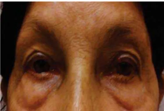
The examination of the eyelids showed severe bilateral eyelid ptosis, absence of palpebral folds in both upper eyelids, palpebral slits of 5 mm in both eyes, distance between the edge of the upper eyelid and unworkable corneal reflex due to severity of the condition in both eyes, distance from the lower margin to the tangent limbo in both eyes, and function of the levator muscle of the upper eyelid valued in 8mm in right eye and 7mm in the left eye (Figure 1).
Macedo HCP, AraújoJRM, LeãoRFC, SilvaGÂR, LeiteCG
Ocular motility was altered by limitations in all movements to look up. The patient was then referred to neurological assessment, which concluded bilateral eyelid ptosis associated to mild dysphagia condition and non-clonic ophthalmoparesis without fatigue or other commemorative signs. Magnetic resonance exams of the brain and chest radiograph were normal.
The bilateral eyelid ptosis condition of myogenic characteristic associated to ophthalmoparesis and dysphagia combined to the age of the patient refers to the diagnosis of oculopharyngeal muscular dystrophy. A positive family history of similar condition corroborates the hereditary, dominant autosomal character of the disease.
The patient was then submitted to bilateral ptosis correction by resection of the aponeuroses of the levators of the upper eyelids, with good aesthetic and functional aspect.
D
ISCUSSIONOculopharyngeal Muscular Dystrophy (OPMD), a disease of dominant autosomal inheritance, is characterized by dysphagia, paresis of the extraocular muscles, proximal paresis of the extremities and progressive bilateral ptosis, forcing the patient to use the front muscles and retroflex the head to compensate the loss of field of vision(3).
Most patients with OPMD are those with French-Canadian ancestry who reside in Quebec.(6) Other cases of this disease are
among the Bukhara Jews immigrants in Israel.(7)
OPMD usually begins insidiously during the fifth or sixth decade of life. These patients develop progressively with myogenic bilateral ptosis. Dysphagia is a prominent characteristic of OPMD, and is initially perceived with the ingestion of solid food, which can later develop into liquid swallowing difficulties.(8)
Figure 1: Severe bilateral eyelid ptosis
Figure 2: Postoperative aspect (After 6 months)
63 Miogenic ptosis in oculopharyngeal muscular dystrophy
Besides ptosis and dysphagia, patients may also have proximal weakness of the limbs in varying degrees of severity. Often there is also an element of extraocular muscle weakness (usually affecting the supraduction), but complete external ophthalmoplegia is rare. The diagnosis of OPMD is based on the history and clinical presentation and can be confirmed by genetic testing. Before the feasibility of such tests, the diagnosis was confirmed by muscle biopsy, which shows vacuoles in the muscle fibers, small and angled fibers, as well as intranuclear inclusion corpuscles.(9)
Aspiration pneumonia and malnutrition are the main causes of death in patients with OPMD, but it does not decrease the life expectancy because they tend to occur in the later stages of the disease.(5)
Regarding the treatment, there is still no cure for OPMD. Current treatment methods are based on improving the signs and symptoms related to eyelid ptosis and dysphagia. The surgical techniques for correction of ptosis in these patients are difficult to be performed. Some authors prefer to advance the levator muscle of the upper eyelid or combine this technique with Muller’s muscle advance in cases of MEPS function preserved.(10)
Another technique also widely used is the primary suspension of the frontal muscle due to the progressive nature of the disease. Fortunately, many patients with OPMD maintain good function of the orbicularis muscle and a good Bell’s phenomenon, which greatly reduces the risk of corneal commitment postoperatively.(5)
C
ONCLUSIONThere is no treatment to cure oculopharyngeal dystrophy. Genetic studies are important not only in the diagnosis but also in the identification of asymptomatic patients and genetic counseling. It is for the health professional to offer biopsychosocial multidisciplinary support in order to minimize the symptoms, such as surgical correction of ptosis, cricopharyngeal myotomy or gastrostomy to relieve dysphagia, as well as physiotherapy and psychological support, promoting better quality of life.
R
EFERENCES1. Lucci LM, Fonseca Junior NL, Sugano DM, Silvério J. Transposição da rima palpebral em ptose miogênica mitocondrial. Arq Bras Oftalmol. 2009;72(2):159-63.
2. Saito FL, Gemperli R, Hiraki PY, Ferreira MC. Cirurgia da ptose palpebral: análise de dois tipos de procedimentos cirúrgicos. Rev Bras Cir Plást. 2010;25(1):11-7.
3. Brais B, Rouleau GA, Bouchard JP, Fardeau M, Tomé FM. Oculopharyngeal muscular dystrophy. Semin Neurol. 1999;19(1):59-66. Review.
4. Chen JJ, Allen RC. Oculopharyngeal muscular dystrophy [Internet]. EyeRounds.org. January 18, 2012. [cited 2015 Feb 3]. Available from: http://webeye.ophth.uiowa.edu/eyeforum/cases/ 147-oculopharyngeal-muscular-dystrophy.htm
5. Brais B, Xie YG, Sanson M, Morgan K, Weissenbach J, Korczyn AD, et al. The oculopharyngeal muscular dystrophy locus maps to the region of the cardiac alpha and beta myosin heavy chain genes on chromosome 14q11.2-q13. Hum Mol Genet. 1995;4(3):429-34.
6. Duranceau AC, Beauchamp G, Jamieson GG, Barbeau A. Oropha-ryngeal dysphagia and oculophaOropha-ryngeal muscular dystrophy. Surg Clin North Am. 1983;63(4):825-32.
7. Blumen SC, Nisipeanu P, Sadeh M, Asherov A, Blumen N, Wirquin Y, et al. Epidemiology and inheritance of oculopharyngeal muscular dystrophy in Israel. Neuromuscul Disord. 1997; 7Suppl 1:S38-40. 8. Díaz de Liaño A, Fernández R L, Yarnóz I C, Artieda S C, González
A G, Artajona R A, et al. Distrofia muscular oculofaríngea: tratamiento quirúrgico. Rev Chil Cir. 2009;61(4):360-5. 9. Rüegg S, Lehky Hagen M, Hohl U, Kappos L, Fuhr P, Plasilov M,
et al. Oculopharyngeal muscular dystrophy - an under-diagnosed disorder? Swiss Med Wkly. 2005;135(39-40):574-86. Review. 10. Burnstine MA, Putterman AM. Upper blepharoplasty: a novel
approach to improving progressive myopathic blepharoptosis. Ophthalmology. 1999;106(11):2098-100.