Vicilins (7S storage globulins) of cowpea
(Vigna unguiculata) seeds bind to chitinous
structures of the midgut of Callosobruchus
maculatus (Coleoptera: Bruchidae) larvae
1Departamento de Bioquímica, Universidade Federal do Rio Grande do Norte,
Natal, RN, Brasil
2Centro de Pesquisa Rene Rachou, Belo Horizonte, MG, Brasil 3Centro Nacional de Recursos Genéticos e Biotecnologia, EMBRAPA,
Brasília, DF, Brasil
4Laboratório de Q uímica e Função de Proteínas e Peptídeos,
Centro de Biociências e Biotecnologia, Universidade Estadual do Norte Fluminense, Campos dos Goytacazes, RJ, Brasil
M.P. Sales1,
P.P. Pimenta2,
N.S. Paes3,
M.F. Grossi-de-Sá3 and
J. Xavier-Filho4
Abstract
The presence of chitin in midgut structures of Callosobruchus macu-latus larvae was shown by chemical and immunocytochemical meth-ods. Detection by Western blotting of cowpea (Vigna unguiculata) seed vicilins (7S storage proteins) bound to these structures suggested that C. maculatus-susceptible vicilins presented less staining when compared to C. maculatus-resistant vicilins. Storage proteins present in the microvilli in the larval midgut of the bruchid were recognized by immunolabeling of vicilins in the appropriate sections with immunogold conjugates. These labeling sites coincided with the sites labeled by an anti-chitin antibody. These results, taken together with those previ-ously published showing that the lower rates of hydrolysis of variant vicilins from C. maculatus-resistant seeds by the insects midgut proteinases and those showing that vicilins bind to chitin matrices, may explain the detrimental effects of variant vicilins on the develop-ment of C. maculatus larvae.
Co rre spo nde nce
M.P. Sales
Departamento de Bioquímica UFRGN
59072-970 Natal, RN Brasil
Fax: + 55-84-211-9208 E-mail: msales@ cb.ufrn.br Research supported by CNPq, CAPES and PRO NEX.
Received January 25, 2000 Accepted September 19, 2000
Ke y wo rds
·Bruchid beetles
·Callosobruchus m aculatus ·Cowpea seeds
·Vigna unguiculata
·Vicilins (7S storage proteins)
·Immunolocalization
·Midgut
Intro ductio n
Cowpea (Vigna unguiculata)is a legume cultivated in most tropical regions of the world (1). In Brazil, cowpea is mostly culti-vated in the northeastern part of the country and is the main protein source for poor popu-lations (2). Cowpea seeds are heavily in-fested with Callosobruchus maculatus (Co-leoptera: Bruchidae) larvae when in storage. Screening of the cowpea germplasm
sug-gested that high levels of trypsin inhibitors in TVu 2027 seeds were responsible for the ob-served resistance to C. maculatus. However, other workers (7-10) did not find a significant correlation between the levels of trypsin or cysteine proteinase inhibitors or even a -amy-lase inhibitors in cowpea seeds and their sus-ceptibility or resistance to infestation with C. maculatus. More recent studies suggest that the resistance of IT81D-1045 seeds to C. macu-latus is due to variant forms of vicilins (7S storage proteins) which are resistant to diges-tion by midgut proteinases and possibly limit food supply to the larvae (11-13). The mech-anism of resistance due to variant vicilins seems to be also linked to their chitin-binding power, a recently discovered property (14-17). This mechanism may be similar to the one attributed to the action of chitin-binding pro-teins (N-acetylglucosamine-specific lectins, chitinases, hevein and antimicrobial pep-tides) which are involved in defense mechan-isms of plants against insects and pathogens (18).
The purpose of the present study was to determine the presence of chitin (or chitin-containing structures) in the midgut of C. maculatus larvae utilizing both chitin and vicilin probes.
Mate rial and Me tho ds
Se e ds
Cowpea (V. unguiculata) seeds of the C. maculatus-susceptible cultivar CE-31 were supplied by Centro de Ciências Agrárias, Universidade Federal do Ceará, Fortaleza, CE, Brazil. C. maculatus-resistant cowpea seeds of the Nigerian line IT81D-1045 were obtained from IITA through Centro Nacio-nal de Pesquisa do Arroz e Feijão-Embrapa, Goiânia, GO, Brazil.
Inse ct
C. maculatus insects were originally
sup-plied by Dr. J.H.R. Santos (Centro de Ciên-cias Agrárias, Universidade Federal do Ceará). Permanent colonies of the insect were established on susceptible and resistant cowpea seeds and reared at 28-30oC and
55-60% relative humidity.
Iso latio n o f vicilins
Vicilins were prepared from cowpea seeds by the procedure of Macedo et al. (19). Ground meal extracted with 50 mM borate buffer, pH 8.0, for 30 min at room tempera-ture was centrifuged (30 min at 8,000 g, 5o
C) and soluble proteins were fractionated by ammonium sulfate precipitation. The 70-90% saturation fraction was dialyzed against wa-ter, freeze-dried and chromatographed on a Sephacryl S-200 column (3 x 40 cm) equili-brated and eluted with the same buffer as used for extraction. The vicilin-rich frac-tions were recovered and chromatographed on a DEAE-Sepharose column (2 x 20 cm) equilibrated with 50 mM Tris-HCl, pH 8.0, and eluted with a NaCl gradient (0 to 1 M) in the same buffer. The vicilin-rich fractions were recovered and submitted to chromatog-raphy on a Sephacryl S-400 column (2.5 x 70 cm) in 0.1 M Tris-HCl, 0.25 M NaCl, pH 8.0. Fractions containing vicilins were dialyzed against water and freeze-dried.
Pro te in de te rm inatio n
The dye-binding method of Bradford (20) was used for protein determination using bovine serum albumin as standard. Occa-sionally, measurement of absorbance at 280 nm was also used.
Antise rum pre paratio n
crude immune sera on a protein A column (protein A bound to Sepharose CL-4B). Pre-immune sera were collected before immuni-zation and used as control.
SD S-PAGE and We ste rn blo tting
Purified vicilins were separated by 13% SDS-PAGE by the method of Laemmli (21) and transferred to nitrocellulose membranes by the method of Towbin et al. (22). The immunoblots were made with the polyclonal antibodies against purified vicilins as de-scribed above. Binding was visualized with a horseradish peroxidase-conjugated second-ary antibody (Bio-Rad Laboratories, Rich-mond, CA, USA) according to manufacturer instructions.
Che m ical te st fo r chitin
Larvae were dissected under magnifica-tion in cold 0.15 M NaCl with the help of tweezers and midguts were separated from the windpipes and Malpighian tubes. Mid-guts were perforated and luminal contents were aspirated and reserved. Midguts were then thoroughly washed to remove remain-ing luminal contents. The presence of chitin in C. maculatus larval midgut and luminal contents was ascertained by the von Wisse-lingh color test (23). This qualitative test detects chitosan produced after treatment of the chitin-containing materials with saturated KOH for 15 min at 160o
C. After reaction, the presence of chitin was observed with a KI/ iodine solution. Controls employing cellu-lose (-) and lobster chitin (+) were used.
Chitin-binding assays
Twenty larvae reared in cowpea seeds (CE-31) were dissected under magnification in cold 0.15 M NaCl with the help of twee-zers. After exposure, midguts were homog-enized with 50 mM sodium phosphate buf-fer, pH 7.6, and centrifuged at 10,000 g. The
pellet was washed with 0.1 N HCl solution to remove diet proteins possibly retained by chitinous structures. Washed pellets were equilibrated with 50 mM phosphate buffer, pH 7.6, before being used for the observa-tion of vicilin binding. Pellets containing 20 midguts were immersed in solutions (2 mg/ ml in 50 mM phosphate buffer, pH 7.6) of both resistant (IT81D-1045) and susceptible (CE-31) cowpea seed vicilins and left to stand for 10 min with gentle shaking. The material was then centrifuged and the two pellets were washed with 50 mM sodium phosphate buffer, pH 7.6, to remove un-bound vicilins. Bound proteins were eluted with soluble chitosan and 0.1 N HCl and analyzed by Western blotting as described before.
Im m uno cyto che m istry
phosphatase and the reaction was amplified with rhodamine. Controls used were midgut sections with or without immunogold and pre-immune sera.
Re sults and D iscussio n
Vicilins consist of multi-subunit combi-nations with molecular masses between 20.1 and 94 kDa (24). Combination of multiple structural genes and extensive post-transla-tional processing results in a high degree of polymorphism for these proteins (25). In legume seeds, vicilins exhibit a considerable amount of sequence homology and micro-heterogeneity and may contribute to plant defense mechanisms (26). The 7S globulins, also known as vicilins, are the major cowpea storage proteins, which are highly heteroge-neous and are also encoded by a multigene family (27). Here cowpea vicilins from the seeds of the cultivars CE-31 and IT81D-1045 were purified by gel filtration through Sephacryl S-200 followed by ion-exchange chromatography on DEAE-Sepharose (data not shown). Three main bands of vicilin polypeptides isolated from resistant and sus-ceptible cowpea seeds were observed under
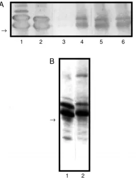
denaturing conditions (SDS-PAGE). Coo-massie blue staining showed that both pro-teins have major molecular masses between 45 and 66 kDa (Figure 1B) in agreement with data previously reported by several groups (19,24,27,28). Sales et al. (12) and Macedo et al. (19) have shown that these proteins are desorbed by different salt con-centrations from hydroxyapatite and DEAE-Sepharose. These findings may reflect the different binding strengths of both vicilins towards the matrices that could be due to differences in isoelectric points and micro-heterogeneity.
The presence of chitin in the midgut of C. maculatus larvae was first shown by a chem-ical test and by in vitro assays. The color test (von Wisselingh test) for the qualitative de-termination of chitin was used to detect the presence of this compound in the luminal fluid and membranous structures of C. macu-latus larvae. After treatment with KOH, the pellets were treated with a KI/iodine solu-tion. The appearance of a brown color was indicative of the presence of chitin. A posi-tive test was confirmed by the addition of 0.1% H2SO4 when the color turned violet.
The von Wisselingh color test for chitin was positive for both the luminal fluid content and membranes of the larval midgut.
In the present study, representative im-munoblot analyses were performed showing the differential binding of vicilins to mem-branous structures of the larval midgut. Re-sistant and susceptible vicilins were found to bind to isolated membranous structures from
C. maculatus larvae and sequentially eluted by chitosan and 0.1 N HCl. Sales et al. (14) reported that when resistant and susceptible vicilins were chromatographed on a chitin column they bound to this matrix. Both vici-lins showed the same pattern of sequential subunit elution by N-acetylglucosamine and chitosan, but subunits from IT81D-1045 were more strongly bound to the chitin matrix. In our assays, resistant vicilins showed a stron-ger binding signal in the eluate from soluble Figure 1 - A, Representative
Western blotting analysis show -ing the differential bind-ing of vi-cilins to isolated membranous structures of larval Callosobru-chus maculatus midgut. Lane 1, Suscept ible vicilins (cont rol); lane 2, resistant vicilins (control); lane 3, susceptible vicilins eluted w ith chitosan; lane 4, resistant vicilins eluted w ith chitosan; lane 5, susceptible vicilins eluted w ith 0.1 N HCl; lane 6, resistant vici-lins eluted w ith 0.1 N HCl. B, SDS-PAGE of purified vicilins. Lane 1, Susceptible vicilins; lane 2, resistant vicilins. The arrow s indicate the 45-kDa molecular mass.
1 2
®
1 2 3 4 5 6
® A
chitosan while for susceptible vicilins the signal was weaker. Both vicilins showed a strong signal in the 0.1 N HCl eluates. These results suggest that membranous structures of the C. maculatus larval midgut have chitin constituents and that the binding pattern for resistant vicilins differs from the pattern ob-served for susceptible vicilins (Figure 1A).
Comparison of the size of larvae grown on susceptible (Figure 2A) and resistant (Fig-ure 2B) cowpea seeds after 18-day infesta-tion showed a substantial inhibiinfesta-tion of the growth of larvae fed on resistant seeds. We utilized 3rd-instar larvae (18 days after egg hatching) that were reared on susceptible (CE-31) cowpea seeds. These larvae weighed 9.0 mg and were 3.0 mm long on average. Larvae at 18 days after egg hatching and not quite fully developed (average mass of 0.9 mg and 1.0 mm in length) were obtained from infested resistant cowpea seeds (IT81D-1045 line). In order to investigate the pres-ence of chitin and associated effects of vici-lins on larval development, we examined midgut tissues of larvae fed on susceptible cowpea seeds and whole larvae that had fed on resistant seeds.
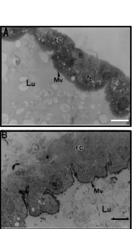
The structure of the larval midgut of C. maculatus allowed to develop for 18 days on cowpea seeds, as seen at low magnification in toluidine blue-stained sections (Figure 3A), was similar to that observed by Vats (29) for
Zabrotes subfasciatus, Callosobruchus annalis, Caryedon serratus and Bruchidius saundersi larvae. This investigator showed that epithelial cells that form the gut wall have a normal polar appearance, with promi-nent nuclei and numerous small translucent vacuoles/droplets towards the apical side. The epithelial cells have microvilli and the lumen contains stained material that may be food particles in the process of being di-gested.
After immunostaining with antibodies to chitin (Figure 4), we found strong immuno-labeling of the apical part of microvilli from the midgut epithelium indicating the
pres-A B
Figure 2 - Inhibitory effect of re-sist ant seeds on t he size (grow t h) of Callosobruchus maculatus larvae. A, Larvae (3rd instar, 18 days old) of insects reared on susceptible (CE-31) seeds. B, Larvae (18 days old) of insect s reared on resist ant (IT81D-1045) seeds. M agnifica-tion: 200X; bar: 500 µm.
Figure 3 - Immunohistochemical detection of vicilins in microsec-tions of the larval midgut of Cal-losobruchus maculatus reared on resistant (IT81D1045) cow -pea seeds. A, M icrosection of the midgut stained w ith toluidine blue. B, M icrosection of the mid-gut immunolabeled w ith anti-vicilin IgG. Lu = Lumen, M v = microvilli, N = nuclei, EC = gut epithelial cells. M agnification: 3,500X; bar: 30 µm.
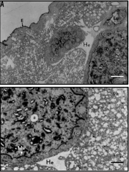
show three cross-sections of the gut wall surrounding the intestinal lumen (Figure 5A). Three cross-sections appear in a single mi-crograph because the gut forms loops when the larvae are fixed and embedded. The epi-thelial cells show microvilli that were strongly labeled after immunostaining with anti-vicilin antibodies. The lumen contains strongly la-beled material possibly corresponding to food particles in the process of being digested. The epidermis was also immunostained. Structures that we interpreted as gut epithe-lial cells did not contain vicilin immunola-beling, indicating that these proteins were not internalized by the cells. The hemolymph surrounding the gut was completely free of immunostaining, suggesting that no altera-tion of the structure of the midgut with dis-ruption of the microvilli or abnormalities in epithelial cells had occurred.
How chitbinding proteins, vicilins cluded, exert their deleterious effects on in-sect development is not known but one may speculate that these effects are mediated by the binding of the proteins to chitin in the peritrophic membrane that lines the midguts of insects. In insects that unequivocally show this membrane, various functions have been attributed to it like protection against bacte-rial and virus invasion and protection against microvillus damage caused by food (30,31). Here we have shown by a qualitative color test that the C.maculatus larval midgut con-tains chitinous structures. In addition, thin sections of the larval midgut showed the presence of immunolabeled sites when an anti-chitin antibody was used. In agreement with the suggestion by Vats (29) and Terra (32), these structures in the midgut of bruchid larvae are not distinct but are localized at the boundaries of the microvilli and apparently do not delimit endo- and ectoperitrophic spaces.
In order to understand the mechanism of action of vicilins, we investigated the bind-ing of these proteins to isolated membrane components of the midgut of C. maculatus
ence of chitin or chitinous structures in the larval midgut. Immunostaining with anti-vicilin IgG, used as a probe for chitin, also showed strong immunolabeling of the apical part of the microvilli, but without apparent staining of the nuclei or epithelial cells. La-beling of vicilins was not much visible in the gut contents of the lumen (Figure 3B).
The structure of the midgut of C. macula-tus larvae allowed to develop for 18 days on resistant cowpea seeds is shown in Figure 5A and B. Sections cut through the midgut
Figure 5 - Immunohistochemical detection of vicilins in microsections of w hole larvae of Callosobruchus maculatus reared on resistant (IT81D-1045) cow pea seeds and immuno-labeled w ith anti-vicilin IgG. Lu = Lumen, M v = microvilli, He = hemolymph, E = epidermis, EC = gut epithelial cells. (A and B: M agnification: 7,800X and 15,600X; bar: 20 µm and 10 µm, respectively).
larvae. Vicilins from resistant seeds reacted most intensely with membrane components and were desorbed from these by soluble chitosan and 0.1 N HCl. Similar results were obtained by Sales et al. (14) who observed that resistant vicilins were more strongly retained by a chitin matrix. Immunocyto-chemical analysis showed that the sites of binding of anti-vicilin IgG are the same as those observed with anti-chitin IgG in the midgut of large larvae reared on susceptible seeds and larval sections of small larvae reared on resistant seeds.
In spite of their very high degree of se-quence homology, vicilins of legume seeds (27) show different digestibility rates when treated with several proteases (33). Sales et al. (12) reported that vicilins from resistant cowpea seeds were more refractory to
diges-tion than vicilins from susceptible cowpea seeds. The antimetabolic effects of resistant or variant cowpea vicilins may be related to these low digestion rates and to their asso-ciation with chitin-containing structures of the insects midgut. The result of both of these phenomena is the impairment of nutri-ent uptake by midgut cells resulting in a lower rate of development of C. maculatus
larvae in seeds of V. unguiculata IT81D-1045.
Ackno wle dgm e nts
We thank Dr. P. Marinho, Department of Biology and Genetics, Federal University of Rio Grande do Norte, for a critical reading of this manuscript.
Re fe re nce s
1. Singh SR & Rachie KO (1985). Cow pea Research, Production and Utilization. John Wiley & Sons, Chichester.
2. M ay PH, Teixeira SM & Santana CA (1988). Cow pea production and economic importance in Brazil. In: Watt EE & Araújo JPP (Editors), Cow pea Research in Brazil. IITA/EM BRAPA, Ibadan, Nigeria, 31-62. 3. Singh BB, Singh SR & Adaji O (1985).
Bruchid resistance in cow pea. Crop Sci-ence, 25: 736-739.
4. Singh BB & Singh SR (1990). Breeding for bruchid resistance in cow pea. In: Fuji K, Gatehouse AM R & Yoshida T (Editors), Bruchids and Legumes: Economy, Ecol-ogy and Coevolution. Kluw er, Dordrecht, 219-228.
5. Gatehouse AM R, Gatehouse JA, Dobie P, Kilminster AM & Boulter D (1979). Bio-chemical basis of insect resistance in Vigna unguiculata. Journal of the Science of Food and Agriculture, 30: 948-958. 6. Gatehouse AM R & Boulter D (1983).
As-sessment of the antimetabolic effects of trypsin inhibitors from cow pea (Vigna un-guiculata) and other legumes on the de-velopment of the bruchid beetle, Calloso-bruchus maculatus. Journal of the Sci-ence of Food and Agriculture, 34: 345-350.
7. Baker TA, Nielsen SS, Shade RE & Singh BB (1989). Physical and chem ical
at-tributes of cow pea lines resistant and sus-ceptible to Callosobruchus maculatus (F.) (Coleoptera: Bruchidae). Journal of Stored Products Research, 25: 1-8.
8. Xavier-Filho J, Campos FAP, Ary M B, Silva CP, Carvalho M M M , M acedo M LR, Le-mos FJA & Grant G (1989). Poor correla-tion betw een the levels of proteinase in-hibitors found in seeds of different culti-vars of cow pea (Vigna unguiculata) and resistance/susceptibility to predation by Callosobruchus maculatus. Journal of Ag-ricultural and Food Chemistry, 37: 1139-1143.
9. Fernandes KVS, Sabelli PA, Barrat DHP, Richardson M , Xavier-Filho J & Shew ry PR (1993). The resistance of cow pea seeds to bruchid beetles is not related to levels of cysteine proteinase inhibitors. Plant M olecular Biology, 23: 215-219. 10. Reis CM , Calvet M M , Sales M P,
Fernandes KVS, Gomes VM & Xavier-Filho J (1997). a-Amylase inhibitors of legume seeds and their involvement in the resis-tance to the bruchid beetles. Arquivos de Biologia e Tecnologia, 40: 413-418. 11. Xavier-Filho J (1991). The resistance of
seeds of cow pea (Vigna unguiculata) to the cow pea w eevil (Callosobruchus macu-latus). M emórias do Instituto Osw aldo Cruz, 86: 75-77.
12. Sales M P, M acedo M RL & Xavier-Filho J
(1992). Digestibility of cow pea (Vigna un-guiculata) vicilins by pepsin, papain and bruchid midgut proteinases. Comparative Biochemistry and Physiology, 103: 945-950.
13. M acedo M RL, Andrade LBS, M oraes RA & Xavier-Filho J (1993). Vicilin variants and the resistance of cow pea (Vigna unguicu-lata) seeds to the cow pea w eevil ( Calloso-bruchus maculatus). Comparative Bio-chemistry and Physiology, 105C: 89-94. 14. Sales M P, Fernandes KVS, Gomes VM &
Xavier-Filho J (1996). Chitin-binding pro-teins from cow pea (Vigna unguiculata) seeds. Brazilian Journal of M edical and Biological Research, 29: 319-326. 15. Firmino F, Fernandes KVS, Sales M P,
Gomes VM , M iranda M RA, Domingues SJS & Xavier-Filho J (1996). Cow pea (Vigna unguiculata) vicilins associate w ith putative chitinous structures in midgut and feces of the bruchid beetles Calloso-bruchus maculatus and Zabrotes subfas-ciatus. Brazilian Journal of M edical and Biological Research, 29: 749-756. 16. Gomes VM , Blanco-Labra A, Sales M P,
M oraes RA, Fernandes KVS, Gomes VM & Xavier-Filho J (1998). Legume seed vici-lins (7S storage proteins) interfere w ith the development of the cow pea w eevil [Callosobruchus maculatus (F.)]. Science of Food and Agriculture, 76: 111-116. 18. Chrispeels M J & Raikhel NV (1991).
Lec-tin, lectin genes and their role in plant defense. Plant Cell, 3: 1-9.
19. M acedo M RL, Fernandes KVS, Sales M P & Xavier-Filho J (1995). Purification and properties of storage proteins (vicilins) from cow pea (Vigna unguiculata) seeds w hich are susceptible or resistant to the bruchid beetle. Brazilian Journal of M edi-cal and Biologiedi-cal Research, 28: 183-190. 20. Bradford M M (1976). A rapid and sensi-tive method for the quantitation of micro-gram quantities of protein utilising the principle of protein-dye binding. Analytical Biochemistry, 72: 248-254.
21. Laemmli UK (1970). Cleavage of struc-tural proteins during the assembly of the head of bacteriophage T4. Nature, 227: 680-685.
22. Tow bin H, Stachelin NT & Gordon J (1979). Electrophoretic transfer of pro-teins from polyacrylamide gels to nitrocel-lulose sheets; procedures and some ap-plications. Proceedings of the National Academy of Sciences, USA, 176: 4350-4354.
23. Roger HJ & Perkins HR (1968). Cell Walls of Filamentous Fungi. Chapter IX. E. & F.N. Spon Ltd., London, 153-160. 24. Pedalino M , Paino-d’Urzo M , Delle Done
G, Grillo S & Rao R (1992). The structure of cow pea (Vigna unguiculata L. Walp) seed storage proteins. Seed Science and Technology, 20: 223-231.
25. Higgins TJV (1984). Synthesis and regula-tion of major proteins in seeds. Annual Review of Plant Physiology, 35: 191-221. 26. Shew ry PR (1995). Plant storage proteins.
Biological Review s, 70: 375-426. 27. Casey R, Domeney C & Ellis N (1986).
Legume storage proteins and their genes. In: M iflin BJ (Editor), Oxford Surveys of Plant M olecular Biology and Cell Biology. Vol. 3. Oxford University Press, Oxford,
1-95.
28. Khan RI, Gatehouse JA & Boulter D (1980). The seed proteins of cow pea (Vigna unguiculata L. Walp). Journal of Experimental Botany, 31: 1599-1611. 29. Vat s LK (1976). Alim ent ary canal in
bruchid larvae (Bruchidae:Coleoptera). Research Bulletin (Science) of the Panjab University Science, 27: 103-106. 30. Peters W (1992). Peritrophic M embranes.
Springer Verlag, Berlin.
31. Walters LL, Irons KP, Guzman H & Tesh RB (1993). Formation and composition of peritrophic membranes in the sand fly, Phlebotomus perniciosus (Diptera: Psy-chodidae). Journal of M edical Entomol-ogy, 30: 179-198.
32. Terra WR (1990). Evolution of digestive system of insects. Annual Review of En-tomology, 35: 181-200.