B cells expressing IL-10 mRNA modulate memory
T cells after DNA-Hsp65 immunization
I.C. Fontoura
1, A.P.F. Trombone
2, L.P. Almeida
1, J.C.C. Lorenzi
1, R.A.M. Rossetti
3, T. Malardo
1,
E. Padilha
6, W. Schluchting
1, R.L.L. Silva
4, A.F. Gembre
1, J.E.C. Fiuza
1, C.L. Silva
1,
A. Panunto-Castelo
5and A.A.M. Coelho-Castelo
1 1Faculdade de Medicina de Ribeirão Preto, Universidade de São Paulo, Ribeirão Preto, SP, Brasil 2
Universidade Sagrado Corac¸ão, Bauru, SP, Brasil
3
Instituto de Ciências Biomédicas, Universidade de São Paulo, São Paulo, SP, Brasil 4
Departamento de Educac¸ão em Saúde, Universidade Federal de Sergipe, Lagarto, SE, Brasil
5
Faculdade de Filosofia, Ciências e Letras de Ribeirão Preto, Universidade de São Paulo, Ribeirão Preto, SP, Brasil 6
Universidade Paranaense, Cascavel, PR, Brasil
Abstract
In DNA vaccines, the gene of interest is cloned into a bacterial plasmid that is engineered to induce protein production for long periods in eukaryotic cells. Previous research has shown that the intramuscular immunization of BALB/c mice with a naked plasmid DNA fragment encoding the Mycobacterium leprae65-kDa heat-shock protein (pcDNA3-Hsp65) induces protection againstM. tuberculosis challenge. A key stage in the protective immune response after immunization is the generation of memory T cells. Previously, we have shown that B cells capture plasmid DNA-Hsp65 and thereby modulate the formation of CD8+memory T cells afterM. tuberculosischallenge in mice. Therefore, clarifying how B cells act as part of the protective
immune response after DNA immunization is important for the development of more-effective vaccines. The aim of this study was to investigate the mechanisms by which B cells modulate memory T cells after DNA-Hsp65 immunization. C57BL/6 and BKO mice were injected three times, at 15-day intervals, with 100mg naked pcDNA-Hsp65 per mouse. Thirty days after immunization, the percentages of effector memory T (TEM) cells (CD4+and CD8+/CD44high
/CD62Llow) and memory CD8+
T cells (CD8+/CD44high/CD62Llow/CD127+) were measured with flow cytometry. Interferong, interleukin 12 (IL-12), and
IL-10 mRNAs were also quantified in whole spleen cells and purified B cells (CD43–) with real-time qPCR. Our data suggest
that a B-cell subpopulation expressing IL-10 downregulated proinflammatory cytokine expression in the spleen, increasing the survival of CD4+TEM cells and CD8+TEM/CD127+cells.
Key words: DNA-Hsp65 vaccine; Memory T cells; B cells
Introduction
DNA vaccines consist of a gene of interest cloned into a bacterial plasmid, which is then further engineered to express the protein for long periods in eukaryotic cells (1). In this way, nucleic acids can be used to induce a specific immune response against a pathogen, offering a wide range of new options in vaccinology (2). In previous studies, the intramuscular immunization of BALB/c mice with a naked DNA fragment encoding Mycobacterium leprae 65-kDa heat-shock protein (pcDNA3-Hsp65) imparted protection againstM. tuberculosischallenge (3,4). The DNA vaccine construct ensures protein production within those cells that capture the plasmid DNA. There-after, the antigens in the cytoplasm become accessible to the proteasomal pathway and can be presented via MHC I to activate CD8+T cells. Cytotoxic T lymphocytes are a
key part of the protective immune response to intracellular pathogens (5). A proportion of the endogenously produced antigen can also be secreted or released upon cell death, and captured by another antigen-presenting cell (APC). Professional APCs can also present antigens derived from the Hsp65 in an MHC II context, activating specific CD4+ T cells, a mechanism called ‘‘cross-presentation’’ (5). Moreover, CpG motifs in the plasmid sequence induce an innate immune response, activated by toll-like receptor 9 (TLR9), which in turn elicits specific T-cell functions (6).
Specific memory lymphocytes are generated during the immune response. Central memory T cells recirculate through the secondary lymphoid organs, as the host immune surveillance mechanism, whereas effector memory T cells (TEM) rapidly differentiate into effector
Correspondence: I.C. Fontoura:<[email protected]>.
cells upon exposure to the antigen for a second time, and thus play an important role in the protective immune response. Therefore, the main goal in vaccine design is the generation of long-lived memory T cells. Some factors that contribute to memory T-cell maintenance are the cytokines interleukin 12 (IL-12) and IL-15, and also IL-7, which plays an essential role in the maintenance of CD8+ memory cells. These cytokines induce the proliferation and antiapoptotic mechanisms of memory T cells, allowing them to survive for extended periods (7).
Although the main function attributed to B cells is antibody production, they are also professional APCs, and can capture, process, and present soluble antigens, leading to the activation of CD4+ T lymphocytes and consequently enhancing the CD8+cytotoxic response (8). A central role of B cells in maintaining CD4+and CD8+ T cells for long periods in mice infected withPlasmodium chabaudi, Listeria monocytogenes, or Lymphocytic cho-riomeningitis virus (LCMV) has previously been demon-strated (9,10). However, their role in memory induction during DNA immunization is not well understood.
We have previously demonstrated that B cells captured the plasmid pcDNA3-Hsp65 (11), presented the expressed protein, and modulated the memory CD8+T cells formation after mice were challenged with M. tuberculosis (12). However, clarifying the specific mechanisms by which B cells induce a protective immune response after DNA immunization is an important step in the development of more-effective DNA vaccines.
Here, we investigated the mechanisms by which B cells modulate memory T cells in the pcDNA3-Hsp65 vaccinated mouse model. Our results showed that a B-cell subpopulation expressing IL-10 mRNA downregulated the expression of proinflammatory cytokines, thus increasing the percentages of CD4+and CD8+memory T cells in the spleen after DNA immunization.
Material and Methods
Mice
Male 6–8-week-old C57BL/6 wild-type (WT) and
B-cell-deficient (BKO;mchain–/–) mice were obtained from Jackson Laboratories (USA) and maintained under specifi c-pathogen-free conditions in the animal house of the Departamento de Imunologia, Faculdade de Medicina de Ribeirão Preto, Universidade de São Paulo. The mice had access to water and sterile foodad libitum, and were maintained under light cycles of 12 h. The protocol used for animal experimen-tation was approved by the Institutional Committee for Animal Use (CETEA-FMRP-USP; process #040/2006).
Immunization
The quadriceps muscle of each mouse was injected three times, at 15-day intervals, with 100mg naked pcDNA3-Hsp65 (DNA-Hsp65) or pcDNA3 (empty vector) in 25% PBS-sucrose, in a total volume of 100mL.
DNA-Hsp65 and the vector were prepared as described previously (4). The mice were euthanized 30 days after the last dose. Other mice were injected with 100mL saline as a control. All experiments were performed with four animals per treatment group.
Phenotyping memory T cells
The spleens were aseptically removed from the immunized mice and the spleen cells were restimulated with 20mg recombinant Hsp65 in RPMI 1640 medium (Invitrogen, USA) containing 10% heat-inactivated fetal bovine serum (FBS) (Gibco BRL, USA), 100 U/mL penicillin, 100 mg/mL streptomycin, and 10 mg/mL genta-micin (Sigma Aldrich, USA) for 24 h. The cells were stained with anti-CD4, anti-CD62L, anti-CD127, anti-CD8 or anti-CD4 antibodies (BD Bioscience, USA). The labeled cells were analyzed with flow cytometry (FACSCantot, BD Bioscience).
Real-time RT-PCR
After stimulation, the spleen cells were collected with 1 mL of TRIzol Reagent (Invitrogen) and their total RNA was extracted according to the manufacturer’s protocol.
On day 30 after immunization, the B cells were separated from the spleens using negative selection (490% CD19+ cells) with an anti-CD43 antibody linked to magnetic beads (MACS MicroBeads System, Miltenyi Biotec, Germany). The B cells were collected with 1 mL of TRIzol Reagent (Invitrogen) and their total RNA was extracted according to the manufacturer’s protocol.
The total RNA was treated with amplification-grade DNase I (Invitrogen). Complementary DNA (cDNA) was reverse transcribed from the mRNA with SuperScript II (Gibco BRL), according to the manufacturer’s instructions. The real-time qPCR reactions were performed with 200 ng cDNA, 0.1mg/mL of each primer (sense and antisense), and Platinum SYBR Green qPCR SuperMix-UDG (Invitrogen), according to the manufacturer’s instruc-tions, on a Rotor-Gene 6000 (Corbett Life Science, Australia). Relative expression was calculated as 2–DDCt (13). The annealing temperature used was 58°C for all genes. The following primers sequences were used: beta-actin: forward 50-AGCTGCGTTTTACACCCTTT-30, reverse 30AAGCCATGCCAATGTTGTCT-50; IL-12 p40: forward 50AGCACCAGCTTCTTCATCAGG-30, reverse 30-GCGCTGGATTCGAACAAAG-50; interferon g (IFN-g): forward 50-GATATCTGGAGGAACTGGCAA-30, reverse 30-GCTCTGCAGGATTTTCATGTC-50; IL-10: forward 50-TGGACAACATACTGCTAACCG-30, reverse 30-GGA TCATTTCCGATAAGGCT-50.
Statistical analyses
Results
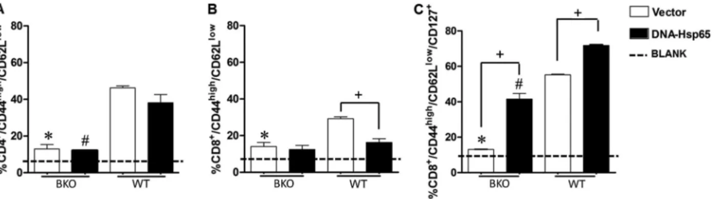
Percentage of memory T cells increased in mouse spleens after DNA immunization
To verify that the absence of B cells plays a negative role in memory generation after DNA-Hsp65 immuniza-tion, the percentages of CD4+and CD8+TEM cells were analyzed in the mouse spleens 30 days after the last vaccination. Our data showed a higher percentage of CD4+/CD44high
/CD62Llowcells in the WT mice than in the BKO mice after immunization (Figure 1A). However, the percentages of CD4+TEM cells did not differ significantly between the groups immunized with pcDNA3-Hsp65 or empty vector in both the WT and BKO immunized mice (Figure 1A). The WT group immunized with empty vector showed a higher percentage of CD8+/CD44high/CD62Llow
cells than the BKO mice immunized with empty vector. However, the percentage of CD8+/CD44high/CD62Llow
cells in the WT and BKO mice immunized with pcDNA3-Hsp65 did not differ significantly (Figure 1B). Because no significant differences were found between the CD8+ TEM subpopulations in the spleens of the WT and BKO mice immunized with pcDNA3-Hsp65, we evaluated the CD8+TEM cells expressing CD127+, a specific marker of memory CD8+T cells (14). We found that when mice were immunized with either DNA-Hsp65 or empty vector, the WT mouse spleens had higher percentages of CD8+ memory cells than the spleens of the BKO mice. In contrast to CD8+TEM, pcDNA3-Hsp65 immunization induced a higher percentage of CD8+TEM/CD127+cells than the empty vector in both the WT and BKO mice (Figure 1C). Taken together, these data suggest that the presence of B cells contributed to the induction of memory
CD4+and CD8+T cells in the spleen after immunization with plasmid DNA.
WT mice displayed reduced proinflammatory cytokine
mRNAs in the spleen
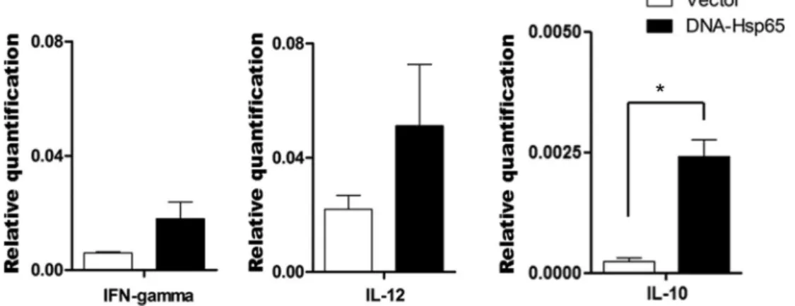
When the transcriptional profiles of the proinfl amma-tory cytokines in the mouse spleens were evaluated 30 days after immunization, we found that DNA-Hsp65 immunization increased the mRNA levels of IFN-gand IL-12 compared with empty-vector immunization in both the WT and BKO mice (Figure 2A and B, respectively). It is noteworthy that transcripts of IFN-gand IL-12 were virtually undetectable in the cells of mice immunized with the empty vector. The WT mice showed lower mRNA expression of IFN-gand IL-12 after DNA-Hsp65 immunization than the BKO mice. In contrast, no significant difference in IL-10 mRNA expression was observed between the WT and BKO groups when the mice were immunized with DNA-Hsp65. IL-10 mRNA expression was only elevated in the WT group immu-nized with the empty pcDNA3 vector (Figure 2C). These data indicate a role for B cells in the regulation of proinflammatory cytokine production in the mouse spleen.
DNA-Hsp65 immunization induced IL-10 mRNA expression by B cells
To clarify the possible mechanisms by which B cells modulate the formation of memory T cells and regulate proinflammatory cytokine expression, the mRNA expres-sion of IFN-g, IL-12, and IL-10 was measured in B cells purified from mouse spleen cells 30 days after immuni-zation. The splenic B cells from mice immunized with
DNA-Hsp65 or empty vector showed similar levels of IFN-g and IL-12 mRNA expression (Figure 3A and B, respectively). However, B cells from the DNA-Hsp65-immunized mice displayed higher levels of IL-10 mRNA than the B cells from the empty-vector-immunized mice. This suggests that DNA-Hsp65 immunization activates a subpopulation of B cells that produces IL-10.
Discussion
Our results suggest that the presence of B cells is necessary to support the formation of memory after DNA immunization. Memory T cells develop after the evolution of the adaptive immune response. This protective response begins after the recognition of the antigen presented by professional APCs to naïve T lymphocytes, which triggers their proliferation and differentiation into effector T cells. After antigen clearance, the immune response is downregulated and most activated cytes undergo apoptosis. The pool of remaining lympho-cytes then differentiates into long-lived memory T cells (15). A previous study showed that as well as presenting antigens, B cells also costimulate T cells through their interaction with CD40 and CD40L on the T-cell surface, enhancing T-cell activation (16). Additional costimulation by their engagement with CD28 induces greater T-cell survival in the effector phase of the immune response, by promoting an increase in antiapoptotic molecules in the activated T cells. This event allows a larger number of the available cells to differentiate into memory cells (17). Our results corroborate these previous reports, because we
observed a higher percentage of CD4+and CD8+T cells in the WT mouse spleen after DNA immunization. These data indicate that B lymphocytes have an important function in promoting the costimulation of T cells, resulting in a higher percentage of memory cells 30 days after immunization. Although no significant difference in the percentage of CD8+ TEM cells was observed between the WT and BKO spleens, there was a two-fold reduction in the percentage of CD8+TEM/CD127+cells in the BKO mice compared with the WT mice over the same period after DNA immunization. CD127 corresponds to the alpha chain in the receptor of IL-7, an important cytokine in the maintenance of memory T cells in the peripheral immune system (18). The CD8+T cells that maintain their CD127 expression after the expansion of the immune response are destined to become memory T cells (7). Therefore, the evaluation of CD127 expression on CD8+ TEM cells allows us to evaluate the specific, long-lived CD8+T cells generated after DNA vaccination.
Our data provide further support for the key role of B cells in improving memory formation by promoting T-cell costimulation, and also demonstrate that DNA immuniza-tion induces a subtype of IL-10-producing B cells that reduces the amount of proinflammatory cytokine mRNAs in the spleen. Although IL-10 is considered a regulatory cytokine, we have shown that this cytokine is required for the formation of memory cells after LCMV infection. In the presence of this cytokine, the resolution of infection occurred later, causing antigen persistence, and thus stimulating the adaptive immune response for a long period. This consequently activated a larger number of Figure 2.Relative expression of cytokine mRNAs in the spleens of wild-type (WT) and B-cell knockout
memory T cells (19). The induction of sufficient immunolog-ical memory depends on the magnitude of the immune response generated against the antigen. Therefore, in an exacerbated inflammatory environment, T cells are highly activated and consequently undergo apoptosis, reducing the memory pool. Under these circumstances, the activa-tion of an IL-10-producing B-cell subpopulaactiva-tion after DNA immunization is important for the modulation of the immune response induced by the DNA vaccine.
Recently, many studies have demonstrated the regulatory role of IL-10-producing B cells (reviewed by Balkwill et al., 20). Although this role has been described in autoimmune diseases, this B-cell subpopulation also inhibited the activation of myeloid cellsin vitro, suggest-ing that these cells have a role in maintainsuggest-ing the homeostasis of the immune system. Although it is difficult to determine the exact phenotype of regulatory B cells, many efforts have been made to investigate the characteristics of these cells. Several studies have demonstrated that their regulatory function could be a transient phenotype of those B cells designated to be
antibody-secreting cells, which is induced when they are stimulated with TLR agonists in the absence of B-cell antigen receptor stimulation. Under these conditions, B cells produce IL-10, leading to the regulation of the immune response (20).
In conclusion, our data suggest the participation of IL-10-producing B cells in the immune response by modulating the induction of the immune response after the presentation of DNA-Hsp65 by impairing the exacerbation of the infl amma-tory response. This may improve the longevity of the antigen derived from Hsp65 in the spleen, thus improving the activation of a protective immune response.
Acknowledgments
We thank Ana Paula Masson and Izaira Tincani Brandão for their help with vaccine construction and the Hsp65 protein used in our experiments, and to Fabiana R. Morais and Walter Turato for their support with all thefl ow-cytometric assays. This study was supported by FAPESP (#2006/05497-2) and CNPq.
References
1. Rajcani J, Mosko T, Rezuchova I. Current developments in viral DNA vaccines: shall they solve the unsolved?Rev Med Virol2005; 15: 303–325, doi: 10.1002/rmv.467.
2. Hellermann GR, Mohapatra SS. Genetic therapy: on the brink of a new future.Genet Vaccines Ther2003; 1: 1, doi: 10.1186/1479-0556-1-1.
3. Bonato VL, Lima VM, Tascon RE, Lowrie DB, Silva CL. Identification and characterization of protective T cells in
hsp65 DNA-vaccinated and Mycobacterium tuberculosis-infected mice.Infect Immun1998; 66: 169–175.
4. Lowrie DB, Tascon RE, Bonato VL, Lima VM, Faccioli LH, Stavropoulos E, et al. Therapy of tuberculosis in mice by DNA vaccination.Nature1999; 400: 269–271, doi: 10.1038/22326. 5. Gurunathan S, Klinman DM, Seder RA. DNA vaccines: immunology, application, and optimization*.Annu Rev Immunol 2000; 18: 927–974, doi: 10.1146/annurev.immunol.18.1.927. Figure 3.Relative expression of cytokine mRNAs in purified B cells from wild-type (WT) mouse spleens
6. Tudor D, Dubuquoy C, Gaboriau V, Lefevre F, Charley B, Riffault S. TLR9 pathway is involved in adjuvant effects of plasmid DNA-based vaccines.Vaccine2005; 23: 1258–1264, doi: 10.1016/j.vaccine.2004.09.001.
7. Nanjappa SG, Walent JH, Morre M, Suresh M. Effects of IL-7 on memory CD8 T cell homeostasis are influenced by the timing of therapy in mice.J Clin Invest2008; 118: 1027–1039. 8. Linton PJ, Harbertson J, Bradley LM. A critical role for B cells in the development of memory CD4 cells.J Immunol 2000; 165: 5558–5565, doi: 10.4049/jimmunol.165.10.5558. 9. Whitmire JK, Asano MS, Kaech SM, Sarkar S, Hannum LG, Shlomchik MJ, et al. Requirement of B cells for generating CD4+T cell memory.J Immunol2009; 182: 1868–1876, doi: 10.4049/jimmunol.0802501.
10. Shen H, Whitmire JK, Fan X, Shedlock DJ, Kaech SM, Ahmed R. A specific role for B cells in the generation of CD8 T cell memory by recombinant Listeria monocytogenes.J Immunol 2003; 170: 1443–1451, doi: 10.4049/jimmunol.170.3.1443. 11. Coelho-Castelo AA, Santos Junior RR, Bonato VL, Jamur
MC, Oliver C, Silva CL. B-lymphocytes in bone marrow or lymph nodes can take up plasmid DNA after intramuscular delivery. Hum Gene Ther 2003; 14: 1279–1285, doi: 10.1089/104303403767740812.
12. Almeida LP, Trombone AP, Lorenzi JC, Rocha CD, Malardo T, Fontoura IC, et al. B cells can modulate the CD8 memory T cell after DNA vaccination against experimental tuberculosis. Genet Vaccines Ther2011; 9: 5, doi: 10.1186/1479-0556-9-5.
13. Livak KJ, Schmittgen TD. Analysis of relative gene expres-sion data using real-time quantitative PCR and the 2(-Delta Delta C(T)) Method. Methods 2001; 25: 402–408, doi: 10.1006/meth.2001.1262.
14. Kaech SM, Tan JT, Wherry EJ, Konieczny BT, Surh CD, Ahmed R. Selective expression of the interleukin 7 receptor identifies effector CD8 T cells that give rise to long-lived memory cells. Nat Immunol2003; 4: 1191–1198, doi: 10.1038/ni1009. 15. Kan A, Hodgkin PD. Mechanisms of cell division as
regulators of acute immune response. Syst Synth Biol 2014; 8: 215–221, doi: 10.1007/s11693-014-9149-3. 16. Constant SL. B lymphocytes as antigen-presenting cells for
CD4+T cell primingin vivo.J Immunol1999; 162: 5695–5703. 17. Boise LH, Minn AJ, Noel PJ, June CH, Accavitti MA, Lindsten T, et al. CD28 costimulation can promote T cell survival by enhancing the expression of Bcl-xL. Immunity. 1995. 3: 87–98.J Immunol2010; 185: 3788–3799. 18. Schluns KS, Lefrancois L. Cytokine control of memory T-cell
development and survival.Nat Rev Immunol2003; 3: 269–279, doi: 10.1038/nri1052.
19. Foulds KE, Rotte MJ, Seder RA. IL-10 is required for optimal CD8 T cell memory following Listeria monocytogenes infection.J Immunol2006; 177: 2565–2574, doi: 10.4049/ jimmunol.177.4.2565.