Belo Horizonte
2013
Mariana Noyma Xavier
MECHANISMS OF BRUCELLA ABORTUS SURVIVAL DURING CHRONIC
INFECTION: THE ROLE OF IL-10 AND PPAR
γ
Belo Horizonte
Escola de Veterinária – UFMG
2013
Tese apresentada à UFMG, como requisito parcial
para a obtenção do título de Doutor.
3
Tese defendida em 25 de junho de 2013, pela comissão examinadora constituída por:
_________________________________________________________
Prof. Renato de Lima Santos
(orientador)
__________________________________________________________
Prof. Sérgio Costa Oliveira
__________________________________________________________
Dr. Andreas J. Bäumler
_________________________________________________________
Dr. Sebastian E. Winter
_________________________________________________________
5
“(...) nem entendo aquilo que entendo: pois estou infinitamente maior do que eu mesma, e nãome alcanço. " (Clarice Linspector)
Agradecimentos
A Deus, por ser a luz que ilumina meu caminho.Ao Frank, por todo o seu apoio e companheirismo. Você e sua família tornaram meus anos nos EUA muito mais especiais.
Ao prof. Renato de Lima Santos, por sua orientação, preocupação com meu crescimento e por ter aberto as portas para um novo mundo de aprendizagem e descobertas.
A Dra. Renée Tsolis, por abrir as portas do seu laboratório e por acreditar nas minhas idéias. Eu não tenho como medir a admiração e o respeito que eu tenho por você como ser humano e como cientista.
A professora Tatiane Paixão, pela confiança e por todos os anos de ensinamento. Você foi um modelo para mim desde quando trabalhamos juntas na iniciação científica.
A Maria Winter, Vydia Atluri, Alanna Spees, Kim Nguyen e Teane Silva, por sua ajuda em todos os experimentos, e por dedicarem parte do seu precioso tempo aos meus projetos.
Ao Dr. Andreas Den Hartigh, pelo treinamento e paciência durante o meu treinamento no laboratório da Dra. Tsolis.
A todos os membros odo laboratório ABRT, especialmente Maria Winter, Sebastian Winter, Marijke Keestra, Jason Mooney, Tobias Kerrinnes, Franziska Faber and Alanna Spees, por fazerem da ida diária ao trabalho uma experiência tão prazerosa. A amizade de vocês foi o meu maior presente durante esta aventura.
7
Summary
RESUMO 10
ABSTRACT 11
INTRODUCTION 12
CHAPTER I: LITERATURE REVIEW 14
1. Brucellosis 14
2. Effects of Brucella on the immune response 14
2.1- The furtive aspect of Brucella spp. 15
2.2- The immune response during Brucella infection 16
3. Role of interleukin 10 (IL-10) during infection
16
3.1- IL-10 during Brucella spp. infection 17
4. Macrophage plasticity during infection 18
5. Role of PPARγ in macrophage activation and metabolism 20
5.1- Metabolic changes in macrophages 20
5.2- The role of
PPAR
20
CHAPTER II: CD4
+T CELL-DERIVED IL-10 PROMOTES BRUCELLA
ABORTUS
PERSISTENCE
VIA
MODULATION
OF
MACROPHAGE
FUNCTION 22
Introduction 22
Material and Methods 23
Results 26
Discussion
48
CAPÍTULO
III:
A
PPAR
-MEDIATED
INCREASE
IN
GLUCOSE
AVAILABILITY SUSTAINS CHRONIC BRUCELLA ABORTUS INFECTION IN
ALTERNATIVELY ACTIVATED MACROPHAGES 50
Introduction 50
Material and Methods 50
Results 53
Discussion
73
CONCLUSION 75
REFERENCES 76
SUPPLEMENTARY MATERIAL 90
SUPPLEMENTARY MATERIAL LIST
Supplementary material and methods
90
Supplementary table 1.
93
Supplementary table 2.
95
Supplementary figure 2.
97
Supplementary figure 3.
98
Supplementary figure 4.
99
Supplementary figure 5.
100
Supplementary figure 6.
101
Supplementary figure 7.
102
Supplementary figure 8.
103
Supplementary figure 9.
104
FIGURE LIST
Figure 1.
Brucella abortus induces IL-10 production by infected organs
during early in vivo infection.
27
Figure 2.
Lack of IL-10 results in lower bacterial survival and increased
pathological changes during early Brucella abortus infection in
vivo.
28
Figure 3.
CD4
+CD25
+T cells are the main producers of IL-10 during early
Brucella abortus in vivo infection.
29
Figure 4.
Expansion of CD4
+CD25
+T cells during Brucella infection.
31
Figure 5.
IL-10 production by macrophages and/or neutrophils is not required
for Brucella abortus long-term persistence in vivo.
33
Figure 6.
Pathology induced by lack of IL-10 production by macrophages and
neutrophls during B. abortus in vivo infection.
34
Figure 7.
IL-10 production during B. abortus infection in mice lacking IL-10
production in T cells.
36
Figure 8.
IL-10 production by T cells is required for Brucella abortus
persistence and for control of inflammatory response in vivo.
37
Figure 9.
IL-10 production by T cells is required for control of Brucella
abortus induced pathology in vivo.
39
Figure 10.
Lack of endogenous IL-10 results in lower Brucella abortus survival
inside macrophages due to bacterial inability to escape the late
endosome.
41
Figure 11. Lack of endogenous IL-10 results in higher NF-
κB activation and
production of pro-inflammatory cytokines by macrophages infected
with B. abortus.
43
Figure 12.
Inability of macrophages to respond to IL-10 results in decreased
persistence of B. abortus in vivo.
45
Figure 13.
Inability of macrophages to respond to IL-10 results in severe acute
B. abortus induced pathology in vivo
46
Figure 14.
Alternatively activated macrophages are more abundant during
chronic brucellosis.
55
Figure 15.
Increased B. abortus survival in AAM during chronic infection
58
Figure 16.
Defects in generation of CAM or AAM affect B. abortus survival in
9
Figure 17.
Increased survival of B. abortus during chronic infection is
dependent on PPAR
63
Figure 18.
B. abortus
infected AAM exhinit a PPAR
-dependent decrease in
glycolytic metabolism
66
Figure 19.
A PPAR
-dependent increase in intracellular glucose availability
promotes survival of B. abortus in macrophages
68
RESUMO
A evasão de resposta imune do hospedeiro é um pré-requisito para doenças bacterianas crônicas. No entanto, os mecanismos subjacentes não são totalmente compreendidos. Neste estudo foi demonstrado que a Brucella abortus, impede a ativação dos macrófagos ao induzir a produção de interleucina-10 (IL-10) por células T CD4+CD25+ durante a infecção aguda. Além disso, falha na produção de IL-10 por células T ou a incapacidade de macrófagos em responder a esta citocina resultou num aumento da capacidade de camundongos em controlar a infecção por B.
abortus, apesar da indução elevada de citocinas pró-inflamatórias, e de grave patologia no
fígado e no baço de camundongos infectados. Apesar dos avanços significativos na compreensão da sobrevivência intracelular de B. abortus in vitro, pouco se sabe sobre o nicho intracelular de B. abortus in vivo. Este estudo demonstra que B. abortus é capaz de sobreviver e replicar preferencialmente em macrófagos alternativamente ativados (AAM), que aumentam em número durante a infecção crônica. O mecanismo desta maior sobrevivência em AAM é uma mudança no metabolismo induzido por “peroxisome proliferator activated receptor gamma” (PPAR ), o que aumenta a disponibilidade de glucose intracelular. A capacidade de transportar glicose foi crucial para persistência, e para aumento da replicação de B. abortus em AAM. Em conjunto, os nossos resultados sugerem que a produção de IL-10 por células T CD4+CD25+ modula a função de macrófagos, a fim de promover infecção persistente por B. abortus. Além disso, tal persistência, também foi determinada por uma mudança na disponibilidade intracellular de nutrientes induzida por PPARy em AAM.
11
ABSTRACT
Evasion of host immune responses is a prerequisite for chronic bacterial diseases; however, the underlying mechanisms are not fully understood. This study demonstrated that Brucella abortus
prevents immune activation of macrophages by inducing CD4+CD25+ T cells to produce
interleukin-10 (IL-10) early during infection. Moreover, either a lack of IL-10 production by T cells or a lack of macrophage responsiveness to this cytokine resulted in an increased ability of mice to control B. abortus infection, while inducing elevated production of pro-inflammatory cytokines, and severe pathology in liver and spleen of infected mice. In spite of the significant advances in understanding intracellular survival of B. abortus at the cellular level, little is known about the chronic intracellular niche of B. abortus in vivo. This study demonstrated that B. abortus is able to survive and replicate preferentially in alternatively activated macrophages (AAM), which increase in numbers during chronic infection. The underlying mechanism to this enhanced survival in AAM is a shift in metabolism induced by peroxisome proliferator activated receptor gamma (PPAR ), which increases the availability of intracellular glucose. The ability to take up glucose was crucial for increased replication of B. abortus in AAM, and for
persistence. Taken together our results suggest that early IL-10 production by CD25+CD4+ T
cells modulates macrophage function in order to promote persistent infection. Additionally, B. abortus persistence was also determined by a shift in intracellular nutrient availability induced by PPAR in AAM.
INTRODUCTION
Brucellosis is a zoonotic bacterial disease caused by bacteria of the genus Brucella, which are able to establish long-term infections in their host (Xavier et al., 2009; Atluri et al., 2011). Human brucellosis, caused most commonly by B. melitensis and
B. abortus, is considered one of the most
important zoonotic diseases worldwide, with more than 500,000 new human cases reported annually (Pappas et al., 2006). The disease is characterized by a long incubation period that leads to a chronic, sometimes lifelong, debilitating infection with serious clinical manifestations such as fever, arthritis, hepatomegaly, and splenomegaly (Corbel, 1997; Atluri et al., 2011).
In vivo, Brucella is found in association with
phagocytic cells, most prominently macrophages, in which a subset of bacteria is able to evade killing in phagolysosomes and replicate successively within an endoplasmic reticulum-associated compartment and a modified autophagosome (Gorvel and Moreno 2002; Starr et al., 2008). The furtive aspect of this pathogen is also exemplified by its ability to evade initial innate immune recognition through toll-like receptors (TLRs) (Andersen-Nissen et al., 2005) as well as by modifications of virulence factors such as lipopolysaccharide - LPS (Lapaque et al., 2006) and flagellin (Terwagne et al., 2013), resulting in a mild pro-inflammatory response that leads to bacterial persistence (Atluri et al., 2011). In spite of the important role of TLR evasion in Brucella
pathogenesis, little is known about subsequent host-pathogen interactions that lead to establishment of chronic infection.
Several pathogens are able to modulate the immune response through induction of regulatory cytokines, such as interleukin-10 (IL-10) (Anderson et al., 2007; Jankovic et
al., 2007; Saraiva and O'garra, 2010). Like
other chronic pathogens, Brucella is able to induce IL-10 in infected cells in vitro and during early in vivo infection, suggesting that the IL-10 pathway could play an
important role in enabling bacterial persistence (Fernandes and Baldwin, 1995; Fernandes et al., 1996; Fernández-Lago et
al., 1996). However, the real impact of
IL-10 during in vivo infection as well as the cell types producing IL-10 or responding to this cytokine during brucellosis is not clear.
Years of research have demonstrated that immune cells can adopt different functional states according to the immune environment around them, and macrophages are no exception (Gordon and Martinez, 2010; Van Dyken and Locksley, 2013). Indeed, an environment rich in Th1 cytokines such as interferon gamma (IFNg) leads to development of classically activated macrophage (CAM) population, characterized by production of nitric oxide (NO) and inflammatory cytokines such as tumor necrosis factor alpha (TNFa) and interleukin 6 (IL-6) (Mosser and Edwards, 2008). Conversely, an environment rich in Th2 cytokines such as interleukin-4 (IL-4) and interleukin-13 (IL-13) promotes the development of alternatively activated macrophages (AAM), which play important roles in allergic inflammation, helminth infection and tissue repair (Reyes and Terrazas, 2007; Shirey et al., 2008; Lawrence and Natoli, 2011).
Interestingly, in addition to their critical influence in establishment of the immune response, CAM and AAM also play important roles in host physiology and metabolism (Chawla, 2010). For instance, development of the AAM phenotype is dependent on peroxisome proliferator activated receptors - PPARs; (Odegaard et
al., 2007), which act downstream of STAT6
signaling to regulate macrophage metabolism.
In this context, it is important to note that a key aspect of Brucella pathogenesis is its interaction with macrophages. Indeed, research focusing on the in vitro Brucella/macrophage interaction has been
critical for the understanding of how B.
abortus survives intracellularly (Gorvel and
13
chronic persistence in vivo do not appear to mediate intracellular replication in cultured macrophages (Hong et al., 2000; Fretin et
al., 2005), suggesting that the different
CHAPTER I: LITERATURE
REVIEW
1 - Brucellosis
Brucellosis is a zoonotic disease caused by Gram-negative bacteria of the genus
Brucella, which are facultative intracelllular
coccobacilli belonging to the α 2-Proteobacteria family (Garrity, 2001). Currently, the genus Brucella is divided into six classical species, according to zoonotic potential and host preference: B. melitensis,
B. abortus, B. suis, B. canis, and B. ovis
(Osterman and Moriyon, 2006). B. melitensis, B. suis, and B. abortus are
considered the most pathogenic species for humans and have small ruminants, pigs and cattle respectively as their preferred hosts (Godfroid et al., 2005). Importantly, two recently identified Brucella species isolated from marine mammals, B. ceti and B.
pinnipedialis, can also cause human
brucellosis (Foster et al., 2007). Moreover,
B. canis, a dog pathogen, has a
comparatively low zoonotic potential, while
B. neotomae and B. ovis, that infect desert
rats and sheep, respectively, are not associated with human disease (Godfroid et
al., 2005; Xavier et al., 2010).
When infecting domestic animals, Brucella spp. targets reproductive organs, such as placenta and mammary glands in females and epididymis in males, resulting in late trimester abortion and infertility (Xavier et
al., 2009; Carvalho Júnior et al., 2012). Brucella spp. transmission within the
reservoir hosts can occur via ingestion or contact with a highly colonized placenta (Atluri et al., 2011). Alternatively, the transmission between natural hosts can occur through ingestion of contaminated milk, or through contact with semen and/or genital secretions during mating (Xavier et
al., 2010; Atluri et al., 2011).
The World Health Organization (WHO) estimates that 500,000 new cases of human brucellosis occur annually, making it one of the most frequently diagnosed zoonoses
worldwide (Pappas et al., 2006). Although brucellosis has been eradicated or efficiently controlled in most developed countries, many areas of the world still have a high prevalence of the disease, including Central and South America, and countries of the Caucasus and Central Asia (Pappas et al., 2006). The economic impact of brucellosis in developing countries is doubled and extremely significant. The human disease causes high morbidity, resource consumption for patient treatment, and inability of infected people to work and provide to their family Additionally, the animal disease affects the livestock, which provides a livelihood to many farmers. Together, these facts have led the WHO to classify the disease a neglected zoonosis (Atluri et al., 2011).
In humans, clinical manifestations of
Brucella infection usually appear between 5
to 60 days after exposure (Young, 1995). During the acute phase of the disease, affected patients present clinical signs such as fever, fatigue, anorexia, mialgia, and joint pain. Frequently, the absence of specific clinical symptoms during initial stage of infection may result in lack of a correct diagnosis, which in turn leads to the development of chronic disease (Young, 1995). Chronic brucellosis, a more severe form of the disease, can be associated with osteo-articular signs and/or colonization of the brain, spleen, liver, genitourinary tract and endocardium (Xavier et al., 2010; Dean
et al., 2012). Treatment of chronic
brucellosis is notoriously difficult, since antimicrobial drugs may not reach the infection foci, such as granulomas and abcesses, caused by the bacteria. As a result, the patient tends to suffer for a long period of months or years with recurrent episodes of fever and weakness (Franco et al., 2007). Importantly, live attenuated vaccines are available for animal brucellosis prevention, however these vaccines are not considered safe for human use (Franco et al., 2007).
15
2.1 – The furtive aspect of Brucella spp.
In the host, the preferential target cells for
Brucella spp. include macrophages,
dendritic cells, and placental trophoblasts (Atluri et al., 2011), in which the bacterium can persist and replicate (Xavier et al., 2010). Occasionally, Brucella spp. can also target B cells (Goenka et al., 2012). Intracellular Brucella survival involves a temporary fusion of the Brucella-containing vacuole (BCV) with the lysosome, and subsequent exclusion of the lysosomal proteins (Starr et al., 2008). Interestingly enough, after this process, the BCV becomes associated with the rough endoplasmatic reticulum, creating the compartment in which intracellular replication of Brucella occurs (Anderson and Cheville, 1986; Pizarro-Cerda et al., 1998; Celli et al., 2003). Once inside the ER-associated compartment, Brucella spp. becomes practically invisible to the immune system (Xavier et al., 2010), fact demonstrated by low production of cytokines and antibodies during the chronic phase of infection (Rodriguez-Zapata et al., 2010; Martirosyan
et al., 2011). Therefore, the initial immune
response becomes key factor for the control of Brucella spp. infection.
The initial detection of pathogenic bacteria by host cells is mediated primarily by innate immunity. This arm of the immune response uses a surveillance system which relies on a pattern of pathogen recognition receptors (PRRs), including membrane-associated toll-like receptors (TLRs) (Iwasaki and Medzhitov, 2004) and cytosolic nucelotide binding and oligomerization domain-like receptors (NLRs) (Franchi et al., 2008). Both TLRs and NLRs function as molecular bar code readers that enable the host to distinguish bacteria from other types of infectious agents by recognizing characteristic combinations of conserved pathogen-associated molecular patterns (PAMPs) (Hoebe et al., 2004). For instance, TLR3, TLR7, and TLR8 recognize nucleic acids and, therefore, are specialized in viral recognition; while TLR1, TLR2, TLR4, TLR5, and TLR6 recognize structures only
present in bacteria (Iwasaki and Medzhitov, 2004).
Previous studies have suggested TLR2, TLR4, TLR6, and TLR9 as the PRRs responsible for sensing different Brucella components, leading to secretion of pro-inflammatory cytokines during early stages of infection in vivo (Weiss et al., 2005; Copin et al., 2007; Macedo et al., 2008; De Almeida et al., 2013). However, the inflammatory response elicited during in
vivo Brucella infection is considerably
milder than the one induced by other proteobacterial pathogens such as
Salomonella, fact that reflects the ability of Brucella spp. to evade the initial detection
by the innate immune system (Barquero-Calvo et al., 2007; Martirosyan et al., 2011). Indeed, Brucella organisms are devoid of many classical structures involved in virulence/immune recognition such as pili, fimbria, capsules and plasmids (Martirosyan
et al., 2011). Moreover, Brucella spp. have
the ability to modify the lipid A fraction of their LPS through the incorporation of a much longer fatty acid residue (C28) when
compared with enterobacterial LPS (C12
-C16), and this modification greatly reduces
the endotoxic properties of Brucella LPS by lowering its TLR4 agonist activity (Lapaque
et al., 2006). Interestingly, Brucella spp.
response resulting in impaired bacterial clearance (Tsolis et al., 2008).
2.2 – The immune response during Brucella
infection
The immune response to Brucella spp. has been characterized most extensively in the murine model. During the initial stage of
Brucella infection in mice, the host response
developed resembles the T helper 1 (Th1) type, with production of interferon gamma (IFN- ) by Th and natural killers (NK) cells, as well as production of interleukin 12 (IL-12) and tumoral necrotis factor alpha (TNF-α) by infected macrophages (Zhan and Cheers, 1995; Zhan et al., 1996; Copin et
al., 2007; Rolán and Tsolis, 2008).
Moreover, in the murine model, both CD4+ and CD8+ T cells contribute to control of the infection, and this is thought to occur via production of IFN- (Araya et al., 1989; Fernandes et al., 1996). Indeed, induction of a Th1 immune response during brucellosis seems to be critical, since mice deficient for IFN- production or mice lacking interferon regulatory factor-1 (IRF-1) are unable to control systemic bacterial replication and succumb to an exacerbated B. abortus infection (Murphy et al., 2001; Ko et al., 2002). Additionally, IL-12 and TNF-α depletion result in increased colony forming units (CFU) counts in spleen from mice infected with B. abortus; however the phenotype observed is not as severe as the one described for absense of IFN- (Zhan and Cheers, 1995; Zhan et al., 1996).
In humans, the initial immune response to
Brucella spp. is also characterized by
elevated levels of pro-inflammatory cytokines linked to Th1 response, such as IL-1 , IL-6, IL-12p40, TNF-α, and IFN- (Rafiei et al., 2006; Rodriguez-Zapata et al., 2010). Additionally, previous studies have demonstrated that mutations in genes encoding the cytokines IFN- , IL-6, TNF-α, and IL-10 contribute to increased susceptibility to human brucellosis (Budak
et al., 2007; Karaoglan et al., 2009).
However, during chronic human brucellosis, the initial Th1 response is dampened and acquires features of Th2 responses, such as
an increase in IL-13 producing T cells (Rafiei et al., 2006).
In spite of its well established furtive behavior, Brucella spp. do rely on an important virulence factor for intracellular survival, the type IV secretion system (T4SS) encoded by the genes virB1-virB12 (O'callaghan et al., 1999; Delrue et al., 2001; Den Hartigh et al., 2008). The critical role of Brucella T4SS is demonstrated by the inability of T4SS deficient mutants to establish in vivo persistence, both in the murine (Hong et al., 2000a; Den Hartigh et
al., 2004; Den Hartigh et al., 2008), as well
as in the caprine infection models (Zygmunt
et al., 2006). Interestingly, previous studies
have demonstrated that the T4SS is required not only for establishment of long-term infection, but also for the induction of Th1 immune response in infected mice (Roux et
al., 2007). This function was confirmed by
the fact that a functional T4SS is necessary for B cell maturation, activation of CD4+ T cells and for initial secretion of IL-12 and IFN- (Rolan and Tsolis, 2007; Rolán and Tsolis, 2008). Moreover, B. abortus
detection by NLRs, leading to ASC-inflammasome mediated production of IL-1 and IL-IL-18, was also shown to be dependent on the type IV secretion system (Gomes et al., 2013).
3 – Role of interleukin 10 (IL-10) during infection
17
Due to its anti-inflammatory functions, IL-10 production has been associated with the control of different auto-immune processes, such as Irritable Bowel Disease and rheumatological disorders (Kuhn et al., 1993; Carter et al., 2012). Interestingly, in the context of infectious diseases, IL-10 production usually results in control of the underlying pathology which can be detrimental to the host, without necessarily affecting pathogen survival (Saraiva and O'garra, 2010).
Initially, the anti-inflammatory role of IL-10 was demonstrated through its ability to regulate both T cell and NK cell function (Fiorentino et al., 1989; Couper et al., 2008). However, current research indicates that these effects are indirect and mediated through direct IL-10 induced regulation of monocytes and macrophage functions (Couper et al., 2008). Indeed, IL-10 influences three important monocyte/macrophage functions: secretion of inflammatory mediators, antigen presentation and phagocytosis (Sabat et al., 2010). In these cells, IL-10 inhibits expression of MHC class II and the co-stimulatory molecules B7-1 and B7-2, in addition to limiting the production of pro-inflammatory cytokines such as IL-6, IL-12, IL-18 and TNF-α, as well as chemokines such as MCP-1, RANTES, IL-8, and MIP-2 (Moore et al., 2001; Couper et al., 2008). Consequently, IL-10 plays an important role in innate immunity, modulating macrophage and dendritic cells effector functions and subsequent T cell activation (Moore et al., 2001; Saraiva and O'garra, 2010).
Macrophages are considered important immune system effector cells, hence are one of the main sources of IL-10 during infection (Siewe et al., 2006; Couper et al., 2008). Macrophage activation takes place through recognition of PAMPs by PRRs, which in turn stimulates the expression of pro-inflammatory cytokines (Iwasaki and Medzhitov, 2004). Therefore, IL-10 production by this cell type is thought to occur through the same mechanism (Siewe
et al., 2006). Indeed, previous studies have
suggested that TLR2 agonists are specialized
in induction of IL-10 expression by antigen presenting cells (APC) (Dillon et al., 2004; Netea et al., 2004). For instance, the TLR2 signaling pathway is crucial for IL-10 production by macrophages stimulated with
Mycobacterium tuberculosis or Yersinia pestis lipopeptides (Sing et al., 2002; Jang et al., 2004). Additionally, significant IL-10
production can occur in response to TLR4 and TLR9 agonists (Boonstra et al., 2006).
If considering IL-10 production by T cells, it was first believed that IL-10 was a product of Th2 clones following protein or antigen stimulation (Fiorentino et al., 1989). However, it is now known that IL-10 is not just produced by Th2 cells, but can also be secreted by most if not all CD4+ T-cell subsets, including Th1 and Th17 (Saraiva and O'garra, 2010). In the context of infectious diseases, regulatory T cells (Tregs) serve as a major source of immunoregulatory IL-10, and may come in several forms depending on their developmental origin (Saraiva and O'garra, 2010; Redford et al., 2011). Naturally occuring T regs are mostly CD25+ and are generated in the thymus, but also peripherally in response to tolerogenic stimuli (Sabat et al., 2010). Thymic and inducible Tregs also express the transcription factor FoxP3 (FoxP3+ Tegs) and can suppress effector responses through the soluble factors transforming growth factor (TGF- ) and IL-10 (Saraiva and O'garra, 2010; Redford et al., 2011). Moreover, several populations of antigen-driven FOXP3- IL-10 producing T cells with regulatory activity that are distinct from naturally occuring Treg cells have been described (Vieira et al., 2004; Saraiva and O'garra, 2010), and are possibly originated along a Th1 cell pathway (Gabrysova et al., 2009). Indeed, FoxP3- IL-10 producing Th1 cells have been shown to play a role in immunomodulation during Leishmania
(Anderson et al., 2007) and Toxoplasma (Jankovic et al., 2007) infections.
3.1 –IL-10 during Brucella spp. infection.
associated with increased susceptibility to human brucellosis (Budak et al., 2007; Karaoglan et al., 2009). The role of IL-10 during Brucella spp. infection has also been studied in the mouse model. Fernández-Lago et al. (1996) have shown that CD-1 mice intravenoysly infected with Brucella
abortus present increased IL-10 levels in
spleen during the first week post-infection. Moreover, in vivo IL-10 neutralization in Balb/C mice intraperitoneally infected with a low (5x10³ CFU) and high (5x10⁶ CFU) dose of Brucella abortus resulted in decreased bacterial survival and increased IFN production during early infection (Fernandes and Baldwin, 1995; Fernandes et
al., 1996). However, this phenotype was not
observed in Brucella infected C57BL/6 mice, despite the fact that splenocytes obtained from this mouse strain had an increased ability to produce IL-10 in response to Brucella abortus infection in
vitro (Fernandes et al., 1996). Interestingly,
anti-Brucella effector functions of IFN activated macrophages were dampened by IL-10 during in vitro infection (Fernandes and Baldwin, 1995; Fernandes et al., 1996). These previous studies suggest an important role of IL-10 in modulating the initial immune response to Brucella abortus, resulting in decreased pathogen survival in some mouse strains, probably due to regulation of macrophage functions.
Goenka et al. (2011) have suggested that B cells play an important role in enabeling
Brucella persistence in the host, since B cell
deficient mice present a decreased ability to control B. abortus infection when compared with control animals. In this study, spleens from Brucella infected B cell deficient mice showed decreased frequency of IL-10 producing cells, indicating that the B cell mediated Brucella persistence could be, at least partially, due to B cell dependent immune modulation through IL-10 production (Goenka et al., 2011). However, this phenotype was only observed around 21 days post-infection, while previous studies have demonstrated that IL-10 levels were increased in Brucella infected mice as soon as the first week of infection (Fernandes and Baldwin, 1995; Fernández-Lago et al.,
1996). Additionally, spleens from B cell deficient and control mice showed no significant differences in IL-10 gene expression at 3 days post- B. abortus infection (Rolán et al., 2009). Taken together, these studies suggest that other cells types could take part in IL-10 production and immune regulation during initial stages of Brucella infection.
4 - Macrophage plasticity during infection
Antigen presenting cells such as macrophages play a key role as first line of defense during infectious processes and as a major link between innate and adaptive immune responses (Benoit et al., 2008). However, in spite of being considered important immune effector cells, macrophages have another significant function – the removal of celular debris in order to clear the interstitial space from foreign bodies and to maintain tissue homeostasis (Mosser and Edwards, 2008). Thus, macrophages are considered a dynamic and heterogenous cell population due to their wide range of functions and the diverse mechanisms which drive their differentiation, tissue distribution and stimuli response (Mosser, 2003; Gordon and Martinez, 2010).
19
macrophages (CAM, M1); alternatively activated macrophages (AAM, M2, M2a); and regulatory macrophages (M2c), according with their function during inflammation (Benoit et al., 2008; Mosser and Edwards, 2008; Gordon and Martinez, 2010).
The term classically activated macrophages is used to describe cells that present evident effector characteristics in response to activation of Th1 or cellular immune response (Mosser and Edwards, 2008). Thus, CAM differentiation occurs when Th1 cytokines such as IFN- and TNF-α are present, resulting in cells with increased bactericidal capacity due to nitric oxid (NO) production as well as secretion of proinflammatory cytokines such as IL-1 , IL-6 and IL-12 (Benoit et al., 2008; Mosser and Edwards, 2008). Normally, the presence of CAM is essential for the control of intracellular pathogens during acute phase of infection, as suggested by the increased susceptibility of IFN and TNFα deficient mice to infection by the intracellular pathogenic bacteria Listeria monocytogenes (Rothe et al., 1993) as well as by the M1 polarization of macrophages during acute
Mycobacterium tuberculosis and Salmonella typhi infection (Ehrt et al., 2001; Benoit et al., 2008). However, an excessive or
prolonged CAM program can be deleterious to the host, via development of severe pathology in affected organs as a consequence of overhelming production of proinflammatory cytokines (Benoit et al., 2008).
As previously described for CAM, the AAM population can also arise in response to innate and/or adaptive immunological stimuli (Mosser and Edwards, 2008). However, while Th1 cytokines are necessary for CAM development, AAM differentiaion usually occurs in the presence of Th2 cytokines, such as interleukin 4 (IL-4) and interleukin 13 (IL-13) (Loke et al., 2007). IL-4 is mostly produced by basophils, mast cells, and CD4+ T cells (Brandt et al., 2000), and is usually present during fungal and parasitic diseases (Kreider et al., 2007). Nevertheless, IL-4 production can also occur
during bacterial infection, exemplified by the detection of this cytokine during
Mycobacterium tuberculosis (Harris et al.,
2007), Francisella tularensis (Shirey et al., 2008), and Yersinia enterocolítica (Tumitan
et al., 2007) infections. When present during
an inflammatory process, IL-4 leads to the development of macrophages with decreased bactericidal capacity, which in turn promotes pathogen persistence. Therefore, it is believed that AAM play an important role in chronic bacterial diseases (Benoit et al., 2008). Moreover, IL-4 is able to convert resident macrophages incells programmed to promote fibrosis and pathology resolution (Mosser and Edwards, 2008). Hence, AAM show increase arginase (Arg1) activity, an enzyme responsible for the conversion of arginine to ornitine, an important precursor of polyamines and collagen used for the production of extracellular matrix (Kreider
et al., 2007). Additionally, studies have
demonstrared that AAM are able to produce significant amounts of the chitinase Ym1 as well as the resistin-like protein Fizz1 (Raes
et al., 2002) and upregulate the expression
of galactose-type C-type lectin CD301 (mMGL1) (Raes et al., 2005). Consequently, increased Arg1, Ym1, Fizz1 and CD301 gene expression and protein levels can be used as biomarkers for the identification of this macrophage subpopulation (Raes et al., 2005; Mosser and Edwards, 2008).
primary function to regulate the immune response and to limit detrimental inflammation (Mosser, 2003). Indeed, IL-10 production by this macrophage subpopulation has been linked to increased persistence of bacterial pathogens such as
Mycobacterium leprae (Sieling and Modlin,
1994), Mycobacterium tuberculosis (O'leary
et al., 2011) and Coxiella burnetti (Ghigo et al., 2001), most likely due to their ability to
modulate the Th1 immune response necessary for the control of intracellular pathogens.
5 - Role of PPARγ in macrophage activation and metabolism
5.1 – Metabolic changes in macrophages
IFN and TLR macrophage activation leads to upregulation of the inducible form of nitric oxide synthase (iNOS) (Everts et al., 2012) and production of reactive oxygen species (ROS) (West et al., 2011). Therefore, ROS and nitric oxide (NO)
production are key functional features of the inflammatory and antibactericidal CAM macrophage, and the metabolic alterations that occur are integral to this process (O'neill and Hardie, 2013). Interestingly, NO competes with oxygen to inhibit cytochrome c oxidase, the terminal electron acceptor of the respiratory chain. This fact prevents the reoxidation of NADH, which in turn limits flux through the tricarboxylic acid (TCA) cycle. Moreover, increased generation of ROS by mitochondria also contributes to reduced macrophage reliance on the TCA cycle and the respiratory chain for energy and ATP production. However, M1 macrophages still need to maintain ATP levels for biosynthesis, as well as to maintain mitocondrial membrane potential and to prevent apoptosis (O'neill and Hardie, 2013). Therefore, decreased TCA flux in CAM leads to ATP production through anaerobic glycolysis and lactate production. Consequently, these cells show elevated expression of the glucose transporter GLUT1 as well as marked switch from expression of the liver isoform of the enzime 6-phosphofructo-2-kinase (encoded by PFKFB1) to the PFKFB3 isoform
(Rodriguez-Prados et al., 2010). This leads to increased glucose uptake and consumption, as well as accumulation of fructose-2,6-bisphosphate which, in turn, increases glycolytic flux (O'neill and Hardie, 2013).
The opposite is true when macrophages are activated by IL-4 and IL-13, which promote the AAM phenotype. This macrophage subpopulation shows a profund increase in the entire program of fatty-acid metabolism, including uptake and oxidation of fatty acids and mitocondrial biogenesis, as well as much lower rates of glycolysis (Vats et al., 2006; Rodriguez-Prados et al., 2010). This shows that while AAM macrophages preferentially utilize glucose, the alternative program of macrophage activation switches over to fatty acid oxidation for energy homeostasis (Vats et al., 2006). Since AAM macrophages are involved in chronic processes and tissue repair, it is possible that the more energy efficient oxidative metabolism is better suited to the long-term roles of this subpopulation (O'neill and Hardie, 2013).
Interestingly, the control of the genetic program for long-term activation is dependent on STAT6 phosphorylation (Martinez et al., 2008; O'neill and Hardie, 2013). As consequence, phosphorylated STAT6 dimerizes and translocates to the nucleus where it induces expression of its target genes, including markers (Arg1, Ym1,
Fizz1, Cd301) and regulators of macrophage
metabolism and alternative activation (i.e.:Ppar , Pparδ and PGC-1 ) (Bensinger
and Tontonoz, 2008). The role of the AAM metabolism regulators will be discussed below.
5.2 – The role of PPAR
21
oxidation and glucose metabolism (Bensinger and Tontonoz, 2008). Interestingly, the PPAR family also plays an important role in the inhibition of inflammatory gene expression (Bensinger and Tontonoz, 2008).
One of the receptors in the PPAR family, PPAR , is a nuclear receptor activated by fatty acids, which has recently been linked to the polarization of macrophage phenotype (Odegaard et al., 2007). Therefore, even though PPAR is best known for its influence in adipocyte development and insulin-resistance (Bensinger and Tontonoz, 2008), it can also have an widespread influence on macrophage biology (Zhang and Chawla, 2004; Tontonoz and Spiegelman, 2008). Interestingly, studies using PPAR –deficient cells have demonstrated that, in the absence of PPAR signaling, macrophages neither appropriately suppress infammatory cytokine production nor acquire an oxidative metabolic program that is associated with the AAM macrophage phenotype (Odegaard
et al., 2007; Bensinger and Tontonoz, 2008).
Previous studies have demonstrated the role of PPAR signalling pathway in medating increased fatty-acid oxidation in AAM macrophages through upregulation of the PPAR coativator 1 (PGC1 ), in a IL-4 and STAT-6 dependent manner (Vats et al., 2006; Bensinger and Tontonoz, 2008). PGC1 is a transcriptional co-activator that promotes oxidative metabolism, notably by
inducing expression of genes involved in fatty-acid oxidation (Wu et al., 1999). Indeed, over-expression of PGC1 in macrophages promotes the M2 phenotype. Moreover, macrophage PGC1 – specific knockdown limits the shift to oxidative phosphorylation and the function of AAM macrophage, and greatly boosts the TLR4 activating effects of LPS (Vats et al., 2006).
An important repressive function of PPAR is to inhibit inflammatory gene expression by several mechanisms, including direct interactions with AP-1 and NF-κB (Chung et
al., 2000) and nucleocytoplasmatic
redistribution of the p65 subunit of NF-κB (Kelly et al., 2004), among others. Indeed, in LPS and IFN activated murine macrophages, PPAR activation repressed the induction of pro-inflammatory genes, including Nos2, Cox2 and Il12 (Ricote et al., 1998; Welch et al., 2003).
In the context of infectious diseases, PPAR induction in macrophages has been implicated in increased persistence of the intracellular pathogens such as Listeria
monocytogenes (Abdullah, Z. et al., 2012)
CHAPTER II: CD4
+T
CELL-DERIVED IL-10 PROMOTES
BRUCELLA ABORTUS
PERSISTENCE VIA MODULATION
OF MACROPHAGE FUNCTION*
Introduction
Persistent bacterial infections have a significant impact on public health (Svetic et
al., 1993). While evasion of host immune
responses is a prerequisite for these chronic infections, the underlying mechanisms are not fully understood. Human brucellosis, caused by the intracellular gram-negative coccobacilli Brucella spp., is considered one of the most important zoonotic diseases worldwide, with more than 500,000 new human cases reported annually (Pappas et
al., 2006). The disease is characterized by a
long incubation period that leads to a chronic, sometimes lifelong, debilitating infection with serious clinical manifestations such as fever, arthritis, hepatomegaly, and splenomegaly (Corbel, 1997; Atluri et al., 2011). Human and animal brucellosis share many similarities, such as persistence in tissues of the mononuclear phagocyte system, including spleen, liver, lymph nodes, and bone marrow (Atluri et al., 2011). Therefore, the use of animal models such as mice has been an important tool to better characterize the immune response to
Brucella infection that leads to long-term
bacterial persistence and chronic disease.
There is general agreement that the initial interferon gamma (IFN- ) mediated Th1 immune response is crucial for the control of
Brucella infection, since absence of IFN-
results in decreased control of bacterial growth (Fernandes and Baldwin, 1995; Fernandes et al., 1996) and IFN- -deficient C57BL/6 mice succumb to overwhelming disease (Murphy et al., 2001). However, the inflammatory response induced by Brucella spp. in vivo is much milder than that observed with pyogenic infections such as salmonellosis, suggesting the stealth of
Brucella as a possible reason for the absence
of early proinflammatory responses (Barquero-Calvo et al., 2007; Martirosyan et
al., 2011). Recent studies have shown that Brucella spp. use both passive and active
mechanisms to evade initial innate immune recognition through toll-like receptors (TLRs) (Andersen-Nissen et al., 2005). Although avoidance of TLR recognition is a key factor in the lack of initial inflammation during Brucella infection, how subsequent interactions of Brucella with the host immune system result in chronic disease is poorly understood.
Interleukin-10 (IL-10) is an immunoregulatory cytokine produced by most T cell subsets, B cells, neutrophils, macrophages, and some dendritic cell subsets (Saraiva and O'garra, 2010). It is suggested that by acting on antigen-presenting cells such as macrophages, IL-10 can inhibit the development of Th1 type responses (Sabat et al., 2010). In the context of infectious diseases, it is believed that the host uses IL-10 to control over-exuberant immune responses to pathogenic microorganisms in order to limit tissue damage (Saraiva and O'garra, 2010). Interestingly, studies using chronic pathogens such as Leishmania major (Belkaid et al., 2001), human cytomegalovirus (Chang and Barry, 2010), or Mycobacterium tuberculosis (reviewed in (Redford et al., 2011)) have demonstrated that the absence of IL-10 leads to a better clearance of these pathogens, with variable degrees of immunopathology. These studies suggest that pathogens have developed mechanisms to take advantage of the host immune-regulation in order to persist for longer periods and establish chronic infection.
Similar to other chronic pathogens, B.
abortus infection induces IL-10 production
(Fernandes and Baldwin, 1995; Fernandes et
al., 1996; Fernández-Lago et al., 1996).
Moreover, IL-10 gene polymorphisms have been associated with increased susceptibility to human brucellosis (Budak et al., 2007). However, questions regarding the impact of IL-10 in B. abortus persistence and * Article accepted for publication in PLOS
23
establishment of chronic infection, as well as the cell types responsible for this cytokine production remain to be answered. Therefore, we used IL-10 deficient mice to determine the role of IL-10 in modulating the initial immune response to Brucella infection. Furthermore, using cell-specific knock-out mice, we elucidated the immunological mechanisms underlying IL-10 induced immune-regulation during brucellosis.
Material and Methods
Bacterial strains, media and culture conditions: Bacterial strains used in this
study were the virulent strain Brucella
abortus 2308 and its isogenic mutant strain
MX2 which has an insertion of
pKSoriT-bla-kan-PsojA-mCherry plasmid (Copin et
al., 2012). For strain MX2, positive clones
were kanamycin resistant and fluorescent, as previously described (Copin et al., 2012). Strains were cultured on tryptic soy agar (Difco/Becton-Dickinson, Sparks, MD) or tryptic soy broth at 37°C on a rotary shaker. Bacterial inocula for mouse infection were cultured on tryptic soy agar plus 5% blood for 3 days (Alton et al., 1975). For cultures of strain MX2, kanamycin (Km) was added to the culture medium at 100µg/mL. All work with B. abortus cells was performed at biosafety level 3.
Bone marrow derived macrophage
infection: Bone marrow-derived
macrophages were differentiated from bone marrow precursors from femora and tibiae of female, 6 to 8 weeks old, C57BL/6J and IL-10-/- mice obtained from The Jackson Laboratory (Bar Harbor) following a previously published procedure (Rolan and Tsolis, 2007). For BMDM experiments, 24-well microtiter plates were seeded with macrophages at concentration of 5 x10⁵ cells/well in 0.5 mL of RPMI media (Invitrogen, Grand Island, NY) supplemented with 10% FBS and 10 mM L-glutamine (RPMI supl) incubated for 48 h at 37°C in 5% CO2. Preparation of the
inoculum and BMDM infection was performed as previously described (Rolan and Tsolis, 2007). Briefly, for inoculum
preparation, B. abortus 2308 was grown for 24 h and then diluted in RPMI supl, and about 5 × 10⁷ bacteria in 0.5 mL of RPMI supl were added to each well of BMDM, reaching multiplicity of infection (MOI) of 100. Microtiter plates were centrifuged at 210 × g for 5 min at room temperature in order to synchronize infection. Cells were incubated for 20 min at 37°C in 5% CO2,
free bacteria were removed by three washes with phosphate-buffered saline (PBS), and the zero-time-point sample was taken as described below. After the PBS wash, RMPI supl plus 50 mg gentamicin per mL was added to the wells, and the cells were incubated at 37°C in 5% CO2. For cytokine
production assays, supernatants from each well were sampled at 0, 8, 24, or 48 h after infection, depending on the experiment performed. In order to determine bacterial survival, the medium was aspirated at the time points described above, and the BMDM were lysed with 0.5 mL of 0.5% Tween 20, followed by rinsing of each well with 0.5 mL of PBS. Viable bacteria were quantified by serial dilution in sterile PBS and plating on TSA. For gene expression assays, BMDM were resuspended in 0.5 mL of TRI-reagent (Molecular Research Center, Cincinnati) at the time-points described above and kept at -80°C until further use. When necessary, 1 ng/mL of mouse rIL-10 (eBioscience, San Diego, CA) or 1 ng/mL of mouse rIFN- (BD Bioscience, San Jose, CA) was added to the wells and kept throughout the experiments. All experiments were performed independently in triplicate at least three times and the standard error for each time point calculated.
RAW-Blue macrophages experiments:
mM L-glutamine (DMEM supl). Preparation of the inoculum and RAW-Blue infection was performed as previously described (Rolan and Tsolis, 2007), using MOI=100. For NF-κB activation assays, supernatant from each well was sampled at 8h and 24h after infection and secretion of the substrate SEAP was detected and measured in a spectrophotometer at 650 nm with QUANTI-Blue (Invivogen, San Diego, CA) according to manufacturer’s instructions. When necessary, 1 ng/mL of mouse rIL-10 (eBioscience, San Diego, CA), 1 ng/mL of mouse rIFN- (BD Bioscience, San Jose, CA),1 µg/mL of anti-mouse IL-10R antibody (R&D Systems, Minneapolis, MN) or anti-mouse IgG isotype antibody control (R&D Systems, Minneapolis, MN) were added to the wells and kept throughout the experiments. All experiments were performed independently in triplicate at least three times and the standard error for each time point calculated.
Ethics Statement: Experiments with mice
were carried out in strict accordance with the recommendations in the Guide for Care and Use of Laboratory Animals of the National Institute of Health and were approved by the Institutional Animal Care and Use Committees at the University of California at Davis (protocol number: 16468).
Animal experiments: Female C57BL/6J
wild-type mice, B6.129P2-Il10tm1Cgn/J;
(IL-10-/-) mice (Goenka et al., 2011) and Il-10 GFP reporter mice (Kamanaka et al., 2006), aged 6-8 weeks, were obtained from The Jackson Laboratory (Bar Harbor). Female and male Il10flox/floxCd4cre+/- (IL-10flox/CD4Cre), and Il10Rflox/floxLysmcre+/-
(IL-10Rflox/LysMCre) aged 6-8 weeks, were reported previously (Pasquali et al., 2010; Pils et al., 2010). Female and male
IL10flox/floxLysMCre+/- (IL-10flox/LysMCre) were generated at UC Davis. For the strains IL-10 flox/CD4Cre, IL-10Rflox/LysMCre and IL-10flox/LysMCre, littermate
Il10flox/floxCd4cre-/-, Il10Rflox/floxLysmcre-/-, and
IL10flox/floxLysMCre-/- mice were used as control, respectively. Mice were held in microisolator cages with sterile bedding and irradiated feed in a biosafety level 3
laboratory. Groups of 3 to 5 mice were inoculated intraperitoneally (i.p.) with 0.2 mL of phosphate-buffered saline (PBS) containing 5 x 10⁵ CFU of B. abortus 2308 as previously described (Rolán and Tsolis, 2008). At 3, 9, 15, 21 and/or 42 days after infection, depending on the experiment performed, the mice were euthanized by CO2 asphyxiation and their serum, livers and
spleens were collected aseptically at necropsy. The livers and spleens were homogenized in 2 mL of PBS, and serial dilutions of the homogenate were plated on TSA for enumeration of CFU. Samples of liver and spleen tissue were also collected for gene expression and histopathology analysis as described below.
ELISA: The presence of IL-10, IL-6 and
TNF-α in BMDM supernatant and in serum samples from C57BL/6, IL-10flox/CD4Cre, IL-10flox/LysMCre and littermate control mice infected with B. abortus 2308 was determined by indirect enzyme-linked immunosorbent assay (ELISA) (eBioscience, San Diego, CA) according to the manufacturer’s instructions. The ELISA test was read at 450 nm with an ELISA microplate reader (MR5000; Dynatech). The sensitivity of the ELISA used was 7.8 pg/mL. Data points are the averages of duplicate dilutions, with each measurement being performed twice.
RT-PCR and real time PCR analysis:
25
levels of Il10, Il6, Ifng and Tnfa were normalized to mRNA levels of the housekeeping gene actin.
Histopathology: Formalin fixed spleen and
liver tissue sections were stained with hematoxylin and eosin, and two veterinary pathologists (MX and TS) performed a blinded evaluation using criteria described in supplementary Table 2. Representative images were obtained using an Olympus BX41 microscope and the brightness adjusted (Adobe Photoshop CS2).
Flow cytometry: Flow cytometric analysis of
IL-10 producing cells was performed in splenocytes from IL-10 GFP reporter mice infected for 3 and 9 days with B. abortus 2308. Briefly, after passing the spleen cells through a 100-μm cell strainer and treating the samples with ACK buffer (0.15 M NH4Cl, 1.0 mM KHCO3, 0.1 mM Na2EDTA
[pH 7.2]) to lyse red blood cells, splenocytes were washed with PBS (Gibco) containing 1% bovine serum albumin (fluorescence-activated cell sorter [FACS] buffer). After cell counting, 4 x 10⁶ cells/mouse were re-suspended in PBS and stained with Aqua Live/Dead cell discriminator (Invitrogen, Grand Island, NY) according to the manufacturer’s protocol. After Live/Dead staining, splenocytes were resuspended in 50 µL of FACS buffer and cells were stained with a cocktail of anti-B220 Brilliant Violet 421 (Biolegend, San Diego, CA), anti-CD3 PE (BD Pharmingen, San Jose, CA), anti-CD11b APC.Cy7 (Biolegend, San Diego, CA), anti-F4/80 Pe.Cy7 (Biolegend, San Diego, CA), anti-Cd11c APC (Biolegend, San Diego, CA). To determine the T cell subset responsible for IL-10 production, cells were stained with a cocktail of anti-CD3 APC.Cy7 (eBioscience, San Diego, CA), anti-CD8 AF700 (BD Pharmigen, San Jose, CA), anti-TCR δ PE (BD Pharmigen, San Jose, CA), anti-CD4 eFluor 450 (eBioscience, San Diego, CA), anti-CD25 Pe.Cy7 (eBioscience, San Diego, CA). The cells were washed with FACS buffer and fixed with 4% formaldehyde for 30 min at 4°C, and resuspended in FACS buffer prior to analysis. Flow cytometry analysis was performed using an LSRII apparatus (Becton
Dickinson, San Diego, CA), and data were collected for 5 x 10⁵ cells/mouse. Resulting data were analyzed using Flowjo software (Treestar, inc. Ashland, OR). Gates were based on Fluorescence-Minus-One (FMO) controls.
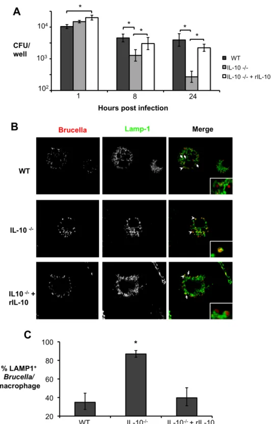
Immunofluorescence microscopy:
Immunofluorescence of Brucella infected BMDM was performed as previously described (Starr et al., 2008). Briefly, B.
abortus MX2 infected BMDM were grown
on 12-mm glass coverslips in 24-well plates were washed three times with PBS, fixed with 3% paraformaldehyde, pH 7.4, at 37°C for 20 min, washed three times with PBS and then incubated for 10 min in 50 mm NH4Cl in PBS in order to quench free
aldehyde groups. Samples were blocked and permeabilized in 10% goat serum and 0.1% saponin in PBS for 30 min at room temperature. Cells were labeled by inverting coverslips onto drops of primary antibodies diluted in 10% horse serum and 0.1% saponin in PBS and incubating for 45 min at room temperature. The primary antibody used was rat anti-mouse LAMP-1 (BD Pharmigen, San Jose, CA). Bound antibodies were detected by incubation with 1:500 dilution of Alexa Fluor 488 donkey anti-rat (Invitrogen, Grand Island, NY) for 45 min at room temperature. Cells were washed twice with 0.1% saponin in PBS, once in PBS, once in H2O and then mounted
in Mowiol 4-88 mounting medium (Calbiochem). Samples were observed on a Carl Zeiss LSM 510 confocal laserscanning microscope for image acquisition (Carl Zeiss Micro Imaging). Confocal images of 1024 × 1024 pixels were acquired as projections of three consecutive slices with a 0.38-μm step and assembled using Adobe Photoshop CS2 (Adobe Systems). For quantification of Brucella MX2 and Lamp1+ compartment colocalization, at least 100 bacteria/sample were counted. All experiments were performed independently in quadruplicate at least two times.
Statistical analysis: Fold changes of ratios
logarithmically prior to statistical analysis. An unpaired Student's t-test was performed on the transformed data to determine whether differences in fold changes between groups were statistically significant (P < 0.05). Significance of differences in histopathology scores was determined by a one-tailed non-parametric test (Mann-Whitney).
Results
Lack of IL-10 production during early B. abortus infection results in lower bacterial survival and increased pathology in vivo
IL-10 has an important role in controlling the immune response induced by different inflammatory processes (Redford et al., 2011). Moreover, Brucella infection has been shown to induce IL-10 production by splenocytes in vitro (Fernandes and Baldwin, 1995) and during intravenous in
vivo infection (Fernandes et al., 1996;
Fernández-Lago et al., 1996). To determine the time-course of IL-10 production during
B. abortus infection, C57BL/6 mice were
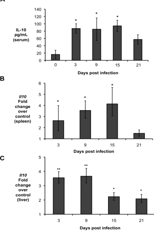
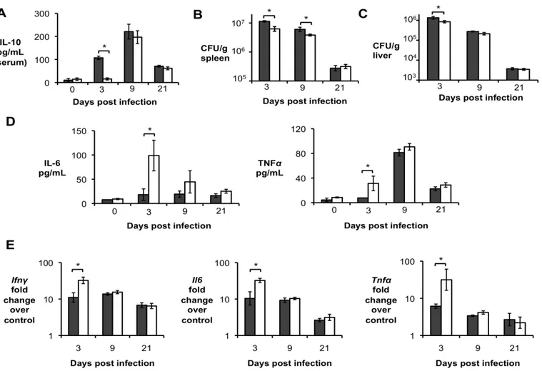
infected intraperitoneally (IP) with 5 x 10⁵ CFU of the virulent B. abortus strain 2308 and IL-10 production was determined at 3, 9, 15, and 21 days post-infection (d.p.i.). Infected mice exhibited significantly higher levels of IL-10 in the serum (Fig. 1A), which was associated with increased IL-10 transcript levels in the spleen (Fig. 1B) and liver (Fig. 1C) as early as 3 d.p.i.
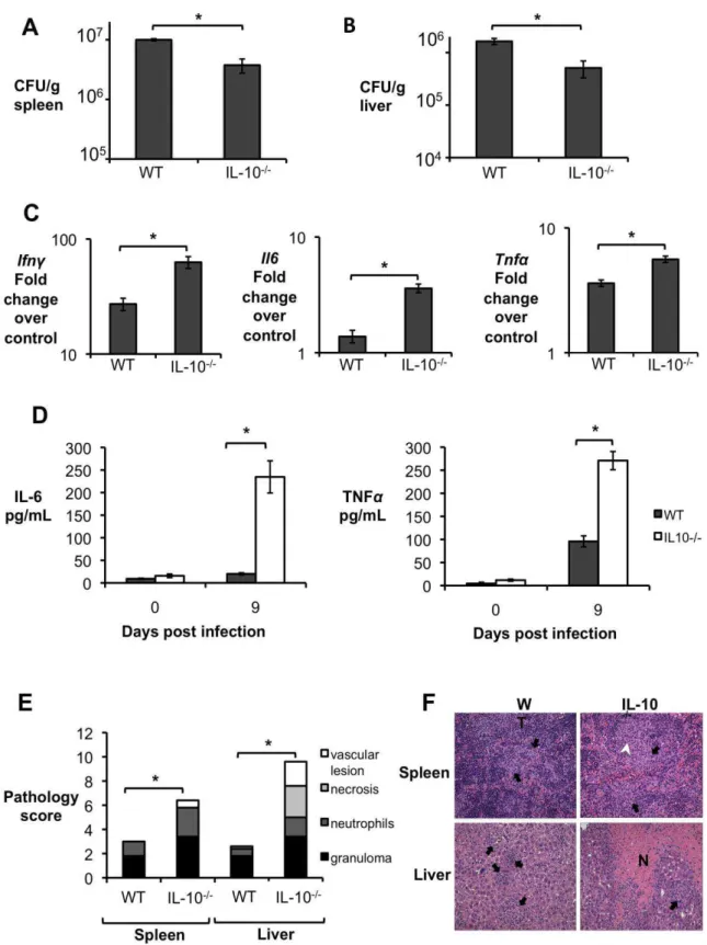
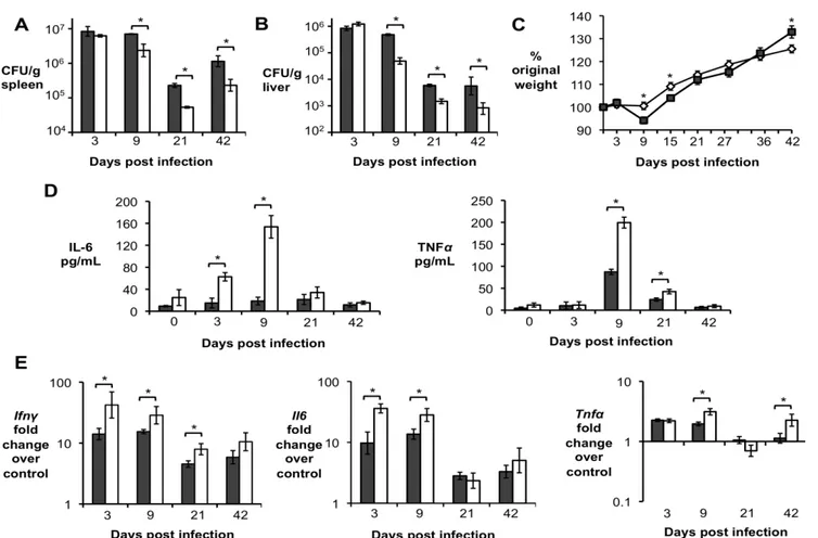
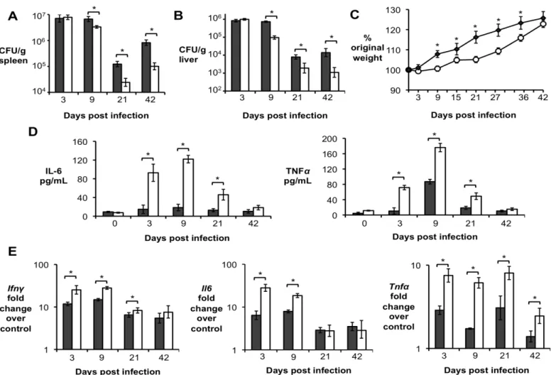
Importantly, significantly increased levels of IL-10 in serum and infected organs was only detected until 15 d.p.i., suggesting a possible regulatory function for this cytokine during acute Brucella infection. To further investigate if IL-10 plays a role in modulating the inflammatory response during acute brucellosis, C57BL/6 wild-type and Il10-deficient mice (IL-10-/-) were infected IP with 5 x 10⁵ CFU of B. abortus 2308 and responses were evaluated at 9 d.p.i.. Interestingly, IL-10-/- mice had significantly lower bacterial survival in both the spleen (Fig. 2A) and the liver (Fig. 2B). IL-10-/- mice also exhibited increased induction of pro-inflammatory cytokines such as IFN- , interleukin-6 (IL-6) and tumor necrosis factor alpha (TNF-α) in infected organs (Fig. 2C) and in serum from infected mice (Fig. 2D).
Figure 1. Brucella abortus induces IL-10 production by infected organs during early in vivo infection. (A) IL-10 levels in serum of C57BL/6 infected with B. abortus for 3, 9, 15, and 21 days
Figure 2. Lack of IL-10 results in lower bacterial survival and increased pathological changes during early Brucella abortus infection in vivo. (A,B) Colonization of spleen (A) and liver (B) of
control or IL-10-/- mice by B. abortus at 9 d.p.i. (C) qRT-PCR analysis of IFN- , IL-6 and TNF-α gene expression in spleens of C57BL/6 and IL-10 -/- 645 mice infected with B. abortus 2308 for 9 days (similar results for liver). (D) IL-6 and TNF-α levels measured by ELISA in serum of C57BL/6 and IL-10
29
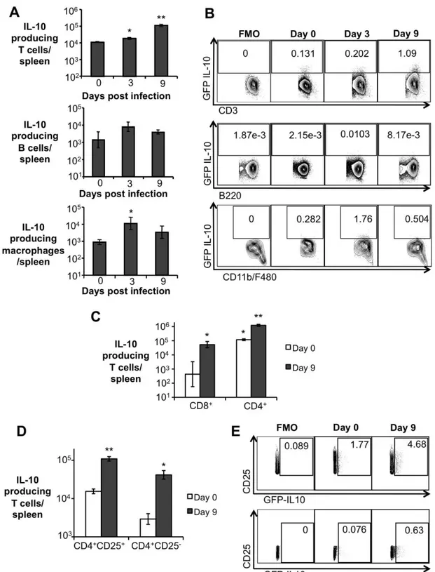
unpaired ttest statistical analysis.(E) Histopathology score of spleen and liver from C57BL/6 and IL10 -/- mice infected with B. abortus 2308 for 9 days. (F) Representative pictures from (E). Black arrow shows microgranulomas, white arrowheads shows neutrophilic infiltrate and upper case N shows areas of coagulative necrosis (x20). n=5. Values represent individual mice (black circles) and geometric mean (black dash). *P<0.05 using Mann-Whitney statistical analysis.Figure 3. CD4+CD25+ T cells are the main producers of IL-10 during early Brucella abortus in vivo
infection. (A) Flow cytometry measurement of IL-10 expression in splenic T cells, B cells, and
Representative data plot of IL-10 expression in splenic T cells (CD3), B cells (B220), and macrophages (CD11b+F4/80+ 659 cells) from C57BL/6 IL-10 GFP-reporter mice. (C) Flow cytometry measurement of IL-10 expression in splenic CD8+ T cells and CD4+ T cells from C57BL/6 IL-10 GFP-reporter mice infected with B. abortus 2308 for 9 days. (D) Flow cytometry measurement of IL-10 expression in splenic CD4+CD25+ T cells and CD4+CD25- T cells from C57BL/6 IL-10 GFP-reporter mice infected with B. abortus 2308 for 9 days. (E) Representative data plot of IL-10 expression in splenic CD4+CD25+ (upper panel) and CD4+CD25- (lower panel) T cells from C57BL/6 IL-10 GFP-reporter mice. Values represent mean ± SEM. n=4. (*) represents P<0.05 relative to uninfected control (day 0), (**) represent P<0.05 relative to day 3 infection for (A), relative to CD8+ day 9 infected for (C) and relative to CD4+CD25- for (D) using unpaired t-test statistical analysis. FMO = fluorescence minus one.
CD4+CD25+ T cells are the main IL-10
producers during early B. abortus infection in vivo
IL-10 can be produced by different T cell subsets, as well as by B cells, neutrophils, macrophages, and some DC subsets (Moore
et al., 2001). To determine the cell types
responsible for IL-10 production during early B. abortus infection, Il10-GFP reporter mice (Kamanaka et al., 2006) were infected IP with 5 x 10⁵ CFU of B. abortus and IL-10 producing cells were identified at 3 and 9 d.p.i. by flow cytometry. A significant increase in the number of IL-10 producing T cells was observed in infected mice at 3 and 9 days post infection, whereas macrophages presented increased production of IL-10 only at 3 d.p.i. Moreover, the number of IL-10 producing B cells, neutrophils, and dendritic cells did not change significantly when compared to uninfected mice (Fig. 3A and 3B, and data not shown). Importantly,
even though a significant increase in the number of IL-10 producing CD8+ T cells was observed (Fig. 3C), a tenfold higher number of IL-10 producing T cells was observed in the CD4+ T cell population (Fig. 3C). No IL-10 production by δ T cells was observed (data not shown).
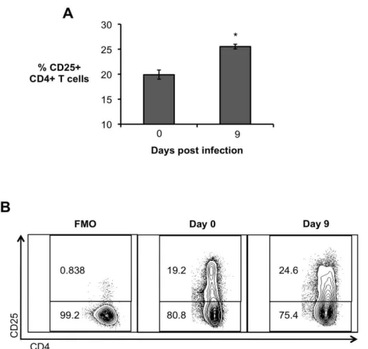
Various IL-10 producing CD4+ T cells have been described, including the CD4+CD25+ subset (Shevach, 2002). Interestingly, an expansion of the CD4+CD25+ T cell population was observed in the spleen of B.
abortus infected mice at 9 d.p.i. (Fig. 4A