A liquid phase blo cking ELISA fo r
the de te ctio n o f antibo die s against
infe ctio us bro nchitis virus
1Departamento de Apoio, Produção e Saúde Animal, Faculdade de O dontologia,
Universidade Estadual Paulista, Araçatuba, SP, Brasil
2Departamento de Microbiologia, Faculdade de Ciências Agrárias e Veterinárias,
Universidade Estadual Paulista, Jaboticabal, SP, Brasil
3Merial Laboratories, Setor de Biologia Aviária, Campinas, SP, Brasil
T.C. Cardoso1,
R.L. Mouro-Sousa2,
C. O liveira3, G. Stringhini1
and A. Augusto-Pinto2
Abstract
A liquid phase blocking ELISA (LPB-ELISA) was developed for the detection and measurement of antibodies against infectious bronchitis virus (IBV). The purified and nonpurified virus used as antigen, the capture and detector antibodies, and the chicken hyperimmune sera were prepared and standardized for this purpose. A total of 156 sera from vaccinated and 100 from specific pathogen-free chickens with no recorded contact with the virus were tested. The respective serum titers obtained in the serum neutralization test (SNT) were compared with those obtained in the LPB-ELISA. There was a high correlation (r2 = 0.8926) between the two tests. The LPB-ELISA represents a single test suitable for the rapid detection of antibodies against bron-chitis virus in chicken sera, with good sensitivity (88%), specificity (100%) and agreement (95.31%).
Co rre spo nde nce
T.C. Cardoso
Departamento de Apoio, Produção e Saúde Animal
Curso de Medicina Veterinária Rua Clóvis Pestana, 793 16015-050 Araçatuba, SP Brasil
Fax: + 55-18-622-6487 E-mail: tcardoso@ fmva.unesp.br Reasearch supported by FAPESP (No. 1997/10877-2).
Received June 18, 1998 Accepted February 26, 1999
Ke y wo rds
·Liquid phase blocking ELISA
·Humoral response
·Infectious bronchitis virus
Infectious bronchitis virus (IBV) infects the respiratory tract, kidneys and oviduct of chicks of all ages causing retarded growth, mortality, reduced egg production and infe-rior egg shell quality (1). For the control of virus infection, broilers are vaccinated at one day of age with live attenuated vaccines. Breeders and egg layers are vaccinated at approximately 8-week intervals with live at-tenuated vaccines and with inactivated vac-cines after they start laying eggs (2).
IBVs contain four structural proteins: S1 and S2, with Mr of 92 K and 84 K, respec-tively, as well as the heterogeneously glyco-sylated membrane polypeptide p23 with an Mr of 34 K, and the nucleocapsid protein with an Mr of 52 K associated with RNA (3).
and slaughter may hamper the interpretation of serological results. A field study on com-mercial broiler flocks without clinical infec-tion favored the use of an indirect and sand-wich ELISA for the serological diagnosis of the IBV humoral response (4-7). Thus, we developed and standardized a liquid phase blocking ELISA (LPB-ELISA), and com-pared its results with those obtained by the SNT for the detection of neutralizing anti-bodies against IBV.
Virus antige n
The Massachusetts strain (Mass 41) of IBV was propagated by infecting 9-11-day-old embryonated specific pathogen-free (SPF) eggs and the allantoic fluid was har-vested as recommended (3). The virus was further purified by previously reported meth-ods (5,6) with some modifications. Briefly, the allantoic fluid collected and pooled from IBV-infected SPF embryonated eggs was clarified by centrifugation at 2,000 g for 20 min at 4o
C and then submitted to 59,000 g
for ultracentrifugation. The viral pellet was resuspended in 3 ml of TNE buffer (1 mM TRIS, 0.15 M NaCl, 1 mM EDTA, pH 7.0) and layered on a continuous 20-55% sucrose gradient (w/v) and centrifuged at 90,000 g
for 10 h at 4o
C. The fractions collected from the gradient which absorbed at 254 nm (viral RNA) and 280 nm (total protein) were pooled and diluted in TNE buffer and the protein concentration was determined by the method of Hartrée (7). The virus infectivity was titrated in SPF embryonated eggs, as recom-mended (1). The same IBV strain, replicated in embryonated SPF eggs and clarified at low-speed centrifugation, was used as non-purified antigen in the LPB-ELISA.
Capture antibo dy
The chicken IBV-specific g-globulin was used as the capture antibody. For this pur-pose, only one group of SPF white Legorns
chicks was used. An inbred C/O Line ob-tained from Merial Laboratories was placed in positive pressure isolation units at one day of age. These ten chicks were vaccinated intra-ocularly (io) with 104.0 CD
50 of puri-fied M41 strain in 50 µl of phosphate-buff-ered solution (PBS) at 2, 6 and 10 weeks of age, respectively. After these procedures, all chicks were again vaccinated intramuscu-larly at two weeks of age with 0.5 mg of inactivated purified M41 strain in 100 µl of PBS mixed with Freunds complete adju-vant (1:1). At 6 and 10 weeks of age these ten chicks were revaccinated intramuscularly with 0.5 mg of inactivated purified M41 strain in 100 µl of PBS mixed with Freunds incomplete adjuvant (1:1). The chicks were bled from the wing vein ten days after the last vaccination. The chicken IBV-specific g-globulin fraction was obtained as described previously (8). The capture antibody (spe-cific g-globulin fraction against purified IBV) was titrated by the SNT.
D e te cto r antibo dy
The detector antibody was prepared by the immunization of three guinea pigs with purified IBV as described in previous ar-ticles (6,7).
Se rum sam ple s
A total of 156 serum samples collected from different vaccinated commercially bred chickens and 100 serum samples from SPF chickens were titrated both by LPB-ELISA and SNT. A positive reference serum was obtained as described previously (6). The negative reference serum consisted of a mix-ture of 10 serum samples collected from SPF chickens.
Se rum ne utralizatio n te st
dilu-tions of the chicken sera were mixed with 100 TCID50 of the Mass 41 IBV strain in microtiter plates (Nunc, Copenhagen, Den-mark) and incubated for 1 h at 37o
C. There-after, an equal volume of chicken embryo-nated kidney cells at 106
cells/ml was added to each well and the plates were further incubated at 37o
C. The reduction in virus-specific cytopathic effect was observed after 48 and 72 h and the virus neutralization titer of the serum calculated by the Spearm-Karber method (6,7).
D e ve lo pm e nt o f LPB-ELISA
Optimal dilutions of all reagents (capture antibody, detector antibody, serum samples and nonpurified virus) were determined us-ing chessboard titration (9). Different cap-ture antibody dilutions were tested against several unpurified viral antigen concentra-tions in order to detect the best discrimina-tion between the positive and the negative reference sera (10).
Applicatio n o f LPB-ELISA
The test was performed as described by McCullough et al. (10) and Araujo et al. (11), with some modifications. The micro-plate (Nunc) wells were coated with the capture antibody diluted 1:250 or 12.5 µg/ well overnight, in 0.05 M carbonate bicarbo-nate buffer, pH 9.6, at 4o
C. After five washings with phosphate-buffered saline solution containing 0.05% Tween 20 (PBST), the plates were blocked with PBS containing 15% skim milk (PBSM). After incubation for 45 min at 37o
C, the plates were ready for use. The sera to be tested, always run in duplicate, were treated with 1% trichloro-acetic acid for protein precipitation before mixing with a fixed concentration of unpuri-fied virus dilution (1:5), also diluted in PBS with 0.5 M NaCl. This liquid phase was executed in separated hemagglutination plates (Nunc) and after incubation at 37oC
for 90 min the virus-sera mixture was trans-ferred to the ELISA plates and incubated at 37o
C for 60 min. The IBV antigen (M41 strain) was stored at -70o
C and used at a concentration of 1:5, which gave an absorb-ance of 1.5 at 492 nm, and the plates were washed as before. An optimal 1:4000 dilu-tion of guinea pig detector anti-IBV serum in phosphate-buffered solution containing Tween and skim milk (PBSTM) was added and the plates were incubated for 60 min at 37o
C. An optimal 1:16,000 dilution of com-mercial rabbit anti-guinea pig IgG conju-gated to horseradish peroxidase (Sigma Chemical Co., St. Louis, MO, USA) in PBSTM was added. After incubation for 60 min at 37o
C, the plates were again washed as described previously. A mixture of 0.006% H2O2 and 0.4 mg o-phenylenediamine/ml, 0.1 M Na2 HPO4 and 0.1 M citric acid buffer, pH 5.0, was used as substrate and chro-mogen. After 15 min of incubation at room temperature, 2 M HCl was added in order to block the enzymatic reaction, and the OD of the plate was read at 492 nm in a Titerteck multiscan reader. On each plate, 22 wells were used for antigen control with no test sera added, and 2 wells were used for the reciprocal serum dilution that inhibited color development in relation to the 22 antigen control wells. The following formula was used for this determination:
T =
(X - A) log Y + (B - X) log Z
B - A
Statistical analysis
The correlation coefficient (r2) between LPB-ELISA and SNT was determined for serological analysis (10). The cutoff point was determined by graphic analysis of the interception of the copositivity and conega-tivity curves of LPB-ELISA and SNT pro-jected on the ordinate axis (11).
Re pro ducibility o f LPB-ELISA
The reproducibility of the LPB-ELISA for antibody detection was determined using the OD values for serum dilutions of nega-tive and posinega-tive controls, tested on twenty different days. These values were subjected
to statistical analysis by the Student t-test. The LPB-ELISA did not detect Newcastle disease, Reovirus or influenza A antibodies (data not shown). The results obtained are discussed below.
The chicken IBV-specific g-globulin used as the capture antibody showed higher SN titer ³log2 8.0. The trichloroacetic acid used for serum precipitation at 1% concentration eliminated the nonspecific reaction of the positive and negative avian serum compo-nents. In order to determine the best dilution for the capture antibody used to provide trapping, g-globulin was titrated in a sand-wich ELISA and the optimal dose was found to be 1:250 or 12.5 µg/well.
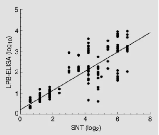
The correlation coefficient between the LPB-ELISA and SNT for a total of 256 serum samples was r2
= 0.8926 (P<0.0005) (Figure 1). The copositivity and conegativity results were determined with the cutoff be-ing ³0.6. Table 1 shows the specificity (100%) and sensitivity (88%) of LPB-ELISA for antibody detection. The agreement be-tween LPB-ELISA and SNT was 95.3%. The reproducibility of LPB-ELISA for anti-body detection had a coefficient of variation of 2.4%.
Antibody responses to the S1, S2, N and M virus proteins were also detected in chicks vaccinated with the inactivated IBV strains. The S1, S2 and N proteins all induced cross-reactive antibodies which were detected by ELISA (1). In poultry, high specificity of serological tests is more important than high sensitivity, since low sensitivity can be com-pensated for by using a larger number of blood samples (4). The LPB-ELISA docu-mented here demonstrated 100% specific-ity; however, when used at the cutoff level ³0.6 reported above, 95.3% agreement was found. In fact, the antigen reacts differently with its specific antibodies, depending on whether it is in the solid or liquid phase, as reported by McCullough et al. (10). With regard to the high agreement found here between the LPB-ELISA and SNT, we may
L
P
B
-E
L
IS
A
(
lo
g10
)
5
4
3
2
1
0
0 2 4 6 8
SNT (log2) Figure 1 - Correlation and linear
regression of antibody titers ob-tained by liquid phase blocking ELISA (LPB-ELISA) (log10) and by serum neut ralizat ion t est (SNT) (log2). The equation for the line is: LPB y = 0.2322x + 0.3419; r 2 = 0.892.
Table 1 - Specificity and sensitivity of LPB-ELISA for the determination of IBV-antibodies in vacci-nated and nonvaccivacci-nated chickens.
aTotal number of serum samples from vaccinated
chickens. bTotal number of serum samples from nonvaccinated SPF chickens. Specificity = 156 - 0/ 156 x 100 = 100% . Sensitivity = 100 - 12/100 x 100 = 88% . Agreement = 156 + 88/256 x 100 = 95.31% .
Serum neutralization test LPB-ELISA
Positive Negative
Positive 156 12
Negative 0 88
Total number of serum 156a 100b
speculate that the high levels of IBV-neutral-izing antibodies were possibly detected by the liquid phase performed in the same way in the SNT (4,9,11-13). The determination of 50% competitive antibody titers in LPB-ELISA by using the mathematical interpola-tion procedure on the basis of a larger num-ber of antigen control wells than those de-scribed (11,13) allowed a more precise esti-mate of these titers, considerably reducing the inter-test variation (2.40%).
The use of chicken IBV-specific g -globu-lin as capture antibody has several advan-tages over the use of rabbit polyclonal anti-serum. Using the polyclonal antibody ampli-fies the nonspecific reactions with the allan-toid fluid proteins, results also found in other studies (6,7,14). In the present study, it was necessary to precipitate the chicken serum proteins, especially the IgM isotype, with 1% trichloroacetic acid to avoid nonspecific protein binding (15). This was confirmed by the results obtained for the positive and nega-tive sera. In fact, it was the first time that LPB-ELISA was applied to chicken sera. This assay has not been used to measure anti-IBV antibodies in broiler chickens as frequently as the commercial indirect ELISA. In spite of the relatively higher correlation coefficients recorded for the indirect ELISA test, compared to the agar-precipitating gel test, HI test and SNT, it is important to
emphasize the different intrinsic properties of each serological test (4).
The LPB-ELISA was compared here with the SNT, especially the liquid phase, where specific antibodies in the test sera effectively block the antigen and prevent it from re-acting in the sandwich ELISA. Thus, the blocking ELISA developed here presented high specificity when compared with values reported by Esterhuysen et al. (9). The LPB-ELISA has been used to measure anti-foot-and-mouth and rabies virus antibodies in cattle and human sera, respectively (9-11,13). Our results showed a significant correlation (r2
= 0.8926) between the LPB-ELISA and SNT, similar to the results obtained in other studies (10,11,13).
Certain types of ELISA, particularly blocking ELISA, may have a number of advantages over the serum neutralization test, but no serological test has indicated virus protection upon analysis. Therefore, the LPB-ELISA is considered a useful tool for routine laboratory diagnosis of IBV antibodies, thereby eliminating the need for cumber-some serological monitoring.
Ackno wle dgm e nts
The authors wish to acknowledge the excellent technical assistance of Merial Labo-ratories in performing the hyperimmune sera.
Re fe re nce s
1. Ignjatovic J & Galli L (1995). Immune re-sponses to structural proteins of avian infectious bronchitis virus. Avian Patholo-gy, 24: 313-332.
2. Box PG, Holm es HC, Finney PM & Froymann R (1988). Infectious bronchitis in laying hens: The relationship betw een haemagglutination inhibition antibody lev-els and resistance to experimental chal-lenge. Avian Pathology, 17: 349-361. 3. Cavanagh D (1983). Coronavirus IBV
gly-copolypeptides: size of their polypeptide moieties and nature of their oligosaccha-rides. Journal of General Virology, 64:
1187-1191.
4. Witt JJ, M ekkes DR, Kouw enhoven B & Verheijden JHM (1997). Sensitivity and specificity of serological tests for infec-tious bronchitis virus antibodies in broil-ers. Avian Pathology, 26: 105-118. 5. Cardoso TC, M ontassier HJ, Galletti M CM
& Pinto AA (1996). Evaluation of indirect ELISA for monitoring antibodies against infectious bronchitis virus. Revista de M
i-crobiologia, 27: 64-69.
6. Cardoso TC, M ontassier HJ, Galletti M CM & Pinto AA (1996). Development and ap-plication of a sandw ich ELISA to measure
chicken antibodies to infectious bronchi-tis virus. Virus Review s and Research, 1: 75-80.
7. Cardoso TC, Sousa RLM , Alessi AC, M ontassier HJ & Pinto AA (1998). A double antibody sandw ich ELISA for rapid diagnosis of virus infections and to meas-ure the humoral response against infec-tious bursal disease on clinical material.
Avian Pathology, 27: 450-454.
anti-sera. Applied M icrobiology,25: 26-36. 9. Esterhuysen JJ, Prehaud C & Thomson
GR (1995). A liquid phase blocking ELISA for the detection of antibodies to rabies virus. Journal of Virological M ethods, 51: 31-42.
10. M cCullough KC, Bruckner L, Schaffiner R, Werner F, Heinz KM & Ulrich K (1992). Relationship betw een the anti FM DV anti-body reaction as measured by different assays and protection in vivo against chal-lenge infection. Veterinary M icrobiology, 30: 99-112.
11. Araujo JP, M ontassier HJ & Pinto AA (1996). Liquid phase blocking sandw ich
enzyme linked immunosorbent assay for the detection of antibodies against foot-and-mouth disease virus in w ater buffalo sera. American Journal of Veterinary
Re-search, 57: 840-843.
12. Cornaglia E, Chretien N, Charara S & Elazhary Y (1994). Detection of porcine respiratory coronavirus and transmissible gastroenteritis virus by an enzyme linked immunosorbent assay. Veterinary M
icro-biology, 42: 349-359.
13. Hamblin C, Barnett ITR & Hedger RS (1986). A new enzyme linked immunosor-bent assay (ELISA) for the detection of antibodies against foot-and-mouth
dis-ease virus. I. Development and method of ELISA. Journal of Immunological M eth-ods, 93: 123-129.
14. M ichalski WP, O’Rourke D & Bagust TJ (1996). Chicken anaemia virus antibody ELISA: problems w ith non specific reac-tions. Avian Pathology, 25: 245-254. 15. De Jonge N, Fillié YE & Deelder AM