Gene expression in IFN-
γ
γ
γ
γ
γ
-activated
murine macrophages
1Laboratório de Imunologia Viral, Instituto Butantan, São Paulo, SP, Brasil 2Max Planck Institute of Immunobiology, Freiburg, Germany
3Basel Institute for Immunology, and University Clinics, Basel, Switzerland C.A. Pereira1, M. Modolell2,
J.R. Frey3 and I. Lefkovits3
Abstract
Macrophages are critical for natural immunity and play a central role
in specific acquired immunity. The IFN-
γ
activation of macrophages
derived from A/J or BALB/c mice yielded two different patterns of
antiviral state in murine hepatitis virus 3 infection, which were related
to a down-regulation of the main virus receptor. Using cDNA
hybrid-ization to evaluate mRNA accumulation in the cells, we were able to
identify several genes that are differently up- or down-regulated by
IFN-
γ
in A/J (267 and 266 genes, respectively, up- and
regu-lated) or BALB/c (297 and 58 genes, respectively, up- and
down-regulated) mouse macrophages. Macrophages from mice with
differ-ent genetic backgrounds behave differdiffer-ently at the molecular level and
comparison of the patterns of non-activated and IFN-
γ
-activated A/J
or BALB/c mouse macrophages revealed, for instance, an
up-regula-tion and a down-regulaup-regula-tion of genes coding for biological funcup-regula-tions
such as enzymatic reactions, nucleic acid synthesis and transport,
protein synthesis, transport and metabolism, cytoskeleton
arrange-ment and extracellular matrix, phagocytosis, resistance and
suscepti-bility to infection and tumors, inflammation, and cell differentiation or
activation. The present data are reported in order to facilitate future
correlation of proteomic/transcriptomic findings as well as of results
obtained from a classical approach for the understanding of biological
phenomena. The possible implication of the role of some of the gene
products relevant to macrophage biology can now be further
scruti-nized. In this respect, a down-regulation of the main murine hepatitis
virus 3 receptor gene was detected only in IFN-
γ
-activated
macro-phages of resistant mice.
Correspondence
C.A. Pereira
Laboratório de Imunologia Viral Instituto Butantan
Av. Vital Brasil, 1500 05503-900 São Paulo, SP Brasil
Fax: +55-11-3726-1505 E-mail: grugel@butantan.gov.br
Research supported in part by Fundação Butantan. C.A. Pereira is a recipient of a CNPq-IA fellowship and of the Swiss National Foundation during part of this research.
Publication supported by FAPESP.
The present address of J.R. Frey is F. Hoffmann-La Roche Ltd., Preclinical CNS Research, PRBG-T, Building 68/452A, 4070 Basel, Switzerland, E-mail:
johann_r.frey@roche.com, and of I. Lefkovits is Institute for Physiology, University of Basel, Vesalianum Vesalgasse 1, 4051 Basel, Switzerland, E-mail: ivan.lefkovits@unibas.ch.
Received December 9, 2003 Accepted August 12, 2004
Key words
•Gene array •mRNA
•Mouse hepatitis virus 3
(MHV3)
•IFN-γ
•BALB/c mouse •A/J mouse •Macrophages
Introduction
The mononuclear phagocyte system
con-stitutes the second major cell population of
the immune system, with varied
morpho-logic forms and functions in most of the
tissues of the organism. Upon stimulation,
these cells undergo striking physiological
changes playing important active roles.
presence on the membranes of the cells
un-der investigation. This was convenient, since
various techniques permitted raising
anti-bodies against the surface molecules, and
the worldwide effort of identifying them led
to the powerful molecular definitions of the
“clusters of differentiation”. Now that we
possess various cDNA libraries, as well as
assays derived from them, we have on hand
a useful tool to probe the expression profile
of transcribed molecules. By clusters of
dif-ferentiation-molecular definition, important
structural entities on the cell membranes
have been identified, and it is expected that
expression profiling will permit us to focus
on intracellular compartments and reveal
meaningful regulatory polypeptides.
A/J mice have been described to be
resis-tant to experimental infection with mouse
hepatitis virus 3 (MHV3), developing a mild
disease that disappears after a few days. On
the other hand, BALB/c mice develop acute
hepatitis after infection and die some days
later (1). Attempts to identify the
mechan-isms involved in the resistance or
suscepti-bility of mouse strains to MHV3 infection
have led different groups to demonstrate the
involvement of virus replication in
macro-phages (2-4), the antiviral state induced by
IFN-
γ
only in macrophages from resistant
animals (1), and the expression of a monokine
with procoagulant activity (5,6).
Studying the molecular basis of the virus
resistance induced by IFN-
γ
in macrophages,
we have recently shown that
down-regula-tion of a viral receptor gene with
conse-quences on the gene product synthesis may
be implicated in the resistance shown by A/
J mice (7). In a previous study, by means of
proteomic analysis of proteins extracted from
A/J or BALB/c macrophages, we were able
to tag several gene products that were
syn-thesized at elevated or diminished levels (8),
indicating that macrophages from resistant
and susceptible strains behave differently at
the molecular level upon IFN-
γ
activation.
The gene profiling technology adopted
for the present study (9-11) using an array
constructed from 1536 individual cDNA
clones allowed us to study the mentioned
defined portion of expressed genes. The
choice of the library is of high relevance to
the problem under study since in our library
there are mainly immunologically relevant
gene probes originating from an
immuno-logically active organ, i.e., the fetal thymus.
Not only can changes be detected at the
quantitative and qualitative level of gene
expression, but it is also possible to identify
them by recognizing their protein product
since the expressed sequence tags have been
established for the individual entities of the
cDNA library used for the evaluation of a
given mRNA sample by hybridization, and
protein products have been analyzed on
2D-SDS gels (10).
By using gene expression profiling to
evaluate the mRNA content of IFN-
γ
-acti-vated macrophages we attempted to provide
detailed information about up- or
down-regu-lated gene products to be in turn linked to the
modulation of biological macrophage
func-tions. As an example using a classical
ap-proach, we previously identified the
regula-tion of the main MHV3 receptor gene
ex-pression in IFN-
γ
-activated macrophages as
a central feature of resistance (7). The gene
expression profiling shown here confirmed
these previous data. Several new regulatory
molecules can be considered for further
scru-tiny.
Material and Methods
Macrophage cultures
de-tached by repeated careful stretching of the
bags, washed once with medium and used in
the experiments. They were cultivated in
Dulbecco’s modified Eagle’s medium
con-taining 10% FCS at a concentration of 10
5cells/well in 96-well plates.
MHV3 replication and receptor expression
Experiments were carried out in order to
evaluate in the macrophages the induction of
anti-MHV3 state and the expression of virus
receptors mediated by exogenous IFN-
γ
.
BALB/c or A/J mouse bone marrow-derived
macrophage cultures were activated for 18 h
with 50 U/ml of IFN-
γ
(Genentech Inc., South
San Francisco, CA, USA) and for the virus
replication assay they were infected with
MHV3 at multiplicity of infection of 0.1.
Cell supernatants were then collected at
dif-ferent times and virus titers determined by a
plaque assay (13). The data, reported as
plaque-forming units per milliliter, were
measured in triplicate cultures. For virus
receptor expression, total cellular RNA was
extracted from IFN-
γ
-activated macrophage
cultures and reverse transcribed and the
cDNAs were submitted to the polymerase
chain reaction containing specific primers.
Samples were then submitted to agarose gel
electrophoresis for visualization (7).
RNA extraction from macrophages and
reverse transcription
Total cellular RNA was extracted from
macrophage cultures (10
7cells/5 ml) on
6-cm diameter Petriperm dishes (Sartorius,
Goettingen, Germany) treated or not for 18 h
with 50 U/ml of IFN-
γ
by the isothiocyanate
method (Trizol reagent; Invitrogen GmbH,
Karlsruhe, Germany). Briefly, 20 ml Trizol
was added to the frozen pellets and
solubili-zation was achieved by passing the lysate
through the pipette. After addition of
chloro-form, vigorous shaking, and standing on ice
for 5 min, the suspension was centrifuged at
12,000
g
for 15 min. The upper aqueous
phase was transferred to a fresh tube and an
equal volume of isopropanol was added. The
RNA precipitate was spun at 12,000
g
for 15
min and the pellet was washed with 75%
alcohol and solubilized in water. Reverse
transcription was performed using reverse
transcriptase SuperScript II (Invitrogen
GmbH) and applying non-radioactive dNTP
components.
Analysis of gene expression
A fetal thymus library was chosen for
this study because: a) the mRNA population
of the fetal thymus encompasses a much
wider spectrum of expressed entities than
any other source of immunocompetent cells,
and, b) the availability of collections of
proteomic data base entries concerning the
“translability” of the gene products into
pro-tein molecules. So, a murine fetal thymus
cDNA library was prepared from fetal
thy-muses of BALB/c mice using cytoplasmic
RNA samples, which were reversely
tran-scribed into cDNAs. The resulting
prepara-tions were amplified using cDNA packaging
into infectious phage particles and, upon
subsequent infection with
Escherichia coli
In order to identify modified gene
ex-pression in IFN-
γ
-activated macrophages the
cDNA was hybridized to the arrayed DNA,
and the relative hybridization intensity was
established by comparing the intensity of
relevant spot pairs using chemoluminescence
as a readout. The procedure was carried out
by labeling the probe with horseradish
per-oxidase and cross-linking with
glutaralde-hyde, followed by the light emitting reaction
of luminol, which produces blue light upon
oxidation. Relative hybridization intensity
was established by comparison to the
inten-sity of control spots.
Nylon sheets with 1536 clones in
dupli-cate were prepared as already described (10).
Briefly, the robot first merged four 96-well
microtiter plates into a single 386-well plate,
and each of the 386-well plates was spotted
on the nylon sheet in duplicate. This was
done with all original microtiter plates. For
good visual orientation the duplicates were
spaced with diagonal, horizontal and
verti-cal orientation. The probe quantitation was
performed via an image analysis system
origi-nally implemented for analysis of protein
spots. The image analysis system is called
Kepler (version 8.0; Large Scale Biology
Corporation, Rockville, MD, USA),
devel-oped at Argonne National Laboratory, and
the spot intensity was modeled as a 3-D
Gaussian curve. No spot normalization was
performed within one hybridization sheet.
Differences in intensity within the
dupli-cates were attributed either to experimental
variations or to robotic deposition of the
spots on the nylon sheet. The nylon sheets
were used to expose X-ray films (30 s to 6
min) and those showing comparable
cyto-chrome c spot intensity (cytocyto-chrome c
con-trol spots served as a measure of
chemilumi-nescence homogeneity) were subjected to
image analysis (Kepler). After signal
quanti-fication, the data were processed for
calcula-tion of arithmetic means. Only those
expres-sion changes based on reliable modeling
were considered. A cut-off limit at 300 units
(since artifacts were present in low intensity
spot modeling) and minimal ratios of 0.5 and
2.0 for up- and down-regulation of genes,
respectively, were applied. As a frame
refer-ence we used the expression of the
elonga-tion factor
α
gene, which is present in six
copies in our ordered library. Each data set
refers to a clonal position for which in most
instances the molecular identity of the cDNA
is known. A/J and BALB/c samples were
hybridized in parallel separately on two
dif-ferent nylon filters, and the repetition was
performed upon stripping on the other filter
(cross-wise). Gene expression analysis was
repeated several times, showing low
experi-mental variation.
Some genes identified as being
differen-tially expressed in IFN-
γ
-activated A/J and
BALB/c mouse macrophages were
submit-ted to database queries in order to investigate
their possible involvement in macrophage
regulatory processes. This topic is addressed
in the discussion of the biological functions
of selected genes.
Results
The approaches used to elucidate the
cel-lular and molecular basis of resistance against
MHV3 infection are indicated in Table 1. A/
J mice were shown to be resistant and BALB/
c mice were shown to be susceptible to
experimental infection with MHV3 (1).
IFN-γ
activation of macrophage cultures from A/
J mice led to a partial restriction of MHV3
growth, in contrast to the BALB/c
macro-phage activation (1). Our control
experi-ments showed at least 10 times lower virus
titers in supernatants of IFN-
γ
-activated A/J
macrophages than in the other cultures (data
not shown). Studies on the expression of
genes coding for virus receptors and on virus
binding to membrane proteins from IFN-
γ
macrophages to restrict MHV3 growth
cor-related with the diminished virus binding
and virus receptor gene expression, leading
us to suggest that these experimental
obser-vations reflect the contribution to the
in vivo
resistance expressed by these mice
follow-ing experimental virus infection (7).
Proteomic studies including
computer-aided image comparison of gels obtained
from 2-D SDS-PAGE of extracted proteins
from IFN-
γ
-activated A/J and BALB/c
mac-rophages revealed the up- and
down-regula-tion of several gene products (Table 1). By
using a similar approach, now focused on
mRNA expression, in the present paper we
report gene expression profiling data
show-ing that several up- and down-regulated genes
were detected in IFN-
γ
-activated
macro-phages from A/J and BALB/c mice (Table
1).
In Table 1 we also present the DNA
hybridization data. The data were selected
by arbitrarily applying a cut-off limit at 300
units and minimal ratios of 0.5 and 2.0 for
up- and down-regulation of genes,
respec-tively. In this way we observed 266 and 58
down-regulated genes (including 175 and 50
signals only detectable in controls) in IFN-
γ
-activated macrophages from A/J and BALB/
c mice, respectively. On the other hand,
IFN-γ
activation of A/J and BALB/c macrophages
led to up-regulation of 267 and 297 genes
(including 174 and 96 signals only
detect-able upon activation), respectively. Selected
up- or down-regulated genes, listed in Tables
2 and 3, respectively, may serve as a basis for
evaluating physiological changes promoted
by IFN-
γ
in macrophages. Table 4 presents a
list of 42 differentially expressed genes and
their function in macrophage biology, as
indicated by recent publications (7,14-55).
One can observe modulation of genes
cod-ing for proteins involved in enzymatic
reac-tions such as adenosine monophosphate
deaminase, pyruvate kinase, acetoacetyl-CoA
synthetase, TNF-
α
converting enzyme, serine
protease inhibitor, or involved in the control
of nucleic acid synthesis and transport such
as DNA ligase, RNA polymerase II, zinc
finger protein or protein synthesis, transport
Table 1. Cellular and molecular basis of resistance against MHV3 obtained by biological assays and expression profiling.
Mouse In vivo Biological assays Proteomic analysis Gene expression profile
strain viral infection
Virus replication Virus receptor Cell lysate (2-D gel spots) (8) mRNA (hybridization spots)
Up-regulation Down-regulation C/IFN-γ ratio Profile Number of spots
A/J Resistant C +++ +++ ∞ Down-regulated 175
>2 (7.13 to 2.02) Down-regulated 91
IFN-γ + - 4 26 0 Up-regulated 174
<0.5 (0.45 to 0.139) Up-regulated 93
BALB/c Susceptible C ++++ ++++ ∞ Down-regulated 50
>2 (4.61 to 2.01) Down-regulated 8
IFN-γ ++++ ++++ 13 16 0 Up-regulated 96
<0.5 (0.49 to 0.108) Up-regulated 20
Data for gene expression profiling in macrophages from A/J or BALB/c mice activated or not with IFN-γin vitro (control, C) and IFN-γ-activated
macrophage cultures (IFN-γ). For the gene expression profile, extracted RNA samples were copied by reverse transcription, and the relative
hybridization intensity was established by comparing the intensity of relevant spot pairs by chemoluminescence. An arbitrary limit of minimal intensity of 300 units and of a minimal ratio of up- and down-regulation of 0.5 and 2, respectively, was utilized. Using these criteria, we identified hybridization spots that, upon IFN-γ activation, decreased (C/IFN-γ >2) or increased (C/IFN-γ <0.5) in intensity, as well as spots below the
thresholds 0.5 and 2.0 in control macrophages (C/IFN-γ = 0) or in IFN-γ-activated macrophages (C/IFN-γ = ∞). The assays are described in the
Table 2. Selected genes up- or down-regulated by IFN-γ in macrophages from A/J mice.
Up-regulated selected genes Up-regulated selected genes Up-regulated selected genes Up-regulated selected genes Up-regulated selected genes
ca A/J C A/J IFN-γ C/IFN-γ Accession Gene
60914 0 5737 0 MMUGTN:023998-30 high-glucose-regulated protein 8
10602 0 3685 0 U50078 guanine nucleotide exchange factor p53
51210 0 3421 0 MMUGTN:037962-2 exportin (nuclear export receptor for tRNAs)
31216 0 3212 0 EM_HUM9:HSRPII140 RNA polymerase II 140-kDa subunit
60804 0 2936 0 NM_013843 zinc finger protein
80111 0 2768 0 EM_MUS:MMGPIP137 gpi-anchored protein
51115 0 2490 0 EM_MUS:MMCOF cofilin
60905 0 2075 0 M92933 lymphocyte common antigen (Ly5)
81211 0 1879 0 EM_MUS:BC003861 hydroxymethylbilane synthase
61202 0 1813 0 MMUGTN:056337-12 platelet-activating factor acetylhydrolase (PAF-AH)
10901 0 1758 0 U87240 lysosomal alpha-mannosidase
51102 0 1293 0 M32599 glyceraldehyde-3-phosphate dehydrogenase
10913 0 1279 0 EM_MUS:MMNPTCC nuclear pore-targeting complex
21003 0 1040 0 X14194 entactin
21112 0 1016 0 AF205079 palladin, actin-associated protein
41216 0 943 0 EM_MUS:AF322193 cleavage and polyadenylation specificity factor 1 (CPSF1)
10604 0 873 0 X53416 filamin, actin-binding protein
11214 0 774 0 EM_MUS:BC006945 DNA polymerase α 2
60214 0 749 0 EM_MUS:BC007133 apoptosis inhibitory protein 5
10810 0 712 0 X84014 laminin-5, α 3B chain
71215 0 619 0 EM_MUS:MM16741 capping protein α 2 subunit
11016 0 492 0 EM_MUS:AB021709 tumor necrosis factor (TNF) α converting enzyme
61204 0 309 0 X57024 glutamate dehydrogenase
70514 2841 20439 0.139 EM_MUS:AF323958 prostaglandin transporter (PGT)
60811 706 4836 0.146 EM_MUS:MMBCATA beta-catenin
10902 531 2829 0.188 MMUGTN:194525-2 serine protease inhibitor
51009 2671 9851 0.271 EM_MUS:AY004877 cytoplasmic dynein heavy chain
60712 978 3430 0.285 J05503 carbamoyl-phosphate synthetase
81208 1316 3951 0.33 EM_MUS:AF210433 glucocorticoid modulatory element binding protein 1 (GMEB-1)
10313 3196 9419 0.34 BC002270 ubiquitin-conjugating enzyme E2 variant 1
71010 5778 13697 0.42 EM_MUS:AF127033 fatty acid synthase
51203 546 1260 0.43 U20780 ubiquitinating enzyme
21004 932 2130 0.44 M64098 high density lipoprotein binding protein (HBP)
10511 2033 4491 0.45 AF143956 coronin-2
70716 18243 40359 0.45 EM_MUS:AB024538 immunoglobulin superfamily containing leucine-rich repeat (ISLRI)
10312 1270 2756 0.46 D16250 bone morphogenetic protein (BMP) receptor
71014 1701 3690 0.46 EM_MUS:MM19604 DNA ligase I
71005 1446 3033 0.48 AF111102 major histocompatibility complex (MHC) class I region
61215 11446 23581 0.49 EM_MUS:MMADAPA1 α-adaptin
Table 2 continued
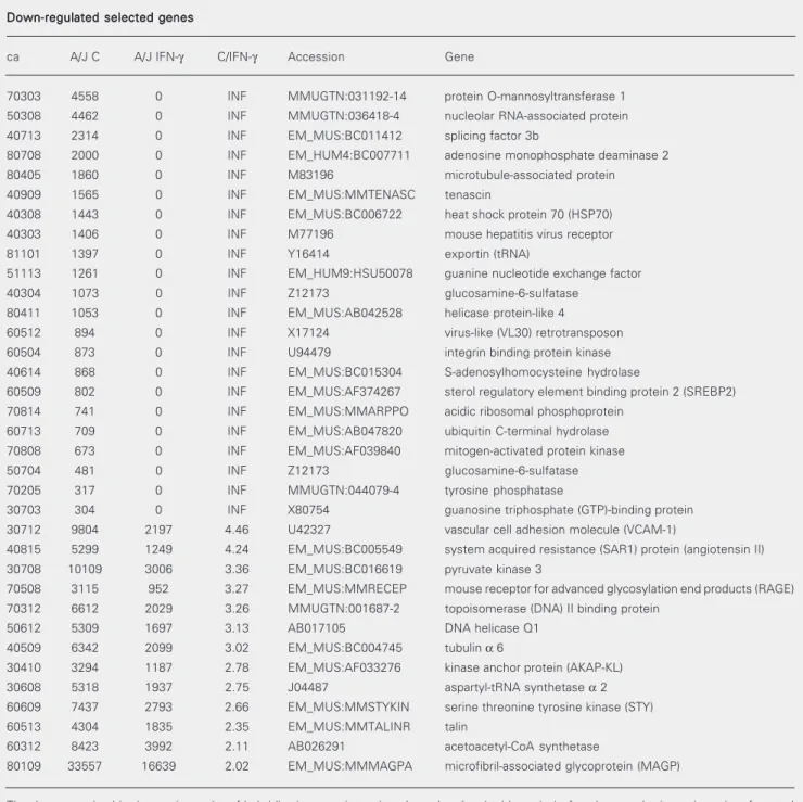
Down-regulated selected genes Down-regulated selected genes Down-regulated selected genes Down-regulated selected genes Down-regulated selected genes
ca A/J C A/J IFN-γ C/IFN-γ Accession Gene
70303 4558 0 INF MMUGTN:031192-14 protein O-mannosyltransferase 1
50308 4462 0 INF MMUGTN:036418-4 nucleolar RNA-associated protein
40713 2314 0 INF EM_MUS:BC011412 splicing factor 3b
80708 2000 0 INF EM_HUM4:BC007711 adenosine monophosphate deaminase 2
80405 1860 0 INF M83196 microtubule-associated protein
40909 1565 0 INF EM_MUS:MMTENASC tenascin
40308 1443 0 INF EM_MUS:BC006722 heat shock protein 70 (HSP70)
40303 1406 0 INF M77196 mouse hepatitis virus receptor
81101 1397 0 INF Y16414 exportin (tRNA)
51113 1261 0 INF EM_HUM9:HSU50078 guanine nucleotide exchange factor
40304 1073 0 INF Z12173 glucosamine-6-sulfatase
80411 1053 0 INF EM_MUS:AB042528 helicase protein-like 4
60512 894 0 INF X17124 virus-like (VL30) retrotransposon
60504 873 0 INF U94479 integrin binding protein kinase
40614 868 0 INF EM_MUS:BC015304 S-adenosylhomocysteine hydrolase
60509 802 0 INF EM_MUS:AF374267 sterol regulatory element binding protein 2 (SREBP2)
70814 741 0 INF EM_MUS:MMARPPO acidic ribosomal phosphoprotein
60713 709 0 INF EM_MUS:AB047820 ubiquitin C-terminal hydrolase
70808 673 0 INF EM_MUS:AF039840 mitogen-activated protein kinase
50704 481 0 INF Z12173 glucosamine-6-sulfatase
70205 317 0 INF MMUGTN:044079-4 tyrosine phosphatase
30703 304 0 INF X80754 guanosine triphosphate (GTP)-binding protein
30712 9804 2197 4.46 U42327 vascular cell adhesion molecule (VCAM-1)
40815 5299 1249 4.24 EM_MUS:BC005549 system acquired resistance (SAR1) protein (angiotensin II)
30708 10109 3006 3.36 EM_MUS:BC016619 pyruvate kinase 3
70508 3115 952 3.27 EM_MUS:MMRECEP mouse receptor for advanced glycosylation end products (RAGE)
70312 6612 2029 3.26 MMUGTN:001687-2 topoisomerase (DNA) II binding protein
50612 5309 1697 3.13 AB017105 DNA helicase Q1
40509 6342 2099 3.02 EM_MUS:BC004745 tubulin α 6
30410 3294 1187 2.78 EM_MUS:AF033276 kinase anchor protein (AKAP-KL)
30608 5318 1937 2.75 J04487 aspartyl-tRNA synthetase α 2
60609 7437 2793 2.66 EM_MUS:MMSTYKIN serine threonine tyrosine kinase (STY)
60513 4304 1835 2.35 EM_MUS:MMTALINR talin
60312 8423 3992 2.11 AB026291 acetoacetyl-CoA synthetase
80109 33557 16639 2.02 EM_MUS:MMMAGPA microfibril-associated glycoprotein (MAGP)
The data, organized in decreasing order of hybridization spot intensity, show the clonal address (ca) of each spot, the intensity units of control non-activated (A/J C) and IFN-γ activated (A/J IFN-γ) macrophage cultures, the respective intensity ratios (C/IFN-γ), the gene accession code
Table 3. Selected genes up- or down-regulated by IFN-γ in macrophages from BALB/c mice.
Up-regulated selected genes Up-regulated selected genes Up-regulated selected genes Up-regulated selected genes Up-regulated selected genes
ca BALB/c C BALB/c IFN-γ C/IFN-γ Accession Gene
71010 0 15880 0 EM_MUS:AF127033 fatty acid synthase
51213 0 12732 0 EM_RO:RNFIBRON fibronectin
60914 0 10402 0 MMUGTN:023998-30 high-glucose-regulated protein 8
60309 0 9452 0 MMUGTN:078718-20 dynactin 4
10312 0 8197 0 D16250 bone morphogenetic protein (BMP) receptor
30913 0 8067 0 EM_MUS:MMPGK1PS1 phosphoglycerate kinase (PGK1-ps1)
71005 0 6849 0 AF111102 major histocompatibility complex (MHC) class I region
10204 0 6600 0 AB021709 tumor necrosis factor (TNF) α converting enzyme
61111 0 6385 0 EM_MUS:BC003329 makorin, ring finger protein 1
80114 0 6322 0 EM_HUM1:AB071698 Np95-like ring finger protein
61014 0 6199 0 EM_MUS:AF210433 glucocorticoid modulatory element binding protein 1 (GMEB-1)
51012 0 5831 0 L07918 GDP-dissociation inhibitor
40509 0 5704 0 EM_MUS:BC004745 tubulin α 6
10411 0 5119 0 EM_MUS:AF322193 cleavage and polyadenylation specificity factor 1 (CPSF1)
40903 0 4969 0 M64085 spi2 proteinase inhibitor (spi2/eb1)
20502 0 4935 0 MMUGTN:033090-1 katanin p60
50405 0 4885 0 EM_MUS:AF230878 transcriptional co-repressor tif1 ß
50710 0 4688 0 EM_MUS:AF035117 rasgap-associated protein p56dok-2 (p56dok-2)
10602 0 4479 0 U50078 guanine nucleotide exchange factor p53
81008 0 4372 0 EM_MUS:MMCYCM cyclophilin
51115 0 4292 0 EM_MUS:MMCOF cofilin
51210 0 4242 0 MMUGTN:037962-2 exportin (nuclear export receptor for tRNAs)
20116 0 4110 0 EM_HUM8:HSLTGFBP4 latent transforming growth factor-ß binding protein-4
60712 778 6314 0.123 J05503 carbamoyl-phosphate synthetase
71014 1056 6742 0.157 EM_MUS:MM19604 DNA ligase I
40515 938 4892 0.192 EM_HUM3:AK056661 vacuolar protein sorting (VPS8)
10902 675 3281 0.206 MMUGTN:194525-2 serine protease inhibitor
60409 987 4319 0.229 AF006010 progestin-induced protein
10710 2156 9020 0.239 MMTFS3 mouse transcription factor s-ii
60909 6776 27783 0.244 MMU72634 rostral cerebellar malformation protein (RCM)
51010 975 3938 0.248 MMUGTN:031192-14 protein O-mannosyltransferase 1
20410 2721 9965 0.273 EM_MUS:MM65KDA 65-kDa macrophage cytosolic protein
10812 573 2092 0.274 MMUGTN:016783-15 phosphoglycerate mutase 1
40303 303 1046 0.290 M77196 mouse hepatitis virus (MHV) receptor
81211 1101 3716 0.296 EM_MUS:BC003861 hydroxymethylbilane synthase
20303 1750 5684 0.308 AF257711 proton-dependent high affinity oligopeptide transporter (PEPT2)
60609 1963 6091 0.322 MMSTYKIN serine threonine tyrosine kinase
21010 1657 5121 0.324 EM_MUS:MMPDIA disulfide isomerase (ERP59)
40104 1546 4398 0.352 U02082 guanine nucleotide regulatory protein
20906 2902 7782 0.373 AF098077 nuclear respiratory factor-1
10313 3984 10144 0.393 BC002270 ubiquitin-conjugating enzyme E2 variant 1
41215 3059 7781 0.393 EM_RO:RN10699 G-protein coupled receptor pH218
30410 1520 3514 0.433 EM_MUS:AF033276 kinase anchor protein (akap-kl)
70508 3199 7160 0.447 MMRECEP receptor for advanced glycosylation end products (RAGE)
10108 3663 8057 0.455 AF155373 nuclear factor κ B subunit
30116 3050 6355 0.48 EM_MUS:BC003887 uridine monophosphate synthetase
30901 8403 17471 0.481 U51167 isocitrate dehydrogenase
and metabolism such as platelet-activating
factor, gpi-anchored protein, ubiquitin
C-terminal hydrolase, vacuolar protein sorting
(VPS8) or in cytoskeleton arrangement and
extracellular matrix such as coronin 2,
entactin, VCAM1, tenascin, fibronectin or
receptor expression such as capping protein
α
2 subunit, growth factor receptor (Gab3),
eph-related receptor tyrosine kinase (MEK4),
mouse hepatitis virus receptor or
phagocyto-sis such as tubulin
α
6, talin, cofilin or in
resistance and susceptibility to infections
and tumor cytotoxicity such as pyruvate
ki-nase, heat shock protein 70, gpi-anchored
protein or in inflammation and cell
activa-tion and differentiaactiva-tion such as apoptosis
inhibitory protein, tyrosine phosphatase,
to-poisomerase II, bone morphogenic protein
receptor. For some genes, such as nuclear
pore targeting complex, splicing factor ß,
cyclophilin, fatty acid synthase,
hydroxy-methylbilane synthase, CPSF1, and 14-3-3
protein ß, the relationship with macrophage
biology is yet to be established. Some of
these investigated genes were found to be
up- or down-regulated only in A/J
macro-phages (10 up-regulated and 8
down-regu-lated), some others only in BALB/c
macro-phages (5 up-regulated and 4
down-regu-lated), some were up- or down-regulated in
cells from both strains (8 up-regulated and 2
down-regulated), one was up-regulated in
A/J macrophages and down-regulated in
BALB/c macrophages, and three were
down-Table 3 continued
Down-regulated selected genes Down-regulated selected genes Down-regulated selected genes Down-regulated selected genes Down-regulated selected genes
ca BALB/c C BALB/c IFN-γ C/IFN-γ Accession Gene
21214 19919 0 INF MMUGTN:003842-11 nicotinamide nucleotide transhydrogenase
50313 5886 0 INF MMUGTN:168786-1 sperm associated antigen
71205 5433 0 INF AF111102 major histocompatibility complex (MHC) class I region
60305 2290 0 INF Y13620 B cell CLL/lymphoma 9 (BCL9)
50308 2262 0 INF MMUGTN:036418-4 nucleolar RNA-associated protein
80203 1752 0 INF M19141 heat shock protein 70 (HSP70)
80714 1555 0 INF EM_MUS:BC005770 beclin 1 (coiled-coil, myosin-like BCL2-interacting protein)
20714 1187 0 INF EM_MUS:BC007483 growth factor receptor bound protein 2-associated protein 1
50604 1184 0 INF Y13622 latent transforming growth factor ß binding protein-4
60804 1141 0 INF NM_013843 zinc finger protein
80611 1138 0 INF EM_MUS:AY013811 protocadherin γ c3
80815 1007 0 INF EM_MUS:MMALPA α catenin
10316 927 0 INF EM_MUS:MM05809 LAF1 transketolase
21203 757 0 IFN AF058797 14-3-3 protein ß
30615 609 0 IFN EM_MUS:MM36220 FK506 binding protein
50613 497 0 INF EM_MUS:BC003300 ATP-binding cassette, sub-family F (GCN20)
81203 496 0 INF EM_MUS:BC007158 procollagen, type I, α 2
80102 444 0 INF NM_011602 talin
50514 334 0 INF EM_MUS:MMMEK4 eph-related receptor tyrosine kinase (MEK4)
80204 17748 8018 2.21 U28322 Krueppel-type zinc finger protein
70308 3839 1775 2.16 NM_031397 bicaudal C homolog 1
30912 8823 4388 2.01 AF073879 myotubularin homologous protein 1
The data, organized in decreasing order of hybridization spot intensity, show the clonal address (ca) of each spot, the intensity units of control non-activated (BALB/c C) and IFN-γ-activated (BALB/c IFN-γ) macrophage cultures, the respective intensity ratios (C/IFN-γ), the gene accession
010511 coronin 2 A/J
*
*
14010913 nuclear pore targeting complex A/J
*
021003 entactin A/J
*
15031216 RNA polymerase II 140-kDa subunit A/J
*
16051102 glyceraldehyde-3P-dehydrogenase A/J
*
*
17060214 apoptosis inhibitory protein A/J
*
*
18060811 ß-catenin A/J
*
19061202 PAF-AH A/J
*
*
20071215 capping protein α 2 subunit A/J
*
*
*
*
21080111 gpi-anchored protein A/J
*
*
*
22030708 pyruvate kinase A/J
*
*
*
23030712 VCAM1 A/J
*
*
24,25040713 splicing factor 3b A/J
*
040909 tenascin A/J
*
*
26060312 acetoacetyl-CoA-synthetase A/J
*
*
27060713 ubiquitin C-t hydrolase A/J
*
*
28070205 tyrosine phosphatase A/J
*
*
*
29,30070312 topoisomerase II binding protein A/J
*
*
31080708 adenosine-MP deaminase A/J
*
*
32020410 65-kDa macrophage cytosolic protein BALB/c
*
*
33,34040104 guanine nucleotide regulatory protein BALB/c
*
35040515 vacuolar protein sorting (VPS8) BALB/c
*
36081008 cyclophilin BALB/c
*
051213 fibronectin BALB/c
*
*
*
*
37020714 growth factor receptor (Gab3) BALB/c
*
*
38021203 14-3-3 protein ß BALB/c
*
030912 myotubularin homologous protein 1 BALB/c
*
*
39050514 MEK4 BALB/c
*
*
010312 BMP receptor A/J BALB/c
*
*
*
40010902 serine protease inhibitor A/J BALB/c
*
*
41,42011016 TNF-α converting enzyme A/J BALB/c
*
*
*
*
43041216 CPSF1 A/J BALB/c
*
051115 cofilin A/J BALB/c
*
*
*
44071010 fatty acid synthase A/J BALB/c
*
071014 DNA ligase 1 A/J BALB/c
*
*
*
45081211 hydroxymethylbilane synthase A/J BALB/c
*
040308 heat shock protein 70 A/J BALB/c
*
*
*
46,47060513 talin A/J BALB/c
*
*
48,49060804 zinc finger protein A/J BALB/c
*
*
*
50,51040303 mouse hepatitis virus receptor A/J BALB/c
*
*
7040509 tubulin α 6 A/J BALB/c
*
*
*
*
52,53070508 RAGE A/J BALB/c
*
*
54,55Inflammation/Differentiation/Cell activation References
Expression*
ca Gene
To be established
Phagocytosis
Enzymatic mediation Nucleic acid synthesis
/Transport
Protein synthesis
/Transport/Metabolism
Cytoskeleton arrangement/Extracellular matrix Receptor expression Resistance/Susceptibility to infection
/Tumor
Table 4. List of 42 differentially expressed genes in IFN-γ-activated A/J and/or BALB/c mouse macrophages and their relationship to key functions in macrophage biology as reported in recent publications.
Gene expression modulated (triangle = increase; inverted triangle = decrease) by IFN-γ activation in A/J and BALB/c mouse macrophages. ca = clonal address;
regulated in A/J macrophages and
up-regu-lated in BALB/c macrophages.
In Figure 1 we present a selected area of
the hybridization filter showing gene
expres-sion profiles of control and IFN-
γ
-activated
macrophages from A/J and BALB/c mice.
The area was chosen so that a pair of
hybrid-ization spots defining a given clonal address
(in this particular case 040303) could be
highlighted. The clonal address corresponds
to the MHV receptor gene. It can be seen that
some dots are larger than others. A
quantita-tive representation of its average relaquantita-tive
intensity is provided in the histogram,
indi-cating that, upon IFN-
γ
activation, A/J mouse
macrophages do not express the MHV
re-ceptor gene, whereas BALB/c mouse
mac-rophages do express this gene. These data
correlate with and confirm our previous
ob-servations made in studies based on
biologi-cal assays (Table 1).
The reproducibility of the results was
adequate for high expressions and less
satis-factory for weak ones. Standard deviations
are not provided because the validity scoring
was based not only on measured values but
also on comparison of subsequent
hybrid-ization upon stripping (other samples and
even other projects). As a result, the scores
for some spots were more reliable than
oth-ers.
Discussion
The experiments planned and executed
in this project are meant to be a continuation
of our earlier efforts, in which, using a
prote-omic approach, we attempted to identify
gene products involved in the regulation of
IFN-
γ
activation of macrophages derived
from mice resistant and susceptible to
exper-imental infection with MHV3 (8). We have
acquired knowledge on nucleic acids and
protein functions while developing
prote-omic/genomic approaches that allow the
iden-tification of the genetic inheritance and the
modulation of gene expression (9-11). Since
there is no definite time point characterizing
gene expression, we have chosen 18 h of
IFN-
γ
macrophage activation since
prote-omic and biological studies are usually done
at this time.
Samples of cDNA preparations from A/J
and BALB/c mice (activated or not with
IFN-
γ
) were hybridized with arrayed cDNA
clones on nylon sheets. Each nylon sheet
040303
A/J A/J + IFN-γ
BALB/c + IFN-γ
BALB/c
BALB/c A/J
IFN-γ
100
75
50
25
0
040303 average relative intensity
+ +
A
B
Figure 1. Selected areas of hy-bridization patterns generated from probes of control and
IFN-γ-activated macrophages from
contained 1536 cDNA clones of fetal
thy-mus origin in duplicate, as well as several
control dots of cytochrome c origin. A
chemi-luminescence readout followed by image
analysis yielded data which indicated the
relative intensity of hybridized entities.
As shown in Table 1, based on the
obser-vation that A/J mice were resistant and
BALB/c mice were susceptible to
experi-mental infection with MHV3, classical
bio-logical assays for the study of the cellular
and molecular basis of resistance of mice to
MHV3 led us to hypothesize and
experimen-tally demonstrate that IFN-
γ
activation could
partially restrict viral multiplication only in
the resistant A/J macrophages and that the
molecular basis of this restriction relied on
the IFN-
γ
induced down-regulation of the
main viral receptor (7). Taken together,
proteomic experiments and gene expression
profiling provided us not only with a
confir-mation of these predictions but with the
possibility of a much deeper comprehension
of the biology of IFN-
γ
activation of
macro-phages. We have demonstrated a panel of
up- and down-regulated genes as well as
identified their relationships to key
func-tions in macrophages.
By expression profiling, we have
evalu-ated the degree of up- or down-regulation of
several genes in macrophages from A/J and
BALB/c mice upon activation by IFN-
γ
. Since
this technology allows us to identify the
expression of genes that are modified upon
activation by IFN-
γ
, we can further examine
the biological role of the gene products and
categorize the possible modulation of
bio-logical functions induced by IFN-
γ
activa-tion in macrophages from both mouse strains.
We have on hand, in terms of hybridization
spots, several identified molecular entities
of the cDNA library, and our
semi-quantita-tive data show the overall influence of
IFN-γ
activation on macrophage gene expression.
Contrary to the proteomic approach, in which
every polypeptide species (if present in
ad-equate amounts, if within the resolution
lim-its of the separation, and if it contains
methi-onine in the amino acid sequence) present in
the 2-D SDS gel matrix shows up on the
radiofluorogram, the hybridization readout
is constrained to the portion of cDNA
mol-ecules present in the library. Among the
1536 clonal entities, there are about one
thousand different molecular clones,
encom-passing about 470 low abundance ones. Since
all of these clones are transcribable and
trans-latable the number is satisfactory for
ad-equate analysis. It represents about 20% of
the messages present in a typical cell (the
lymphocyte being the model) (56). Note that
a cell has altogether about 40,000 mRNA
molecules, some present in a relatively large
copy number, others with only 3-5 copies
per cell. There are approximately 5,000
dif-ferent mRNA molecules in a cell (56). Our
experiments revealed only those up- and
down-regulated which have members
pres-ent among the 1536 pres-entities tested.
In Figure 1 we show the hybridization
patterns of a selected area of the gene
ex-pression array obtained from control and
IFN-
γ
-activated A/J and BALB/c
macro-phages, with the duplicated gene spots
cor-responding to the main MHV receptor (clonal
address 040303) highlighted, as well as its
histograms of average relative intensity. One
can observe that, as predicted by classical
assays performed in the past (7), there is a
down-regulation of this receptor gene only
in IFN-
γ
-activated macrophages from
resis-tant A/J mice. Also, as previously shown (7),
the receptor gene expression in IFN-
γ
-acti-vated BALB/c macrophages was high,
al-though its basal expression in control
macro-phages was found to be not always
reproduc-ible, possibly due to variations in the
physi-ological state of the cells.
bio-logical functions induced by IFN-
γ
activa-tion in macrophages. As it is preliminarily
shown in Table 4, we could identify genes
coding for proteins participating in processes
of macrophage biology like enzymatic
reac-tions, nucleic acid synthesis and transport,
protein synthesis, transport and metabolism,
cytoskeleton arrangement and extracellular
matrix, receptor expression, phagocytosis,
resistance/susceptibility to infection or
tu-mors and inflammation or cell
differentia-tion. The data indicate that the overall gene
regulation by IFN-
γ
can be quite different in
macrophages originating from mice with
dif-ferent genetic backgrounds and this panel of
IFN-
γ
-regulated genes may serve as a
start-ing point for general and specific studies of
macrophage biology.
This paper reveals a large assembly of
genes differentially expressed in
macro-phages of two murine genetic backgrounds
(A/J and BALB/c) upon IFN-
γ
activation.
These data will turn out to be very useful for
general studies of macrophage biology and
can be an alternative strategy to confirm
hypothetical as well as already defined
fea-tures of macrophages, such as that of MHV
receptor gene modulation upon IFN-
γ
acti-vation.
Acknowledgments
The Basel Institute of Immunology was
founded and supported by F. Hoffmann-La
Roche and Co. Ltd., Basel, Switzerland.
References
1. Lucchiari MA, Martin JP, Modolell M & Pereira CA (1991). Acquired immunity of A/J mice to mouse hepatitis virus 3 infection. Depend-ence on interferon gamma synthesis and macrophage sensitivity to interferon gamma. Journal of General Virology, 72: 1317-1322. 2. Arnheiter H, Baechi T & Haller O (1982). Adult mouse hepatocytes
in primary monolayer culture express genetic resistance to mouse hepatitis virus type 3. Journal of Immunology, 129: 1275-1281. 3. Pereira CA, Steffan AM & Kirn A (1984). Interaction between mouse
hepatitis viruses and primary cultures of Kupffer and endothelial liver cells from resistant and susceptible inbred mouse strains. Journal of General Virology, 65: 1617-1620.
4. Lamontagne L, Descoteaux JP & Jolicoeur P (1989). Mouse hepati-tis virus 3 replication in T and B lymphocytes correlate with viral pathogenicity. Journal of Immunology, 142: 4458-4465.
5. Dindzans VJ, Skamene E & Levy GA (1986). Susceptibility/resis-tance to mouse hepatitis virus strain 3 and macrophage procoagu-lant activity are genetically linked and controlled by two-non-H-2 linked genes. Journal of Immunology, 137: 2355-2360.
6. Fingerote RJ, Abecassis M, Phillips MJ, Rao YS, Cole EH, Leibowitz J & Levy GA (1996). Loss of resistance to MHV3 infection after treatment with corticosteroid is associated with induction of macro-phage PCA. Journal of Virology, 70: 4275-4282.
7. Vassão RC, De Franco MT, Hartz D, Modolell M, Sippel AE & Pereira CA (2000). Down-regulation of Bgp1a viral receptor by interferon
γ is
related to the antiviral state and resistance to mouse hepatitis virus 3 infection. Virology, 274: 278-283.
8. Pereira CA, Lucchiari MA, Modolell M, Kuhn L & Lefkovits I (1993). An attempt to identify gene products related to the induction of an antiviral state in macrophages resistant and sensitive to IFN-gamma. Research in Virology, 144: 479-486.
9. Lefkovits I, Kettman JR & Frey JR (2001). Global analysis of gene expression in cells of the immune system. I. Analytical limitations in
obtaining sequence information on polypeptides in two-dimensional gel spots. Electrophoresis, 21: 2688-2693.
10. Frey JR, Nguyen C, Houlgatte R et al. (2001). Global analysis of gene expression in cells of the immune system. II. Cell free transla-tion products and high-density filter hybridizatransla-tion data. Electropho-resis, 21: 2694-2702.
11. Lefkovits I, Kettman JR & Frey JR (2001). Proteomic analysis of rare molecular species of translated polypeptides from a mouse fetal thymus cDNA library. Proteomics, 1: 560-573.
12. Munder PG, Modolell M & Wallach DFH (1971). Cell propagation on films of polymeric fluorocarbon as a mean to regulate pericellular pH and pO2 in cultured monolayers. FEBS Letters, 15: 191-196.
13. Pereira CA, Mercier G, Oth D & Dupuy JM (1984). Induction of natural killer cells and interferon during mouse hepatitis virus infec-tion of resistant and susceptible inbred mouse strains. Immunobiol-ogy, 166: 35-42.
14. Schuller S, Neefjes J, Ottenhoff T, Thole J & Young D (2001). Coronin is involved in uptake of Mycobacterium bovis BCG in hu-man macrophages but not in phagosome maintenance. Cell Micro-biology, 3: 785-793.
15. Gronski Jr TJ, Martin RL, Kobayashi DK, Walsh BC, Holman MC, Huber M, Van Wart HE & Shapiro SD (1997). Hydrolysis of a broad spectrum of extracellular matrix proteins by human macrophage elastase. Journal of Biological Chemistry, 272: 12189-12194. 16. Ishida T, Matsuura K, Setoguchi M, Higuchi Y & Yamamoto S
(1994). Enhancement of murine serum amyloid A3 mRNA expres-sion by glucocorticoids and its regulation by cytokines. Journal of Leukocyte Biology, 56: 797-806.
18. Horie T, Dobashi K, Iizuka K, Yoshii A, Shimizu Y, Nakazawa T & Mori M (1999). Interferon-gamma rescue TNF-alpha-induced apop-tosis mediated by up-regulation of TNFR2 on EoL-1 cells. Experi-mental Hematology, 27: 512-519.
19. Lapteva N, Ando Y, Nieda M, Hohjoh H, Okai M, Kikuchi A, Dymshits G, Ishikawa Y, Juji T & Tokunaga K (2001). Profiling of genes expressed in human monocytes and monocyte-derived dendritic cells using cDNA expression array. British Journal of Haematology, 114: 191-197.
20. Tselepis AD, Karabina SA, Stengel D, Piedagnel R, Chapman MJ & Ninio E (2001). N-linked glycosylation of macrophage-derived PAF-AH is a major determinant of enzyme association with plasma HDL. Journal of Lipid Research, 42: 1645-1654.
21. Witke W, Li W, Kwiatkowski DJ & Southwick FS (2001). Compari-sons of CapG and gelsolin-null macrophages: demonstration of a unique role for CapG in receptor-mediated ruffling, phagocytosis, and vesicle rocketing. Journal of Cell Biology, 154: 775-784. 22. Coelho PS, Klein A, Talvani A, Coutinho SF, Takeuchi O, Akira S,
Silva JS, Canizzaro H, Gazzinelli RT & Teixeira MM (2002). Glycosyl-phosphatidyl inositol-anchored mucin like glycoproteins isolated from Trypanosoma cruzi trypomastigotes induce in vivo leukocyte recruitment dependent on MCP-1 production by IFN-gamma-primed macrophages. Journal of Leukocyte Biology, 71: 837-844. 23. Duncan JR, Potter CB, Cappellini MD, Kurtz JB, Anderson MJ &
Weatherall DJ (1983). Aplastic crisis due to parvovirus infection in pyruvate kinase deficiency. Lancet, 2: 14-16.
24. Nansen A, Christensen JP, Ropke C, Marker O, Scheynius A & Thomsen AR (1998). Role of interferon-gamma in the pathogenesis of LCMV-induced meningitis: unimpaired leukocyte recruitment, but deficient macrophage activation in interferon-gamma knock-out mice. Journal of Neuroimmunology, 86: 202-212.
25. Peng HB, Spiedcker M & Liao JK (1998). Inducible nitric oxide: An autoregulatory feedback inhibitor of vascular inflammation. Journal of Immunology, 161: 1970-1976.
26. Harkonen E, Virtanen I, Linnala A, Laitinen LL & Kinnula VL (1995). Modulation of fibronectin and tenascin production in human bron-chial epithelial cells by inflammatory cytokines in vitro. American Journal of Respiratory Cell and Molecular Biology, 13: 109-115. 27. Bergstrom JD, Wong GA, Edwards PA & Edmond J (1984). The
regulation of acetoacetyl-CoA synthetase activity by modulators of cholesterol synthesis in vivo and utilization of acetoacetate for cholesterogenesis. Journal of Biological Chemistry, 259: 14548-14553.
28. Glockzin S, von Knethen A, Scheffner M & Brune B (1999). Activa-tion of the cell death program by nitric oxide involves inhibiActiva-tion of the proteasome. Journal of Biological Chemistry, 274: 19581-19586. 29. Xaus J, Comalada M, Valledor AF, Cardo M, Herrero C, Soler C, Lloberas J & Celada A (2001). Molecular mechanisms involved in macrophage survival, proliferation, activation and apoptosis. Immu-nobiology, 204: 543-550.
30. Simoncic PD, Lee-Loy A, Barber DL, Tremblay ML & McGlade CJ (2002). The T cell protein tyrosine phosphatase is a negative regula-tor of Janus family kinases 1 and 3. Current Biology, 12: 446-453. 31. Chiou WF, Chou CJ & Chenm CF (2001). Camptothecin suppresses
nitric oxide biosynthesis in RAW 264.7 macrophages. Life Sci-ences, 69: 625-635.
32. Nikolajeva V, Eze D, Kamradze A, Indulena M & Muiznieks I (1996). Protective effect of adenylate deaminase (from Penicillium lanosoviride) against acute infections in mice. Immunopharmacolo-gy, 35: 163-169.
33. Shinomiya H, Hagi A, Fukuzumi M, Mizobuchi M, Hirata H & Utsumi
S (1995). Complete primary structure and phosphorylation site of the 65-kDa macrophage protein phosphorylated by stimulation with bacterial lipopolysaccharide. Journal of Immunology, 154: 3471-3478.
34. Kikuchi H, Fujinawa T, Kuribayashi F, Nakanishi A, Imajoh-Ohmi S, Goto M & Kanegasaki S (1994). Induction of essential components of the superoxide generating system in human monoblastic leuke-mia U937 cells. Journal of Biochemistry, 116: 742-746.
35. Vestal DJ, Buss JE, McKercher SR, Jenkins NA, Copeland NG, Kelner GS, Asundi VK & Maki RA (1998). Murine GBP-2: a new IFN-gamma-induced member of the GBP family of GTPases isolated from macrophages. Journal of Interferon and Cytokine Research, 18: 977-985.
36. Luo W & Chang A (2000). An endosome-to-plasma membrane pathway involved in trafficking of a mutant plasma membrane ATPase in yeast. Molecular Biology of the Cell, 11: 579-592. 37. Jun CD, Yoon HJ, Kim HM & Chung HT (1995). Fibronectin activates
murine peritoneal macrophages for tumor cell destruction in the presence of IFN-gamma. Biochemical and Biophysical Research Communications, 206: 969-974.
38. Wolf I, Jenkins BJ, Liu Y, Seiffert M, Custodio JM, Young P & Rohrschneider LR (2002). Gab3, a new DOS/Gab family member, facilitates macrophage differentiation. Molecular and Cellular Biol-ogy, 22: 231-244.
39. Nandurkar HH & Huysmans R (2002). The myotubularin family: novel phosphoinositide regulators. International Union of Biochem-istry and Molecular Biology Life, 53: 37-43.
40. Gould SE, Day M, Jones SS & Dorai H (2002). BMP-7 regulates chemokine, cytokine, and hemodynamic gene expression in proxi-mal tubule cells. Kidney International, 61: 51-60.
41. Hamerman JA, Hayashi F, Schroeder LA, Gygi SP, Haas AL, Hampson L, Coughlin P, Aebersold R & Aderem A (2002). Serpin 2a is induced in activated macrophages and conjugates to a ubiquitin homolog. Journal of Immunology, 168: 2415-2423.
42. Kwak JY, Park SY, Han MK, Lee HS, Sohn MH, Kim UH, McGregor JR, Samlowski WE & Yim CY (1998). Receptor-mediated activation of murine peritoneal macrophages by antithrombin III acts as a costimulatory signal for nitric oxide synthesis. Cellular Immunology, 188: 33-40.
43. Rovida E, Paccagnini A, Del Rosso M, Peschon J & Dello Sbarba P (2001). TNF-alpha-converting enzyme cleaves the macrophage colony-stimulating factor receptor in macrophages undergoing acti-vation. Journal of Immunology, 166: 1583-1589.
44. Matsui S, Matsumoto S, Adachi R et al. (2002). LIM kinase 1 modulates opsonized zymosan-triggered activation of macrophage-like U937 cells. Possible involvement of phosphorylation of cofilin and reorganization of actin cytoskeleton. Journal of Biological Chem-istry, 277: 544-549.
45. Khan Z & Francis GE (1987). Contrasting patterns of DNA strand breakage and ADP-ribosylation-dependent DNA ligation during granulocyte and monocyte differentiation. Blood, 69: 1114-1119. 46. Breoler M, Dorner B, More SH, Roderian T, Fleischer B & von Bonin
A (2001). Heat shock proteins as “danger signals”: eukaryotic Hsp60 enhances and accelerates antigen-specific IFN-gamma pro-duction in T cells. European Journal of Immunology, 31: 2051-2059. 47. Panjwani NN, Popova L & Srivastava PK (2002). Heath shock pro-teins gp96 and hsp70 activate the release of nitric oxide by APCs. Journal of Immunology, 168: 2997-3003.
49. Greenberg S, Burridge K & Silverstein SC (1990). Colocalization of F-actin and talin during Fc receptor-mediated phagocytosis in mouse macrophages. Journal of Experimental Medicine, 172: 1853-1856. 50. Heyninck K, De Valck D, Vanden Berghe W, Van Criekinge W,
Contreras R, Fiers W, Haegeman G & Beyaert R (1999). The zinc finger protein A20 inhibits TNF-induced NF-kappa B-dependent gene expression by interfering with an RIP- or TRAF2-mediated transacti-vation signal and directly binds to a novel NF-kappa B-inhibiting protein ABIN. Journal of Cell Biology, 145: 1471-1482.
51. Shin JN, Kim I, Lee JS, Koh GY, Lee ZH & Kim HH (2002). A novel zinc finger protein that inhibits osteoclastogenesis and the function of tumor necrosis factor-associated factor 6. Journal of Biological Chemistry, 277: 8346-8353.
52. Siffert JC, Baldacini O, Kuhry JG, Wachsmann D, Benabdelmou-mene S, Faradji A, Monteil H & Poindron P (1993). Effects of Clostridium difficile toxin B on human monocytes and macrophages: possible relationship with cytoskeletal rearrangement. Infection and
Immunity, 61: 1082-1090.
53. Rammes A, Roth J, Goebeler M, Klempt M, Hartmann M & Sorg C (1997). Myeloid-related protein (MRP) 8 and MRP14, calcium-bind-ing proteins of the S100 family, are secreted by activated mono-cytes via a novel, tubulin-dependent pathway. Journal of Biological Chemistry, 272: 9496-9502.
54. Schmidt AM, Hori O, Cao R, Yan SD, Brett J, Wautier JL, Ogawa S, Kuwabara K, Matsumoto M & Stern D (1996). RAGE: a novel cellular receptor for advanced glycation end products. Diabetes, 45: S77-S80.
55. Ohgami N, Nagai R, Ikemoto M, Arai H, Kuniyasu A, Horiuchi S & Nakayama H (2001). CD36, a member of the class b scavenger receptor family, as a receptor for advanced glycation end products. Journal of Biological Chemistry, 276: 3195-3202.