Received on 15 May 2008; revised 10 September 2008.
Address for correspondence: Dr. Lessandra Michelim, Laboratório de Biotecnologia Vegetal e Microbiologia Aplicada. Instituto de Biotecnologia da Universidade de Caxias do Sul. R. Francisco G. Vargas 1130, Petrópolis. Zip code: 95001-970. Caxias do Sul, RS, Brazil. Phone: (54) 32146750. Fax: (54) 32187237. Cellphone: (54) 991229469. E-mail: lessandra@gmail.com.
The Brazilian Journal of Infectious Diseases 2008;12(5):423-429. © 2008 by The Brazilian Journal of Infectious Diseases and Contexto Publishing. All rights reserved.
Comparison of PCR-Based Molecular Markers for the Characterization of
Proteus mirabilis
Clinical Isolates
Lessandra Michelim, Gabriela Muller, Jucimar Zacaria, Ana Paula Longaray Delamare, Sérgio Olavo Pinto da Costa and Sergio Echeverrigaray
Institute of Biotechnology, University of Caxias do Sul; Caxias do Sul, RS, Brazil
Proteus mirabilis is one of the most important pathogens associated with complicated urinary tract infections (acute pyelonephritis, bladder infections, kidney stones) and bacteremia, affecting patients with anatomical abnormalities, immunodeficiency, and long-term urinary catheterization. For epidemiological purposes, various molecular typing methods, such as pulse-field gel electrophoresis (PFGE) or ribotyping, have been developed for this pathogen. However, these methods are labor intensive and time-consuming. We evaluated the discriminatory power of several PCR-based fingerprinting methods (RAPD, ISSR, ERIC-PCR, BOX-PCR and rep-PCR) for P. mirabilis clinical isolates. Typing patterns and clustering analysis indicated that RAPD, BOX-PCR and ERIC-PCR differentiated P. mirabilis strains from Escherichia coli, Hafnia alvei, and Morganella morganii. With the exception of rep-PCR, the methods gave medium to high discriminatory efficiency in P. mirabilis. In general, the results obtained with RAPD, BOX-PCR and ERIC-PCR were in good agreement. We concluded that a combination of ERIC-PCR and BOX-PCR results is a rapid and reliable alternative for discrimination among P. mirabilis clinical isolates, contributing to epidemiological studies.
Key-Words: Proteus mirabilis, molecular markers, fingerprinting, PCR.
Proteus mirabilis (Enterobacteriaceae) is a Gram-negative rod-shaped bacterium, frequently found in soil, water and the intestinal tract of many animals, including humans. This dimorphic bacterium can undergo morphological and physiological changes in response to environmental and growth conditions. These modifications lead to its most peculiar characteristic, swarming behavior, a process in which short vegetative swimming cells differentiate to long, highly flagellated forms referred to as swarmer cells [1].
Proteus mirabilis is not a common cause of urinary tract infections in normal hosts, occasionally involved in uncomplicated cystitis or pyelonephritis. However, it is one of the most important pathogens associated with complicated urinary tract infections (acute pyelonephritis, bladder infections, and kidney stones) and bacteremia, affecting patients with anatomical abnormalities, immunodeficiency, and long-term urinary catheterization [1-3]. Proteus mirabilis virulence is associated with several virulence factors, including hemolysin, swarming, adhesins, proteases, and ureases [4-6]. Expression of most of these factors is coordinately upregulated during swarming [6-8].
Because of the increasing clinical relevance of P. mirabilis [3], the selection of efficient molecular fingerprinting methods is of great epidemiological importance. Bacterial genotyping opened new opportunities for epidemiological studies, allowing the identification of clinical and environmental
isolates, evaluation of their relationships, monitoring of clone dissemination, and characterization of bacterial populations within more or less restricted environments [9]. Among PCR-based molecular markers, RAPD (random amplified polymorphic DNA), and repetitive sequence-based PCR genomic fingerprinting have been found to be particularly efficient for bacterial analysis [9-13]. Repeated sequences ERIC (enterobacterial repetitive intergenic consensus sequence), REP (repetitive extragenic palindromic sequence), and BOX (repetitive intergenic sequence elements of Streptococcus) have been specifically designed for prokaryotic fingerprinting.
Ribotyping and PFGE (pulsed-field gel electrophoresis) are efficient for Proteus characterization at the species level [14] and for identification of individual strains of P. mirabilis [15,16]. However, these methods are laborious, expensive, and time consuming, limiting their application in routine clinical laboratories [9]. RAPD, a PCR-based method, has been used with success in the identification of clinical isolates of P. mirabilis [17] and P. penneri [18]. More recently, the tandem tetramer microsatellites (GACA)4 and (CAAT)4, also known as intergenic single sequence repeats (ISSR), have given a high degree of discrimination for P. mirabilis [19].
We evaluated and compared the efficiency of five PCR-based molecular markers for the characterization of P. mirabilis clinical isolates, in order to select informative markers for epidemiological studies, and to monitor P. mirabilis populations within hospital environments.
Material and Methods
Bacterial Isolates
by routine procedures for the different sample sources, and were identified by conventional microscopic and biochemical tests: Gram staining, motility, swarming behavior, indole production, phenylalanine dehydrogenase, ornithine decarboxylase, gas production from glucose, H2S production, urease, tryptophan deaminase, lysine decarboxylase, and citrate and lactose utilization. Antibiotic resistance was evaluated by the disc-diffusion method and analyzed as described by CLSI document M100-S17 [20].
Bacterial isolates were maintained on trypticase soybean agar (TSA), and permanent stocks were conserved on TSBG (tryptone soy broth with 15% glycerol) at –80ºC. For DNA analysis, single colonies were transferred to 1ml of LB (Luria Broth) and incubated at 37ºC for 18 h.
PCR Fingerprinting
DNA samples were prepared as described by Lu [4], with some modifications. Briefly, single colonies of each isolate were inoculated on LB medium and grown overnight at 37°C. Cells were collected by centrifugation at 13,000 x g for 5 min, and ressuspended in 100mL of extraction buffer (1% Triton-X-100, 100mM Tris-HCl pH 8.3, and 1mM EDTA). The samples were incubated for 20 min in a boiling water bath (100°C) and centrifuged at 13,000 xg for 5 min. The supernatant was transferred to a new tube containing 180mL of ultrapure Milli-Q (Millipore) water. Samples were aliquoted and conserved at -80°C.
For RAPD, ERIC, BOX and REP analysis, 2 mL of the DNA samples were transferred to 23 mL of amplification mix containing: 20mM Tris-HCl pH 8.4, 50mM KCl, 7mM MgCl2, 0.25% Triton-X-100, 8mM dNTPs, 1 mM of each primer (ERIC and REP) or 1.5 mM of the primer for RAPD and BOX, and 1.25U of Taq Polymerase (Invitrogen). DNA amplification was conducted on a MJ Research thermocycler programmed for an initial denaturation step at 92ºC (4 min), followed by 40 cycles of denaturation for 1 min at 94ºC, annealing for 1 min at the appropriate temperature (RAPD and REP- 40ºC, ERIC- 48ºC, and BOX- 50ºC), extension for 5 min at 72ºC, and a final extension for 5 min at 72ºC. Samples were maintained at 4ºC until electrophoretic separation of amplification products.
The primers used were: ERIC-1R ATGTAAGCTCCTGGGGATTCAC-3’) and ERIC-2 AAGTAAGTGACTGGGGTGAGCG-3’), and REP-PCR-1R IIIICGICGICATCIGGC-3’), and REP-PCR-2I (5’-ICGICTTATCIGGCCTAC-3’), described by Versalovic et al. [21], BOX-A1R (5’-CTACGGCAAGGCGACGCTGACG-3’) previously used for several bacterial species [11,12,22], and RAPD and ISSR (Table 2).
The amplification reaction for ISSR markers included 2 mL of DNA samples and 23 mL of a PCR mix, including 20mM Tris-HCl pH 8.4, 50mM KCl, 3mM MgCl2, 2% formamide, 0.75mM of each dNTP, 1 mM of each primer and 1.5U of Taq Polymerase (Invitrogen). For the DNA amplification, the reaction mixture was denatured for 5 min at 92ºC, followed by 40 cycles at 94ºC (1 min), 48ºC to 50°C (45s) and 72ºC (2 min), with a final extension for 5 min at 72ºC.
The amplification products were electrophoresed in 1.5% agarose gels in Tris-borate buffer (0.089M Tris, 0.089M boric acid, 0.002M EDTA). Lambda EcoRI/HindIII was used as a molecular size standard. The gels were stained with ethidium bromide (10mg/mL), visualized on a UV light transilluminator, and documented with the UVITEC system. Image analyses were carried out using Labimage software.
The amplifications with the five methods were repeated three times (independent cultures and DNA extractions) to evaluate reproducibility, with two replications of each isolate per round. Only well defined and reproducible amplification products (presence and intensity) were scored and used in statistical analyses.
Statistical Analysis
Similarity Jaccard’s coefficients, Pearson’s correlation between distance matrices, and cluster analysis (unweighted pair-group method with average linkage - UPGMA) were performed using the SSCP 10.1 software package. Bootstrap analysis was done with the Winboot program. The discriminatory index (DI) was calculated from the relative frequencies of the different profiles obtained by a given primer or method, and was calculated using Simpson’s diversity index [23], as follows:
Where N is the total number of isolates and n
jis the number
of isolates belonging to the jth type.
Results
RAPD Typing
Initially, a set of 10 decameric RAPD primers (Table 2) were selected from the 60 primers of kits A, X and Z of Operon Techn., based on the number, quality and polymorphisms of amplification products, using three arbitrarily-chosen P. mirabilis isolates (IBPro 101, IBPro102 and IBPro120). Applied to all the isolates, these primers generated 188 amplification products, varying between 300 and 2,445 bp. Considering just the 29 Proteus isolates, 86 bands were identified, of which 51 (59%) exhibited some degree of polymorphism. Each decameric primer amplified from 3 to 14 segments, of which 25 to 80% were polymorphic.
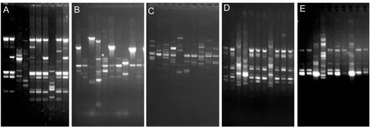
Thirty-five Proteus-specific amplification products were identified that can be used to design Proteus-specific SCAR primers (Table 2). An example of RAPD profiles showing three Proteus-specific bands of 2113bp, 831bp and 431bp, and several polymorphic bands, is shown in Figure 1. Considering all the amplification products, RAPD markers allowed the discrimination of almost all the isolates, except for three isolates obtained from patient 10 (IBPro 111, 112 and 116), two isolates (IBPro 121 and IBPro 122) from hemocultures of patient 18, and isolates IBPro 102 and 131, obtained from foot secretions and a skin biopsy of patient 9 (Figure 2). These isolates showed the same antibiotic resistance patterns (Table 1) and Dienes types (data not shown).
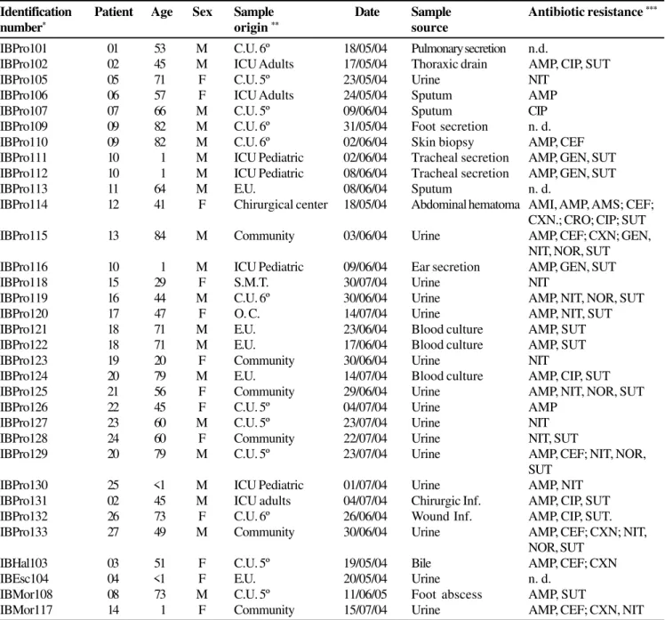
Table 1. List of isolates, their origin, sample source, and antibiotic resistance.
As expected, E. coli, M. morgani, and H. alvei were clearly differentiated from each other and from the P. mirabilis isolates (Figure 2).
ISSR Fingerprinting
Seven ISSR (intergenic single sequence repeats) primers were evaluated against three isolates of P. mirabilis (IBPro 101, IBPro102 and IBPro120). The primers used were (AC)8T, (AG)8A, (GA)8T, (AG)8YT, (GATA)4, (GACA)4, and (GTGC)4. No amplification was obtained with primer (AG)8YT; while primers
(GATA)4 and (GTGC)4 produced a smear. The other four primers generated well-defined amplification products (Figure 1). Applied to all the isolates, the selected primers generated 49 scorable bands, varying from 185 to 2,715 bp, 38 (77.5%) of these bands were polymorphic within P. mirabilis. The main problem observed with ISSR markers was low reproducibility.
Considered together, the four ISSR primers allowed discrimination of all the isolates. However, the three outgroup species included in the analysis were clustered together with Proteus isolates.
Identification Patient Age Sex Sample Date Sample Antibiotic resistance ***
number* origin ** source
IBPro101 01 53 M C.U. 6º 18/05/04 Pulmonary secretion n.d.
IBPro102 02 45 M ICU Adults 17/05/04 Thoraxic drain AMP, CIP, SUT
IBPro105 05 71 F C.U. 5º 23/05/04 Urine NIT
IBPro106 06 57 F ICU Adults 24/05/04 Sputum AMP
IBPro107 07 66 M C.U. 5º 09/06/04 Sputum CIP
IBPro109 09 82 M C.U. 6º 31/05/04 Foot secretion n. d.
IBPro110 09 82 M C.U. 6º 02/06/04 Skin biopsy AMP, CEF
IBPro111 10 1 M ICU Pediatric 02/06/04 Tracheal secretion AMP, GEN, SUT
IBPro112 10 1 M ICU Pediatric 08/06/04 Tracheal secretion AMP, GEN, SUT
IBPro113 11 64 M E.U. 08/06/04 Sputum n. d.
IBPro114 12 41 F Chirurgical center 18/05/04 Abdominal hematoma AMI, AMP, AMS; CEF;
CXN.; CRO; CIP; SUT
IBPro115 13 84 M Community 03/06/04 Urine AMP, CEF; CXN; GEN,
NIT, NOR, SUT
IBPro116 10 1 M ICU Pediatric 09/06/04 Ear secretion AMP, GEN, SUT
IBPro118 15 29 F S.M.T. 30/07/04 Urine NIT
IBPro119 16 44 M C.U. 6º 30/06/04 Urine AMP, NIT, NOR, SUT
IBPro120 17 47 F O. C. 14/07/04 Urine AMP, NIT, SUT
IBPro121 18 71 M E.U. 23/06/04 Blood culture AMP, SUT
IBPro122 18 71 M E.U. 17/06/04 Blood culture AMP, SUT
IBPro123 19 20 F Community 30/06/04 Urine NIT
IBPro124 20 79 M E.U. 14/07/04 Blood culture AMP, CIP, SUT
IBPro125 21 56 F Community 29/06/04 Urine AMP, NIT, NOR, SUT
IBPro126 22 45 F C.U. 5º 04/07/04 Urine AMP
IBPro127 23 60 M C.U. 5º 23/07/04 Urine NIT
IBPro128 24 60 F Community 22/07/04 Urine NIT, SUT
IBPro129 20 79 M C.U. 5º 23/07/04 Urine AMP, CEF; NIT, NOR,
SUT
IBPro130 25 <1 M ICU Pediatric 01/07/04 Urine AMP, NIT
IBPro131 02 45 M ICU adults 04/07/04 Chirurgic Inf. AMP, CIP, SUT
IBPro132 26 73 F C.U. 6º 26/06/04 Wound Inf. AMP, CIP, SUT.
IBPro133 27 49 M Community 30/06/04 Urine AMP, CEF; CXN; NIT,
NOR, SUT
IBHal103 03 51 F C.U. 5º 19/05/04 Bile AMP, CEF; CXN
IBEsc104 04 <1 F E.U. 20/05/04 Urine n. d.
IBMor108 08 73 M C.U. 5º 11/06/05 Foot abscess AMP, SUT
IBMor117 14 1 F Community 15/07/04 Urine AMP, CEF; CXN, NIT
* IBPro – Proteus mirabilis; IBEco- Escherichia coli; IBMor- Morganella morganii; IBHal- Hafnia alvei. ** C.U. - Care Unit 5th or 6th floor;
O.C.-Obstetric center; I.C.U.- Intensive Care Unit; C.C.- Chirurgical center; E.U.- Emergency Unit ** *AMI- Amicacin; AMP- Ampicillin;
Primer Primer sequences Total number Total number Number of Simpson’s diversity
of bands of bands in Proteus polymorphic bands index (DI)
in Proteus
RAPD
OPA10 5' GTGATCGCAG 3' 23 5 3 0.756
OPA11 5' CAATCGCCGT 3' 31 12 9 0.899
OPD20 5' ACCCGGTCAC 3' 9 3 2 0.333
OPX13 5' ACGGGAGCAA 3' 23 13 9 0.921
OPX15 5' CAGACAAGCC 3' 15 8 2 0.563
OPZ04 5' AGGCTGTGCT 3' 11 7 1 0.335
OPZ08 5' GGGTGGGTAA 3' 15 10 8 0.884
OPZ10 5' CCGACAAACC 3' 16 7 5 0.627
OPZ19 5' GTGCGAGCAA 3' 26 14 7 0.847
OPZ20 5' ACTTTGGCGG 3' 19 7 5 0.945
188 86 51 0.998
ISSR 1 (AC)8T 15 15 15 0.953
ISSR 2 (AG)8A 12 12 5 0.829
ISSR 3 (GA)8T 6 6 2 0.458
ISSR 6 (GACA)4 16 16 16 1.000
49 49 38 1.000
ERIC-PCR ERIC 1R and ERIC 2 22 10 9 0.970
BOX- PCR BOX- A1R 20 12 10 0.980
REP-PCR REP-PCR 1R and 2I 14 4 2 0.621
Figure 1. Examples of the profiles obtained using the five PCR methods. A. RAPD OPA11, B. ERIC-PCR, C. ISSR 6, D. BOX-PCR, E. REP-PCR. Samples (from left) IB Pro101 and 102, IB Hal 103, IB Eco 104, IB Pro 105 to IB Pro 107, IB Mor 108, and IB Pro 109 and 110.
Table 2. Primer sequences.
Table 3. Pearson product-moment correlation coefficient between similarity values obtained with genetic fingerprinting methods.
BOX REP RAPD ISSR ERIC
BOX - 0.712** 0.848** 0.054ns 0.565**
REP - 0.817** 0.143* 0.670**
RAPD - 0.066ns 0.693**
ISSR - 0.161*
ERIC
Repetitive-PCR Fingerprinting
As can be observed in Table 2 and Figure 1, ERIC-PCR and BOX-PCR resulted in detection of 10 and 12 amplification products in Proteus isolates, respectively. Nine of the 10 ERIC markers, and 10 of the 12 BOX markers exhibited some degree of polymorphism, being useful as discriminant markers. Three bands (ERIC-515bp, BOX-1199bp and BOX-402bp) were characteristic of P. mirabilis. As occurred with RAPD analysis, the control species (E. coli, M. morganii, and H. alvei) were clearly discriminated from each other, and from the Proteus isolates (Figure 2). Despite the low number of amplification
products obtained, ERIC-PCR and BOX markers allowed separating the 29 Proteus isolates into several groups (Figure 2). Moreover, these markers confirmed the identity of isolates IBPro 121 and 122 (patient 18), IBPro 102 and IBPro 131 (patient 9), and IBPro 111, 112 and 116 isolated from tracheal and ear secretions of patient 10.
REP-PCR using primers 1R and 2I yielded 14 amplification products, which allowed the separation of the four bacterial species included in our analysis. A very low number of bands was obtained in P. mirabilis (four bands) with just two polymorphic products (769bp and 641bp), and a Proteus-specific product of 1,220 bp.
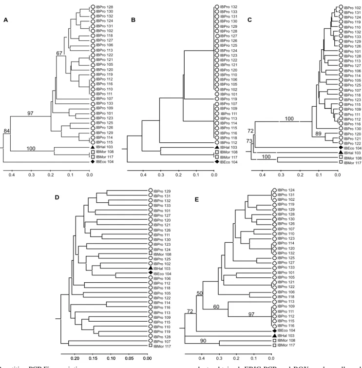
Figure 2. Dendrograms obtained for Proteus mirabilis and outgroup species using different PCR fingerprinting methods. A. BOX, B. REP-PCR, C. RAPD, D. ISSR and E. ERIC-PCR.
IBPro 130 IBPro 132 IBPro 124 IBPro 131 IBPro 102 IBPro 118 IBPro 127 IBPro 106 IBPro 113 IBPro 122 IBPro 121 IBPro 105 IBPro 120 IBPro 119 IBPro 112 IBPro 116 IBPro 110 IBPro 111 IBPro 107 IBPro 133 IBPro 109 IBPro 101 IBPro 123 IBPro 125 IBPro 126 IBPro 129 IBPro 114 IBPro 115 IBHal 103 IBMor 108 IBMor 117 IBEco 104 0.0 0.1 0.2 0.3 0.4 84 IBPro 128 97 100 67 A IBPro 132 IBPro 133 IBPro 131 IBPro 130 IBPro 129 IBPro 128 IBPro 127 IBPro 126 IBPro 125 IBPro 124 IBPro 123 IBPro 122 IBPro 121 IBPro 120 IBPro 110 IBPro 106 IBPro 105 IBPro 102 IBPro 101 IBPro 119 IBPro 107 IBPro 109 IBPro 111 IBPro 113 IBPro 114 IBPro 115 IBPro 116 IBPro 118 IBPro 112 IBHal 103 IBMor 108 IBMor 117 IBEco 104 0.0 0.1 0.2 0.3 0.4 B IBPro 102 IBPro 131 IBPro 124 IBPro 119 IBPro 110 IBPro 132 IBPro 133 IBPro 129 IBPro 126 IBPro 101 IBPro 128 IBPro 113 IBPro 127 IBPro 106 IBPro 114 IBPro 105 IBPro 125 IBPro 107 IBPro 118 IBPro 123 IBPro 115 IBPro 109 IBPro 111 IBPro 112 IBPro 116 IBPro 130 IBPro 120 IBPro 121 IBPro 122 IBEco 104 IBHal 103 IBMor 108 IBMor 117 0.0 0.1 0.2 0.3 0.4 100 72 73 100 89 IBPro 129 IBPro 131 IBPro 132 IBPro 133 IBPro 101 IBPro 127 IBPro 120 IBPro 121 IBPro 126 IBPro 111 IBPro 130 IBPro 123 IBPro 124 IBMor 108 IBPro 125 IBPro 102 IBHal 103 IBEco 104 IBPro 106 IBPro 112 IBPro 118 IBPro 105 IBPro 122 IBPro 114 IBPro 116 IBPro 113 IBPro 109 IBPro 115 IBPro 110 IBPro 119 IBPro 128 IBPro 107 IBMor 117 0.00 0.05 0.10 0.15
0.20 0.15 0.10 0.05 0.00
Comparison of Methods
As can be observed in Table 2, the discriminatory index (Simpson’s index), which represents the probability that two randomly chosen isolates will be distinguished by a given method, varied from 0.621 for REP-PCR to 1.000 for ISSR, with high values for RAPD, BOX and ERIC. Among RAPD primers, OPZ20, OPX13, OPA11, OPZ08 and OPA19 gave the highest DI values (Table 2). A high level of variation in the discriminatory indexes was also observed among ISSR primers; ISSR 1, 2 and 6 (0.829 to 1.000) were more discriminant than ISSR3 (0.458).
Comparison of the similarity values obtained with the five DNA fingerprinting methods that we used gave high and significant correlations between RAPD and BOX, RAPD and ERIC, and BOX and ERIC (Table 3). REP similarity values correlated with those obtained using BOX, RAPD, and ERIC; but these correlations should be interpreted carefully due to the low number of amplification products obtained with REP. No correlation or low correlations were observed between ISSR similarity values and those obtained with the other methods.
Most clusters found in the dendrograms obtained by BOX, ERIC, and RAPD fingerprinting techniques were similar (Figure 3). Specifically, bacterial isolates classified as E. coli, H. alvei and M. morganii formed individual clusters with more than 75% confidence, well separated from P. mirabilis isolates. Moreover, P. mirabilis isolates obtained from the same patient (patients 9, 10 and 18) from different data and/or sample sources and exhibiting the same antibiotic resistance and Dienes behavior, were genetically identical or very similar.
Discussion
Studies on the molecular epidemiology of infection due to Proteus species have employed a variety of methods, including ribotyping, PFGE, RAPD, and tandem-repeat microsatellite fingerprinting [14-19]. We showed that RAPD markers vary in their discriminatory ability. Some primers (OPZ20, OPX13, OPA11, OPZ08 and OPA19) showed high discrimination indices. The use of three primers (OPA11, OPX13 and OPZ8) allowed the characterization of all of the P. mirabilis isolates. The efficiency of RAPD markers for Proteus fingerprinting was previously reported by Binden et al. [17], in an epidemiological investigation of P. mirabilis from pregnant women and their neonates, and by Hoffman et al. [18] in a study of clinical isolates of P. penneri. In general, the relatively low reproducibility of RAPD typing limits its application to large-scale inter-laboratory studies. However, in our study RAPD showed high reproducibility between replications (within and between gels), which, associated with its high discrimination ability, makes this one of the most suitable methods for local Proteus epidemiological studies.
Among the primers selected for ISSR analysis, three primers showed high discriminatory power and allowed discrimination of all the isolates. Our data corroborate the conclusions reported by Cieslikowski et al. [19], who showed (GACA)4
and (CAAT)4 to be informative primers, and indicates that other primers, such as (AC)8T and (AG)8A, could be useful in P. mirabilis studies. However, despite the large size of the primers, ISSR markers applied to P. mirabilis gave low reproducibility, and were not suitable for identification to the genus level.
The repetitive-DNA markers ERIC-PCR, and particularly BOX-PCR, were more informative than rep-PCR, which amplified only four bands in P. mirabilis. ERIC and BOX-PCR amplified 22 bands in Proteus,of which 19 were polymorphic. Repetitive-DNA markers have been used with success in the identification of a large number of Gram-negative bacteria, including Escherichia coli [12], Salmonella [10], Aeromonas [13], Burkholderia [11], Vibrio [24], among others.
Proteus mirabilis isolates obtained from the same patient (patients 9, 10 and 18) using different data and/or sample sources and exhibiting the same antibiotic resistance and Dienes behavior, exhibited identical or very similar RAPD, ERIC-PCR and BOX-PCR patterns, indicating that these molecular markers can be used to check for self contamination or strain persistence in a given patient.
In summary, we found that RAPD, ERIC-PCR and BOX-PCR markers have a high discriminatory ability, allowing the genetic typing of clinical P. mirabilis isolates, which should prove useful for epidemiological studies of this bacterium.
Acknowledgements
The authors thank the Foundation of the University of Caxias do Sul for financial support, and CNPq for student fellowships.
References
1. Mobley H.L.T., Belas R. Swarming and pathogenicity of Proteus mirabilis in urinary tract. Trends Microbiol 1995;3:280-4. 2. Chow A.W., Taylor R.R., Yoshikawa T.T., Guze, L.B. A nosocomial
outbreak of bacteria on struvite crystal habit and its importance in urinary stone formation. J Crystal Growth 1979;104:475-84. 3. O’Hara C.M., Brenner F.W., Miller J.M. Classification, identification, and clinical significance of Proteus, Providencia, and Morganella. Clin Microbiol Rev 2000;13:534-46. 4. Wassif C., Cheek D., Belas, R. Molecular analysis of
metalloproteases from Proteus mirabilis. J Bacteriol
1995;177:5790-8.
5. Rozalski A., Sidorczyk Z., Kotelko K.Potential virulence factors of Proteus bacilli. Microbiol Mol Biol Rev 1997;61:65-89. 6. Fraser G.M., Claret L., Furness R., Gupta S., Hugues, C.
Swarming-couples expression of the Proteus mirabilis hpmBA haemolysin operon. Microbiol 2002;148:2191-201.
7. Mobley H.L.T., Island D., Hausinger R.P. Molecular biology of microbial ureases. Microbiol Rev 1995;59:451-80.
8. Walker K.E., Moghaddame-Jafari S., Lockatell C.V., et al. ZapA, the IgA-degrading metalloprotease of Proteus mirabilis, is a virulence factor expressed specifically in swarmer cells. Molec Microbiol 1999;32:825-36.
9. Olive D.M., Bean P. Principles and applications of methods for DNA-based typing of microbial organisms. J Clin Microbiol
1999;37:1661-9.
11. Coenye T., Spilker T., Martin A., LiPuma J.J. Comparative assessment of genotyping methods for epidemiologic study of Burkholderia cepacia Genomovar III. J Clin Microbiol
2002;40:3300-7.
12. Seurinck S., Verstraete W., Siciliano S.D. Use of 16S-23S rRNA intergenic spacer region PCR and repetitive extragenic palindromic PCR analyses of Escherichia coli isolates to identify nonpoint fecal sources. Appl Environ Microbiol 2003;69:4942-50. 13. Szczuka E., Kaznowski A. Typing of clinical and environmental
Aeromonas sp. strains by random amplified polymorphic DNA PCR, repetitive extragenic palindromic PCR, and enterobacterial repetitive intergenic consensus sequence PCR. J Clin Microbiol
2004;42:220-8.
14. Pignato S., Giammanco G.M., Grimont F., et al. Molecular characterization of the genera Proteus, Morganella, and Providencia by ribotyping. J Clin Microbiol 1999;37:2840-7. 15. Pfaller M.A., Mujeeb I., Hollis R.J., et al. Evaluation of the
discriminatory powers of the Dienes test and ribotyping as typing methods for Proteus mirabilis. J Clin Microbiol 2000;38:1077-80. 16. Sabbuba N.A., Mahenthiralingam E., Stickler D.J. Molecular epidemiology of Proteus mirabilis infections of the catheterized urinary tract. J Clin Microbiol 2003;41:4961-5.
17. Bingen E., Boissinot C., Desjardins P., et al. Arbitray primed polymerase chain reaction provides rapid differentiation of Proteus mirabilis isolates from pediatric hospital. J Clin Microbiol 1993;31:1055-9.
18. Hoffmann G., Gajdos G., Czako M., et al. Diversity among clinical isolates of Proteus penneri detected by random amplified polymorphic DNA analysis. Zentbl Bakteriol
1998;288:351-60.
19. Cieslikowski T., Gradecka D., Mielczarek M., Kaca, W. Tandem tetramer-based microsatellite fingerprinting for t y p i n g o f P r o t e u s m i r a b i l i s s t r a i n s. J C l i n M i c r o b i o l
2 0 0 3; 4 1 : 1 6 7 3 - 8 0 .
20. CLSI. Performance standards for antimicrobial susceptibility testing, 17th informational supplement. CLSI/NCCLS document
M100-S17. Wayne, PA: National Committee for Clinical Laboratory Standards, 2007.
21. Versalovic J., Koeuth T., Lupski R.J. Distribution of repetitive DNA sequences in eubacteria and application to fingerprinting of bacterial genomes. Nucleic Acids Res 1991;9:6823-31. 22. Versalovic J., Schneider M., de Brujin F.J., Lupskin, J.R. Genomic
fingerprinting of bacteria using repetitive sequence-based polymerase chain reaction. Methods Mol Cell Biol
1994;5:25-40.
23. H u n t e r P. R . , G a s t o n M . A . N u m e r i c a l i n d e x o f t h e discriminatory ability of typing systems: an application of Simpson´s index of diversity. J Clin Microbiol 1988; 26: 2465-2466.