Cátia Sofia Vicente Gomes
Licenciada em Biologia
Cues for Cancer Stem Cells Origin
Dissertação para obtenção do Grau de Mestre em Genética Molecular e Biomedicina
Orientador: Dora Maria Tuna de Oliveira Brites
Investigadora Coordenadora e Professora Catedrática Convidada
Faculdade de Farmácia, Universidade de Lisboa
Co-orientador: Ana Sofia Iria Azeredo Falcão de Jesus
Investigadora Auxiliar (Ciência 2007)
Faculdade de Farmácia, Universidade de Lisboa
Júri:
Presidente: Prof. Doutora Margarida Casal Ribeiro Casto Caldas Braga Arguente: Doutora Maria Margarida Fonseca Rodrigues Diogo
Vogal: Prof. Doutora Dora Maria Tuna de Oliveira Brites
iii Cues for cancer stem cells origin
Copyright Cátia Sofia Vicente Gomes, FCT/UNL, UNL
v
Part of the results discussed in this thesis were presented in the following publications/
communications:
Torrado E, Gomes C, Santos G, Brites D, Falcão AS. Directing mouse embryonic neurosphere differentiation towards nerve cell lineages. Experimental Neurology 2012 [Submitted];
Gomes C, Santos G, Torrado E, Falcão AS, Brites D. Applying Neural Stem Cell Biology To Brain Tumour Research: New Cues For Gliomagenesis In The Elderly. 26ª Reunião do Grupo de Estudos do Envelhecimento Cerebral e Demência, Tomar, 29-30 June, 2012 [Poster comunication];
vii
AGRADECIMENTOS
As minhas primeiras palavras de agradecimento são, naturalmente, dirigidas à Professora Doutora Dora Brites. Muito tenho a agradecer pela oportunidade que me foi dada de realizar a minha tese de mestrado sob a orientação da Professora. Sem dúvida que a confiança que demonstrou ao permitir-me desenvolver este trabalho me levaram sempre a tentar responder da melhor forma aos desafios. O rigor e a qualidade que me foram incutidos como ferramentas chave para um bom trabalho, foram uma constante ao longo deste ano. E, isso não seria possível sem a preciosa e frequente ajuda da Professora. Muito obrigada por todo o tempo dispensado com o meu trabalho e pela orientação na procura de respostas às questões que foram surgindo. A oportunidade de trabalhar neste grupo e todos os conhecimentos que me foram transmitidos contribuíram de forma muito positiva para a minha formação profissional e serão essenciais no meu futuro, por isso, mais uma vez, obrigada.
À Doutora Ana Sofia Falcão, co-orientadora deste trabalho, agradeço toda a disponibilidade, apoio e dedicação revelados ao longo da execução da tese. As dúvidas, questões e barreiras não teriam sido ultrapassadas sem a tua ajuda, procura e encorajamento. Tenho a agradecer-te a constante preocupação na realização do trabalho e na qualidade com que estava a ser encaminhado. Obrigada por todos os ensinamentos e paciência com uma aluna de mestrado que pouco tinha contacto com a investigação. Quero ainda agradecer-te a simpatia e carinho com que sempre fui tratada e que contribuíram para que a inicial “distância” entre co-orientadora/orientanda, fosse sendo diminuída ao longo deste ano.
À Professora Doutora Margarida Castro Caldas, orientadora interna desta tese, quero expressar o meu agradecimento pela disponibilidade e prontidão no esclarecimento das minhas dúvidas.
À Professora Doutora Alexandra Brito e ao Professor Doutor Rui Silva quero agradecer todo o apoio, esclerecimento de dúvidas e sugestões feitas ao longo deste ano e que contribuíram para a realização do meu trabalho.
À Professora Doutora Adelaide Fernandes e à Doutora Ana Rita Vaz, o meu muito obrigada pelo carinho e disponibilidade com que sempre me brindaram. Adelaide, a ti quero agradecer todo o apoio em alturas menos fáceis e a transmissão de conhecimentos que me encaminharam na direcção correcta. Também tu me levas a acreditar que é possível voar mais alto! A ti Rita, tenho a agradecer todo o encorajamento, o esclarecimento de dúvidas e as palavras amigas quando nem tudo corria como pretendia. As tuas conversas e convívio com o “rapazes pequenos” levaram à construção desta amizade, obrigada “tia” .
viii
foi realizado quase lado-a-lado (pelo menos numa fase inicial), agradeço-te a “partilhada” preocupação na procura de bons resultados e possíveis soluções para as barreiras que fomos encontrando.
Às meninas que partilharam comigo este ano de trabalho, dentro e fora do CPM, um obrigada muito especial.
Carolina, todos os momentos deste caminho e a chegada a esta meta foram sempre partilhados contigo, “mana”. O facto de termos feito este percurso de uma forma tão próxima fez com que o apoio e ajuda fosse constante em todas as situações. Quero agradecer-te o facto de estares sempre presente, a confiança que fomos constuíndo, as palavras de incentivo e encorajamento, etc, etc, etc. Sem dúvida que foste “parte essencial” desta etapa, dando-lhe um contributo enorme. Conseguímos tornar os longos dias de trabalho mais divertidos, pois muitas foram as vezes em que partilhámos a mesma bancada, ou por ajuda ou simplesmente por companhia. Por isto, e tudo o resto, um especial “beijinho borboleta” pela pessoa especial e amiga que te tornante, e por me teres sempre feito acreditar que seria possível chegarmos juntas até aqui.
Andreia e Inês, as minhas “doutorazinhas”, a vocês quero agradecer essa boa disposição e disponibilidade para ajudar. Andreia, muito mais importante que a ajuda nas formatações … tenho a agradecer-te a confiança e amizade que fomos construindo ao longo deste ano. O teu apoio e palavras de incentivo foram uma enorme contribuição para a execução deste trabalho. Inês, a minha “mana”, apesar de inicialmente teres-me sido apresentada como “o feitio difícil“ tenho a dizer-te que nunca o achei… Para mim és uma pessoa amiga e sempre pronta a ajudar. A tua organização ensinou-me também que essa é uma ferramenta essencial. Obrigada por tudo meninas . A ti Filipa, quero agradecer a forma como fui recebida. Sem dúvida que a tua simpatia e característica vontade de ajudar e integrar, são importantes quando chegamos a um espaço novo. Obrigada pelo apoio e encorajamento, bem como os momentos de boa disposição. Cláudia, a nossa entrada no CPM teve quase a mesma data (embora a tua para um voo mais alto!) fez com que nos ajudássemos e partilhássemos novos conhecimentos. A tua simpatia e palavras amigas, bem como esse espirito de convívio, foram importantes ao longo deste ano. Desejo-te muita sorte para este teu novo percurso académico.
Aos restantes membros, bolseiros, com quem partilhei, não só a falta de luz natural na nossa sala de trabalho, como também o convívio e boa disposição. Obrigada por me fazerem acreditar que o local de trabalho pode ter o conforto necessário a tantas horas de estudo. Um obrigada especial ao André e ao Duarte! André, nem sempre o entrar “sorrateiramente” numa sala é suficiente para “pregar belos sustos”…depois ensino-te a técnica infalível Obrigada pelo convívio e boa disposição com que sempre me abordaste! Duarte, a ti quero agradecer o facto de quase adivinhares quando precisava de uma palavra amiga. A proximidade que foi ocorrendo levou a que te tornasses numa pessoa em quem sei que posso confiar!
ix Nádia, um obrigada especial pelo encorajamento em momentos menos bons, pela alegria quando tudo correu como pretendi e pelas palavras de incentivo e conversas que tanto contribuíram para a execução da minha tese. Dentro e fora do mundo académico sei que és uma pessoa especial e presente. Obrigada!
Miguel, um obrigada muito especial por estares sempre presente, pelo teu carinho, apoio, opinião e preocupação pelo meu sucesso e bem-estar. O facto de também tu teres o “bichinho” da ciência faz com que percebas que por vezes o trabalho consegue absorver-nos de uma forma quase completa… E por isso quero agradecer-te a paciência e compreensão que sempre demonstraste. Por vezes tenho vontade de te rifar…mas não o faço…tenho medo que não voltes a “sair à casa” Gosto muito de ti!
À minha família, um obrigada pelo apoio nesta etapa da minha vida.
Um agradecimento muito especial vai para as minhas manas (e cunhados, claro ), sem a vossa ajuda nada disto seria possível. Cláudia quero agradecer-te por todas as palavras amigas e o apoio incondicional a que sempre me habituaste. A tua presença constante e opinião nas minhas decisões têm-me levado sempre a seguir o caminho correcto. Vânia, a minha mana gémea, apesar das escolhas académicas nos terem levado para percursos distintos, o teu apoio e preocupação para que tudo corresse como pretendia foram essenciais para este trabalho e para encurtar a distância a que fomos sujeitas. Aquilo que sentimos umas pelas outras e a ligação que existe entre as três é algo que não consigo explicar em palavras… Obrigada pelas manas maravilhosas que são… ADORO-VOS.
Um obrigada cheio de carinho vai para o meu sobrinho lindo. Rodrigo, as tuas palavras, ainda perceptíveis apenas para alguns adultos , os convites para brincar e telefonemas só para dar “bilinhos” foram essenciais para que este percurso fosse sempre acompanhado por um brilho especial. Espero ter compensado os meus fins-de-semana de trabalho, em falta no mundo da brincadeira, com aqueles que reservava para fazer corridas em pistas de carros e jogar à bola. Usando as tuas palavras “a tia Cátia adoda o Godigo”!
Às minhas estrelinhas da sorte…
xi
ABSTRACT
Neural stem/progenitor cells (NSPC) can differentiate into neurons and glial cells in the central nervous system. Interestingly, NSPC biology is being applied to the study of human brain tumours, since these cells share some common features with glioma cells. However, it is not known the developmental stage with more similarities to glioma cells, or the most susceptible to malignant transformation.
We aimed to identify the stage(s) in the NSPC differentiation process towards astrocytes where cells acquire phenotype characteristics comparable to glioma cells.
NSPC that were obtained from E15 mouse brain, were grew as neurospheres (NS) and induced to astroglial differentiation until 7 days in vitro (DIV). After the cellular characterization of NS and
differentiating cells, tumour-related factors were evaluated and their behavior compared to the one of GL261 mouse glioma cells.
Astroglial differentiation led to a decrease in progenitor cells, as expected. Multidrug resistance-associated protein 1 expression decreased and autophagy marker increased with differentiation. The vascular endothelial growth factor (VEGF), matrix metalloproteinases and S100B protein increased until 2/3 DIV, while the 1 DIV cells showed the highest migratory potential towards the chemotactic VEGF or GL261-conditioned media.
Comparison of data with glioma cells characteristics point to the first and second days of NSPC differentiation to astrocytes as the stages closing matching GL261 cells, and likely the most vulnerable to malignancy transformation.
Keywords: Astrocytes, Neural progenitor cells, Neural stem cells, Glioma cells, Gliomagenesis,
xiii
RESUMO
As células estaminais/progenitoras neurais (CEPN) podem diferenciar em neurónios e células da glia no sistema nervoso central. A biologia das CEPN tem sido aplicada ao estudo dos tumoures cerebrais humanos, uma vez que estas células partilham algumas características com as células de glioma. Contudo, não é conhecido o estadio do desenvolvimento mais semelhante às células de glioma, ou o mais susceptível à transformação maligna.
O nosso objectivo é identificar o(s) estadio(s), no processo de diferenciação das CEPN em astróctios, no(s) qual(is) as células adquirem características fenotípicas comparadas às células de glioma.
As CEPN, obtidas de cérebros de embriões de ratinho no 15º dia de gestação, foram cultivadas como neuroesferas e induzidas à diferenciação astroglial até 7 dias in vitro (DIV). Após a
caracterização celular das neuroesferas e células em diferenciação, foram avaliados determinados factores tumorais e o seu comportamento comparativamente às células de linha celular de glioma de ratinho (GL261).
A diferenciação astroglial levou ao decréscimo das células progenitoras, como esperado. A expressão da proteína associada à resistência a multidrogas decresceu, enquanto a autofagia aumentou ao longo da diferenciação. O factor de crescimento endotelial vascular (VEGF), metaloproteinases e a proteína S100B revelaram um aumento da expressão até 2/3 DIV. Ainda, o fenótipo correspondente a 1 DIV em condições de diferenciação foi o que apresentou maior potencial migratório para o VEGF ou para o meio proveniente das células de glioma.
A comparação dos dados obtidos para os vários factores, levou-nos a sugerir o primeiro e segundo dias das CEPN em condições de diferenciação em astrócitos, como os estadios mais próximos das células de glioma, e por consequente, mais vulneráveis a transformação maligna.
Palavras-chave: Astrócitos, Células estaminais neurais, Células progenitoras neurais, Células de
xv
INDEX
ABBREVIATIONS ... xxi
I. INTRODUCTION...1
1. Neural Stem Cells (NSC) ...3
1.1. NSC in central nervous system development ...4
1.2. NSC in the adult brain niches ...5
1.3. Therapeutic potential of NSC ...7
1.4.In vitro culture of NSC: the neurosphere assay ... 10
2. Neural stem cells, tumour stem cells and brain tumours: dangerous relationships? ... 13
2.1. Gliomas – an overview ... 14
2.2. Cancer stem cell hypothesis ... 16
2.3. Cues for glioma origin ... 19
3. Aims ... 22
II. MATERIAL AND METHODS ... 23
1. Reactives ... 25
1.1. Cell cultures media ... 25
1.2. Supplements and chemicals ... 25
1.3. Antibodies ... 25
2. Equipment ... 26
3. Methods ... 26
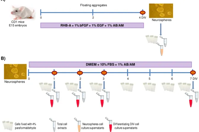
3.1. Cell cultures and treatments ... 26
3.1.1 Primary neurosphere culture ... 26
3.1.2 NS in vitro astroglial differentiation ... 27
3.1.3 GL261 mouse glioma cell line ... 28
3.2. Immunocytochemistry ... 29
3.3. Proliferative potential... 30
3.4. Western blot assay ... 30
3.5. Gelatin Zymography ... 31
3.6. ELISA ... 31
3.7. Migration Assay ... 31
INDEX
xvi
III. RESULTS ... 33
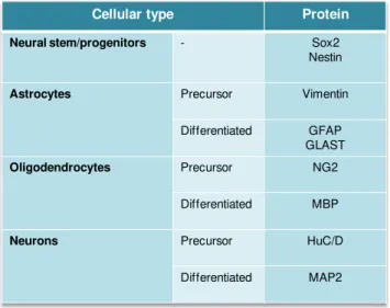
1. Characterization of NS and of the different developmental phenotypes ... 35
1.1. Differentiation of NS into astrocytes leads to a reduction in the undifferentiated cell markers Sox2 and nestin ... 35
1.2. Differentiation of NS into astrocytes leads to a reduction of BrdU, a marker of proliferating cells ... 37
1.3. Differentiation of NS into astrocytes leads to a reduction in the early astrocytic progenitor marker vimentin and an increase in the astrocytic markers GFAP and GLAST ... 38
1.4. Differentiation of NS into astrocytes leads to an highly decrease in the oligodendroglial markers NG2 and MBP ... 38
1.5. Differentiation of NS into astrocytes leads to a highly decrease in the neuronal markers HuC/D and MAP2 ... 40
2. Comparison between tumour-related factors in glioma cells and in the successive developmental phenotypes from neurospheres ... 42
2.1. Cells from NS reveal levels of Mrp1 expression very similar to those of glioma cells ... 42
2.2. Cells differentiated during 24 h from NS are those exhibiting autophagic levels most similar to the ones observed in glioma cells ... 43
2.3. Glioma cells evidence higher levels of VEGF and VEGFR-2 expression than NS or differentiating astrocytes ... 44
2.4. MMP-9 and MMP-2 expression levels in glioma cells are similar to the ones in 7 DIV differentiating astrocytes ... 46
2.5. Evaluation of S100B protein expression by NS/differentiating astrocytes and and its comparison with glioma cells ... 47
2.6. Migratory potential of NS and differentiating astrocytes are higher than that of glioma cells ... 47
IV. DISCUSSION ... 49
Future perspectives ... 59
xvii
FIGURE INDEX
I. INTRODUCTION...1
Fig. I.1 – Classical view of neural stem cells hierarchy. ...3 Fig. I.2 – Gradual transformation of radial glial cells into astrocytes-like cells, from the embryonic period to adulthood. ...5 Fig. I.3 – The adult niches, cell types and stem cell lineage. ...7 Fig. I.4 – Schematic representation of nanoparticles delivery by neural stem cells inside a brain tumour ... 10 Fig. I.5 – Principal feactures and phenotypes of neurospheres. ... 12 Fig. I.6 – Cell types and associated tumours of the central nervous system ... 14 Fig. I.7 – Schematic representation of the relationship between neural stem cells, neural progenitor cells, cancer stem cells and brain tumours ... 18 Fig. I.8 – Shared features by neural stem cells and glioma cells ... 21
II. MATERIAL AND METHODS ... 23
Fig. II.1 - Schematic representation of the experimental model, involving the preparation of primary neurosphere culture and the induction of astroglial differentiation ... 28 Fig. II.2 – Schematic representation of the migration assay ... 32
III. RESULTS ... 33
FIGURE INDEX
xviii
Fig. III.11 – Comparison of the migratory potential between neurospheres plus the astrocytes differentiation stages and the glioma cells towards VEGF and GL261-conditioned media ... 48
IV. DISCUSSION ... 49
xix
TABLE INDEX
I. INTRODUCTION...1
Table I. 1 – Astrocytoma grades ... 15
II. MATERIAL AND METHODS ... 23
xxi
ABBREVIATIONS
AB/AM Antibiotic antimycotic solution
AD Alzheimer’s disease
ALS Amyotrophic lateral sclerosis
BBB Blood brain barrier
bFGF Basic fibroblast growth factor
Bmi-1 B-cell-specific Moloney murine leukemia virus integration site 1
BMP Bone morphogenetic proteins
BrdU 5-Bromo-2’-Deoxyridine
BSA Bovine serum albumin
CEPN Células estaminais/progenitoras neurais
CNS Central nervous system
CSC Cancer stem cells
DAPI 4',6-diamidino-2-phenylindole
DIV Days in vitro
Dlx2 DLX2 gene (protein coding) distal-less homeobox 2
DMEM Dulbecco’s modified Eagle’s medium
DNA Deoxyribonucleic acid
DNAse I Deoxyribonuclease I
E Embryonic day
ECM Extracellular matrix
EGF Epidermal growth factor
ELISA Enzyme-linked immunosorbent assay
FBS Fetal bovine serum
FGF Fibroblast growth factor
FITC Fluorescein isothiocyanate
GBM Glioblastoma multiforme
GFAP Glial fibrillary acidic protein
GLAST Glutamate aspartate transporter
HBSS Hanksʼ balanced salt solution without Ca2+ and Mg2+
LC3 Microtubule-associated protein light chain 3
MAP2 Microtubule-associated protein 2
MBP Meylin basic protein
MMP Matrix metalloproteinases
Mrp1 Multidrug resistence-associated protein 1
NEP Neuroepithelial progenitor cells
NG2 Neural/glial antigen 2
ABBREVIATIONS
xxii
NPC Neural precursor cells
NS Neurospheres
NSC Neural stem cells
NSPC Neural stem/progenitor cells
PD Parkinson’s disease
PDGF Platelet-derived growth factor
PDGFRα Platelet-derived growth factor receptor α
PDL Poly-D-lysine
PFA Paraformaldehyde
PI Propidium iodide
PMSF Phenylmethylsulfonyl fluoride
PTEN Phosphatase and tensin homolog
RGC Radial glial cells
RT Room temperature
SDS Sodium dodecyl sulphate
SDS-PAGE Sodium dodecyl sulfate-polyacrilamide gel electrophoresis
SGZ Subgranular zone
Sox2 SRY (sex determining region Y)-box 2
SVZ Subventricular zone
TMZ Temozolomide
VEGF Vascular endothelial growth factor
VEGFR-2 Vascular endothelial growth factor receptor 2
VZ Ventricular zone
I. INTRODUCTION
3
1. Neural Stem Cells
The term “stem cell” was originally proposed by Heackel in 1868 as cited by Breunig et al.
(2011). Stem cells are a class of undifferentiated cells with the ability to differentiate into specialized cell types. They can also generate new stem cells by self-renewal and can be classified according to their sources: embryonic stem cells (that arise from the blastocyst phase of embryonic development) and adult stem cells (that arise from adult tissue). Stem cells can give rise to several cell types with more limited self-renewal and proliferation ability, including neural stem cells (NSC).
NSC only became popular in the early 1990s (Breunig et al., 2011) and are described as a brain
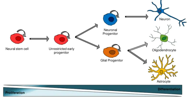
population with the ability of self-renewal, capable to maintain a pool of neural stem-like cells (extensive proliferative potential). These cells can also differentiate into more restricted precursor cells, designated by neural precursor cells (NPC) that are able to produce the three major cell types that compose the central nervous system (CNS): neurons, astrocytes and oligodendrocytes (Gage, 2000; Siebzehnrubl et al., 2011; Temple, 2001) (Fig. I.1).
NSC present a promising therapeutic tool for brain disorders, as they might be used to replace virtually any type of neuron lost from neurodegenerative disords such as Parkinson’s disease (PD), Alzheimer’s disease (AD) and amyotrophic lateral sclerosis (ALS) (Breunig et al., 2011; Parish et al.,
2008). Interestingly, NSC show tropism for brain tumours, namely gliomas and thus, they can be used as carriers for anti-tumourigenic drugs (Aboody et al., 2000; Noble, 2000). However, if in one hand
NSC may have a therapeutic potential, on the other hand it is suggested that these cells might generate brain tumours, due to their high proliferative potential. Hence, it is very important to understand the mechanisms by which NSC generate the diversity of their resulting progeny (Breunig
et al., 2011; Vescovi et al., 2006).
Fig. I.1 – Classical view of neural stem cells hierarchy. The normal neural stem cell production of progenitor cells, which subsequently generates the three differentiated cell types of the central nervous system: neurons, oligodendrocytes and astrocytes (trilineage potential). Along the differentiation process, the proliferative potential decrease, while the number of differentiated cells increase.
Unrestricted early progenitor
Glial Progenitor Neuronal Progenitor
Oligodendrocyte Neuron
Astrocyte Neural stem cell
I. INTRODUCTION
4
1.1. NSC in central nervous system development
The CNS of mammals is a highly complex structure made up of a huge number of neurons, glial cells and synapses, all linked by extremely heterogeneous anatomical and functional relationships. This complex and heterogeneous cellular population derives from NSC or primary progenitors (Bonfanti and Peretto, 2007; Merkle and Alvarez-Buylla, 2006). Very early in the mammalian development, the CNS begins with the development of the neuroectoderm, which forms the neural plate [at embryonic day 7.5 (E7.5) in mice] and then folds, giving rise to the neural tube (at E8.5 in mice) (Conti and Cattaneo, 2010; Merkle and Alvarez-Buylla, 2006; Temple, 2001). Within this primitive neural structure, a complex and heterogeneous population of NSC and primary progenitors can be found, the neuroepithelial progenitor cells (NEP). NEP are radially elongated and contact with both the apical (ventricular) and basal (pial) surfaces of the embryonic brain (Merkle and Alvarez-Buylla, 2006) expressing neural precursor markers such as Sox1 [SRY (sex determining region Y)-box 1] and Nestin, due to their “stemness” properties (Conti and Cattaneo, 2010). During the development course, these cells undergo both symmetric and asymmetric types of division. On the first stage of neural development, NEP undergo symmetrical divisions to expand the neural stem cell pool (self-renewal and proliferative potential), while in the second stage, they initiate asymmetrical division to generate a stem cell (that remains in the ventricular zone - VZ) and a daughter cell, that migrates radially outward to its final position in the brain (intermediate progenitor) (Farkas and Huttner, 2008; Merkle and Alvarez-Buylla, 2006).
In a later stage, NEP also originate the radial glial cells (RGC) (around E9.5 in mice) (Bentivoglio and Mazzarello, 1999; Bonfanti and Peretto, 2007; Conti and Cattaneo, 2010) which are the principal primary progenitors of the mammalian embryonic forebrain (and early postnatal brain). Similarly to NEP, RGC divide in the VZ and maintain contact with the pial surface via a radially projecting basal process. Thus, it is thought that NEP transform directly into RGC, which are the main cell type in the developing brain. They are considered an important transient population once RGC function both as neural progenitors and a scaffold for migrating immature neurons (Conti and Cattaneo, 2010). RGC express glial/astroglial markers as the glutamate aspartate transporter (GLAST), the glial fibrillary acidic protein (GFAP) and the brain lipid binding protein (BLBP) (Farkas and Huttner, 2008; Merkle and Alvarez-Buylla, 2006) and have astroglial cells anatomical-like features, such as endfeet on blood vessels, intermediate filaments, and glycogen granules (Merkle and Alvarez-Buylla, 2006). RGC can undergo symmetrical proliferative or asymmetrical neurogenic divisions to generate neurons (the functional unit of the nervous system), as well astrocytes and oligodendrocytes. These cells provide a critical support role for optimal neuronal functioning and survival but its differentiation potential is less broad than that of NEP (Merkle and Alvarez-Buylla, 2006; Zhao et al., 2008). Hence, NEP and RGC comprise the first group of stem and progenitor cells
5
The SVZ, a region just above the ventricular zone (VZ), remains the stem character in the adult brain. Particularly, once RGC and SVZ astrocytes share many properties, it is though that both belong to the same lineage. Moreover, RGC of the neonatal lateral ventricular wall occupy the same region as the astrocytic stem cells of the adult SVZ, and they act as multipotent NSC either in vitro or in vivo.
SVZ cells also retain a radial process and express GFAP, suggesting that RG cells may directly transform into SVZ astrocytes, in adult brain. In summary, current evidences suggest that NSC gradually transform from NEP to RGC and from these to astrocytes-like cells (Merkle and Alvarez-Buylla, 2006).
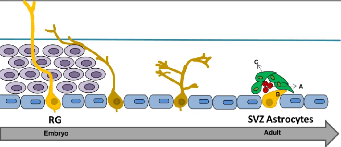
Fig. I.2 – Gradual transformation of radial glial cells into astrocytes-like cells, from the embryonic period to adulthood. Fate of radial glial (RG) cells from development (left) to adulthood (right). Different degrees of yellow indicate progressive maturation from RG to subventricular zone (SVZ) astrocyte-like cells, also called neurogenic SVZ astrocytes. In the embryo, RG cells behave as multipotent stem cells. In brain parenchyma they transform into SVZ astrocytes through a transient unipolar form which has lost contact with both pia mater and ventricular surface (in grey: ependyma). The SVZ is composed by type B cells – SVZ astrocytes stem cells (yellow), type C cells – transient-amplifying cells (green) and type A cells - neuroblasts (red) , such as described in the next section. Hence, it is thought that RG cells (in the embryo) disappear and originate SVZ astrocytes (in the adult). Adapted from Bonfanti and Peretto (2007).
1.2. NSC in the adult brain niches
Neurogenesis in the adult mammalian CNS was first described in the 1960s (Siebzehnrubl et al., 2011; Till and Mc, 1961). Adult neurogenesis is maintained by NSC that persist in the adult
mammalian brain and undergo self-renewal and have multipotency capacity generating neurons and macroglia (astrocytes and oligodendrocytes). Interestingly, it is thought that this adult neurogenesis might be related with malignant processes giving rise to adult brain tumours (Riquelme et al., 2008).
Two germinal regions are found in the adult mammalian brain: the SVZ of the forebrain lateral ventricle (Fig. I.3 A) and the subgranular zone (SGZ) of the hippocampal dentate gyrus (Fig. I.3 D) (Doetsch, 2003; Riquelme et al., 2008; Siebzehnrubl et al., 2011). These two zones are known as
neurogenic niches for adult stem cells because their microenvironment regulate and support self-renewal, activation and differentiation (features of stem-like cells) (Doetsch, 2003; Riquelme et al.,
2008). Also, they have several architectural elements that contribute to the adult neurogenesis such as extensive cell-cell interactions, proximity to the cerebrospinal fluid of the lateral ventricle, close
Embryo Adult
RG
SVZ Astrocytes
B C
I. INTRODUCTION
6
association with blood vessels, rich extracellular matrices and specialized basal lamina (Doetsch, 2003). Therefore, they are exposed to a variety of growth factors (Tavazoie et al., 2008). Recent data
suggest that a major property of several stem cell niches is the intimate association with endothelial cells, which regulate stem cell self-renewal and differentiation (Riquelme et al., 2008).
In the SVZ, three types of neural progenitors can be identified, type A, B and C cells (Fig. I.3 B). Type B cells are radial glia-like cells located in the subependymal layer. Type B cells are also designated as SVZ astrocytes stem cell progenitors, because they express vimentin, nestin and GFAP, which divide and generate transient amplifying progenitors [type C cells; GFAP-/ distal-less homeobox 2 (Dlx2)+] that originate neuroblasts [type A cells; GFAP-/ Dlx2+/ doublecortin (Dcx)+] (Fig. I.3 C) (Riquelme et al., 2008). Neuroblasts migrate along the rostral migratory stream to the olfactory
bulb, where they differentiate into granule cells and interneurons (Tavazoie et al., 2008; Zhao et al.,
2008).
The SGZ is a brain region composed by radial glia-like cells also designated by type I cells, which express nestin, GFAP and Sox2 [SRY (sex determining region Y) – box2]. These cells proliferate and generate type II cells which differentiate to granule neurons. Type I and II cells are thus considered the neural progenitor cells of the SGZ (Siebzehnrubl et al., 2011; Zhao et al., 2008) (Fig.
I.3 E/F).
In summary, adult NSC are not a population of fully undifferentiated cells; instead, they are a subset of cells that exhibit common features with differentiated astrocytes, such as the expression of GFAP but, concomitantly, they also exhibit certain RGC properties. Adult NSC are present specifically in the SVZ and SGZ brain regions (Doetsch, 2003; Duan et al., 2008). Following the evolutionary
process during brain embryo development, such as the one described above, RGC are the in vivo
primary precursors of neurons and glia, and postnatally, radial glia transition into astrocytes persists through type B cells (SVZ) and type I cells (SGZ) niches (Merkle et al., 2007; Riquelme et al., 2008).
In addition to their role as stem cells, within adult neurogenic niches, these astrocyte-like cells work as sensors and regulators of the microenvironment. They envelop and contact all cell types and structures in the niches, including blood vessels and the basal lamina, allowing them to integrate diverse signals from many sources. Moreover, these cells are commonly associated via gap junctions and they have the ability to form a syncytium, which may allow them to propagate signals locally or throughout the entire niche, controlling activation and differentiation of stem cells (Riquelme et al.,
2008).
To understand the generation of neurons from cells with astrocytic properties, it is necessary to refer some signalling pathways. SVZ astrocytes are adjacent to the ependymal cell layer expressing the protein Noggin that may promote SVZ neurogenesis by antagonizing signalling of the bone morphogenetic proteins (BMP). In SGZ neurogenesis, hippocampal astrocytes promote the differentiation of adult hippocampal progenitor cells into immature neurons. Lie and al. (2005), proved that the blockade of the Wnt signalling pathway inhibit the neurogenic activity of astrocytes in vitro and
SGZ neurogenesis in vivo, suggesting that hippocampal astrocytes may act through Wnt signalling
7
Fig. I.3 – The adult niches, cell types and stem cell lineage. A) Frontal schematic representation of the adult
mouse brain showing the location of the subventricular zone (SVZ). B) Schematic representation showing the cell types and their organization in the SVZ. Ependymal cells (blue dark) line the lateral ventricle. Groups of neuroblasts (red) travel through tunnels formed by the processes of SVZ astrocytes (salmon). Focal clusters of rapidly dividing Type C cells (green light) are scattered along the network of chains of neuroblasts. SVZ astrocytes occasionally extend a process to contact the lateral ventricle. C) SVZ astrocytes (Type B cell) act as stem cells in this region and divide to generate transit-amplifying (Type C cell), which in turn divide to generate the neuroblasts (Type A cell) that migrate to the olfactory bulb. D) Frontal schematic representation showing the location of the SGZ in the hippocampus. The SGZ lies between the granular cell layer and the hilus. E) Schematic representation showing the cell types and their organization in the SGZ. SGZ astrocytes (Type I cell, orange) are in close proximity to blood vessels (endothelium, grey). Endothelial cells are likely an important source of signals for neurogenesis. F) SGZ astrocytes divide to generate intermediate precursors (Type II cell, blue), which generate granule neurons (green dark). Dcx (doublecortin), Dlx2 (distal-less homeobox 2), GFAP (glial fibrillary acidic protein). Adapted from Doetsch (2003); Riquelme et al. (2008); Vescovi et al. (2006).
1.3. Therapeutic potential of NSC
The physiological loss of tissue homeostasis during life gives rise to a progressive and extensive decline in the physical and cognitive performance. This loss could be aggravated by pathological factors triggering the development of disorders such as PD, AD and ALS. Apart from neurodegenerative disorders, the brain tumours are another serious pathology that dramatically affects the quality of life and life expectancy in patients (Artegiani and Calegari, 2012; Goldman and Windrem, 2006).
For many CNS diseases, particularly in cancer, treatment options are very limited. Surgical intervention is restricted by the local limited accessibility, as well as by the high risk of disturbing vital normal brain functions. Also, the use of systemic chemotherapeutics might not be effective due to the
Subventricular zone (SVZ)
Subventricular zone (SVZ)
Type B cell
GFAP +
Type C cell
GFAP – Dlx2 +
Type A cell
GFAP – Dlx2 + Dcx + Ependymal cells SVZ Astrocyte D en ta te gyr us
Subgranular zone (SGZ)
Type I cell
GFAP + Type II cell Granule neuron SGZ Astrocyte Endothelium (blood vessels)
Subgranular zone (SGZ)
I. INTRODUCTION
8
largely impermeable blood-brain barrier (BBB) (Joo et al., 2012). Similarly to the chemotherapy,
radiation therapy is a common modality for the treatment of various brain tumours; however it is clear that these therapeutic regimens may themselves produce injury, being often associated with significant cognitive impairment (Joo et al., 2012; Noble, 2000). Hence, stem cell-based therapies
have been lately proposed and might represent a plausible alternative strategy in several of these and other disorders (Bonnamain et al., 2012; Pluchino et al., 2005). NSC display a strong tropism for
tissue lesions and seem to migrate towards critical sites to release molecules aimed at preventing cell death and facilitating regeneration of targeted cell populations. Moreover, this migratory capacity makes these cells of particular interest as therapeutic delivery vehicles directly into the lessoned area using genetically engineered stem cells (Bonnamain et al., 2012; Noble, 2000). Thus, NSC therapeutic
potential can be addressed in two different ways - through endogenous NSC or, in the other hand, through the transplantation of these cells, depending on the type of brain injury (Pluchino et al., 2005).
Endogenous adult NSC, existing mainly in neurogenic niches (namely SVZ and SGZ, as described in the previous section) may endure neurogenesis and gliogenesis in response to several different injuries such as those occurring during inflammatory, ischemic, or traumatic events, acting as part of an “intrinsic” brain self-repair process during adulthood (Pluchino et al., 2005; Taupin, 2006). It is
believed that these insults may trigger a cascade of cellular and molecular signals, mediated by the release of soluble mediators (cytokines, chemokines, metalloproteases, adhesion molecules, etc) capable of supporting neurogenesis and gliogenesis that, in turn, favours brain regeneration (Chang et al., 2012; Pluchino et al., 2005). Nevertheless, the intrinsic signals are not sufficient to promote
proliferation and differentiation of NSC. Therefore, regeneration could be triggered by the stimulation of endogenous repair mechanisms at sites of degeneration leading NSC to secrete a plethora of trophic factors able to protect and prevent neural cell damage, and to re-establish the functional interactions between neural and glial cells (De Feo et al., 2012; Taupin, 2006). Studies revealed that
in the SVZ, newly generated neuronal cells migrate partially through the rostro-migratory stream to the sites of nerve cell degeneration (Arvidsson et al., 2002). For instance, recent studies report that
selective-serotonin reuptake inhibitors, such as fluoxetine, stimulate proliferation of NSC and increase the number of cells with neuronal features. It was shown that fluoxetine promotes both proliferation and neuronal differentiation of NSC and exerts protective effects in NSC, suggesting its therapeutic usage in several neurodegenerative diseases, such as AD and PD, considering its actions on NSC (Chang et al., 2012). Despite the generation of new neuronal cells at the sites of degeneration, this is
insufficient to promote functional recovery after neurological injuries. This failure results from the low number of new neurons generated, or even because they are non-functional (Joo et al., 2012; Taupin,
2006).
The transplantation of NSC appears as another type of cellular therapy based mainly on the ability of transplanted NPC to migrate and adapt their behaviour and fate to the CNS microenvironment, and to promote neuroprotection via different and articulated strategies encompassing not only cell replacement, but also the so-called “bystander” effect (a mode of action named “therapeutic plasticity”) (Colleoni and Torrente, 2008; De Feo et al., 2012; Pluchino et al.,
9
Huntington´s disease or stroke, suggest that intracerebral transplantation of these cells directly at the site of the lesion would be the more appropriated strategy to facilitate tissue regeneration (Bonnamain
et al., 2012). Neural stem/progenitor cells (NSPC) can spontaneously differentiate in vivo under the
influence of the microenvironment, in cells with the desired phenotypes. In fact, it was recently shown that undifferentiated human NSPC have the capability to survive and differentiate into neurons and glial cells after xenotransplantation in the rat spinal cord (Mothe et al., 2011). On the other hand, this
therapeutic strategy has also been considered for tumour treatment because the behaviour of NSC although capable of being influenced by the tumour signals, have the ability to target the primary tumour mass, tumour outgrowths and distant tumour pockets (Colleoni and Torrente, 2008; De Feo et al., 2012). The transplanted NSC might carry some genes/factors of interest, such as genes encoding
proteins that induce differentiation of neoplastic cells and/or their signal-transduction mediators, cell cycle modulators, apoptosis-promoting agents, anti-angiogenesis factors, immune-enhancing agents and oncolytic factors (Colleoni and Torrente, 2008). The signals and factors that might influence the tumour tropism of NSC and their interaction within the tumour environment are currently under investigation. However, it is speculated that soluble factors (overexpressed by tumour cells) may be an important signal for the long-range attraction of NSC from distant sites. Recent studies indicate that tumour-upregulated vascular endothelial growth factor (VEGF) and stromal cell-derived factor-1 (SDF-1) act as soluble chemotactic factors inducing tropism of NSC (mainly to glioma). The presence of these chemoattractant factors may allow NSC to communicate with each other and facilitate the observed migration (Colleoni and Torrente, 2008; Joo et al., 2012). Particularly in brain tumours, over
the last years, studies about the potential use of NSC as a therapeutic tool for glioblastoma have been reported. For instance, in 2000, Benedetti et al. reported that NSC genetically modified to produce
interleukin-4 (IL-4) that could promote tumour regression and prolonged survival in mice injected intracranially with the GL261 mouse glioma cell line (Benedetti et al., 2000). Later, Ehtesham et al.
(2002) demonstrate that the inoculation of NSC transduced with tumour necrosis factor-related apoptosis-inducing ligand (TRAIL) (a member of the tumour necrosis factor protein super family) into human glioma drove to potent induction of tumour cell apoptosis and consequently to a highly significant decrease in tumour volume.
Nowadays, due to the limited delivery of drugs to brain tumours (hindered primarily by BBB), drug delivery nanosystems are being studied, such as nanoparticles (NP), which include nanospheres and nanocapsules, micelles, dendrimers, nanocrystals, and nanogolds (Roger et al., 2011). The
therapeutic agents (such as anticancer drugs), can be encapsulated into the NP (Lopes et al., 2011)
and then carried by them (Garcion et al., 2006; Huynh et al., 2009; Roger et al., 2011). Thus, NP can
protect therapeutic agents from chemical or enzymatic degradation and allow their sustained and controlled release (Roger et al., 2011), being these drug-loaded NP administered by systemic or direct
delivery to the CNS. Thus, due to the special tropism of NSC, NP and stem cells to glioma cells, these are promising tools to treat brain tumours (Ferreira et al., 2008). NP have been shown to enter inside
the target cells via passive transporter (Banerji and Hayes, 2007) or active endocytosis (Lorenz et al.,
2006; Rejman et al., 2004). Once inside the cells, NP are usually transported to the endo-lysosomal
I. INTRODUCTION
10
Inside NSC, NP can be administered directly into the brain via a intratumoural or contralateral injection
into the tumour mass (Fig. I.4) (Roger et al., 2011).
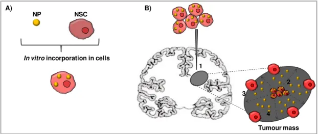
Fig. I.4 – Schematic representation of nanoparticles delivery by neural stem cells inside a brain tumour.
A) Incorporation of nanoparticles (NP) containing anticancer drugs into neural stem cells (NSC). B) Delivery strategy: 1. Intratumoural injection of NSC loaded with NP into the tumour mass. 2. Migration of NSC loaded with NP. 3. NSC distribution at the border between normal brain parenchyma and the tumour mass. 4. Release of the therapeutic agent. Adapted from Roger et al. (2011).
The stimulation of endogenous neural progenitor and stem cells, and the transplantation of adult-derived neural progenitor and stem cells may represent valid strategies for the treatment of a broad range of CNS diseases and injuries. However, their clinical application is limited by both ethical and logistical problems such as their isolation and their immunological compatibility in allogenic transplantation (Colleoni and Torrente, 2008; Taupin, 2006).
1.4. In vitro culture of NSC: the neurosphere assay
NSC sources are located in inaccessible areas within the brain, thus severely limiting their clinical utility. Hence, it is highly desirable and urgent to find an alternate source of neural cells (Sun et al., 2011; Suzuki et al., 2004). Working on an attempt to find a method of expanding NSPC, several
groups contributed to the discovery of two culture systems for the maintenance and expansion of NSC: neurospheres (NS, in suspension) and adhesive substrate-bound (adherent) cultures. Both systems are good culture methods; however, although the adherent culture is richer in NSC and grow significantly faster than NS, it could only maintain robust growth during 6-7 passages, while NS could be maintained for more than 10 passages (Sun et al., 2011).
“NS”, the culture system used in the present thesis, was performed for the first time by Reynolds and Weiss (1992). They have demonstrated that cells from embryonic or adult nervous system can be cultivated and propagated in vitro as NS from single NSC, suggesting their potency for
self-renewal (Zhao et al., 2008). NS agglomerate and form spherical clusters and can be expanded in
long term suspension cultures, in the presence of growth factors (Bez et al., 2003; Breier et al., 2010).
These cellular structures represent three dimensional heterogeneous clusters of proliferating cells, B)
NSC NP
In vitro incorporation in cells A)
1
2
4 3
11
including stem cells, committed progenitors, and differentiated cells such as neurons, astrocytes and oligodendrocytes (Bonnamain et al., 2012; Breier et al., 2010; Garbossa et al., 2012).
The morphological and functional heterogeneity of these free-floating structures is observed and evaluated through several factors as size, apoptosis, phagocytosis, proliferation/self-renewal, differentiation and migration. Spheres of different sizes could be generated from cells plated at the same time and under identical culture conditions, resulting in larger NS (with a dark core) and/or smaller ones (more translucent) (Bez et al., 2003). The size of NS influences their cell activity in that
the highest cell activity (i.e., mitosis, protein synthesis) occurs at the periphery of the agglomerates more accessible to nutrients and oxygen, while necrosis, and low or absent mitotic and transcriptional activity, are typical of the inner layers where nutritional exchanges are more difficult. Moreover, results of propidium iodide (PI)/Hoechst staining (to detect cellular death), showed some PI+ cells on the core of the big floating-free structures (Fig. I.5A) (Bez et al., 2003; Sun et al., 2011).
NSC derived from NS are not synchronized and can be in any phase of the cell cycle. Besides nestin (Fig. I.5C1) and Sox2 (Fig. I.5D1), markers of NPC, NS also express MAP2 (microtubule-associated protein 2, mature neuron marker) and GFAP, showing that the culture is also composed of mature neural populations (Sun et al., 2011). As expected, nestin+ undifferentiated cells are located in
the periphery of the sphere, while differentiated neuronal (βIII-tubulin+) and glial (GFAP+) cells (Fig.I.5B) reside in the center, probably due to a growth factor gradient from the outside to the inside of the sphere (Breier et al., 2010). Cell proliferation might be the most important feature of NS, and
both the epidermal and fibroblast growth factors (EGF and FGF, respectively) have a crucial role in the NSPCs cell cycling maintenance. The self-renewal and proliferation can be shown and quantified by the presence of immature cells (nestin+) and cycling cells [5-Bromo-2’-Deoxyiridine (BrdU+)] in the outer layer of the cluster. This double immnunofluorescence staining shows that BrdU+ cells are also
nestin+ cells (Fig. I.5E4) (Sun et al., 2011). In the same way that growth factors maintain the
proliferative characteristics, their withdrawal drive cells to stop proliferating and start differentiating and expressing neurotrophins. This, will cause a 50% decrease in BrdU labelling and nestin expression, and an increase in the number of GFAP+and β-tubulin III+ cells (Schwindt
et al., 2009). With growth
factors withdrawal, neural progenitor cells migrate radially out of the sphere onto a given extracellular matrix (ECM), and thereby differentiate into cells expressing neural and glial markers which form the migration area, while the zone pattern inside the sphere disappears. After some time of differentiation, βIII-tubulin+ cells are found at the edge of the sphere while nestin+ and GFAP+ cells are
heterogeneously distributed throughout the sphere (Breier et al., 2010; Schwindt et al., 2009).
Additionally, the dissociation of NS also favours differentiation of dissociated cells into O4+ oligodendrocytes, GFAP+ astrocytes or βIII-tubulin (TuJ1)+ neurons (Bez
et al., 2003; Sun et al.,
I. INTRODUCTION
12
k
Fig. I.5 – Principal feactures and phenotypes of neurospheres. A) Dead cells (red) in the core of the
neurosphere (NS) are labeled with propidium iodide (PI) (red) and nuclei are counterstained with Hoechst 33342 (blue). Scale bar represents 30 µm. B) NS with a central core of glial fibrillary acidic protein (GFAP) positive glia (green) surrounded by nestin (red) positive neural stem cells at the sphere edge. Scale bar represents 50 µm. C1) and D1) NS contain nestin-positive (red) and Sox2-positive [SRY (sex determining region Y)-box 2 (green)] cells, respectively. C2) and D2) Nuclei were stained with DAPI (4',6-diamidino-2-phenylindole, blue). C3) and D3) merge of images C1+C2 and D1+D2, respectively. Scale bar represents 100 µm in the series C and 20 µm in the series D. E1-4) Staining for nuclei (DAPI), proliferation (BrdU, red) and nestin (green). Scale bar represents 30 µm. Figure A and E are from Sun et al. (2011), B is from Lu et al. (2010), and C and D from Wang et al. (2007).
As referred above, the characteristic migration of these cluster cells is easily assessed because upon growth factors withdrawal, NPC start leaving the sphere in a 90o angle and their travel distance
over time can be measured through a phase contrast microscope. The process of migration is regulated by intracellular, as well as extracellular stimuli. The migration of human NPC out of the neurosphere is controlled by the mitogen MAPK (mitogen activated protein kinase) ERK1/2 (Extracellular signal-regulated protein kinases 1 and 2) –dependent and –independent pathways. Moreover, human neural progenitor cell migration is preserved on collagen, fibronectin and poly-L lysine matrices, indicating a crucial role of the ECM in neural migration not only in vivo, but also in vitro
(Breier et al., 2010).
Overall, the importance of having a reliable model of NSPC is related to the fact that these cells can serve as a valuable source of cell type-specific somatic precursors for neural transplantation, thus
C1 C2
A
B
D1 C1
C2
C3
D2
D3
13
offering a potential starting point for therapy of neurodegenerative diseases. In addition, this model can also be a useful tool for testing neurotoxic substances for their abilities to interfere with basic process of brain development, such as proliferation, migration, differentiation and apoptosis, a procedure that was called by developmental neurotoxicity testing (Breier et al., 2010).
2. Neural stem cells, tumour stem cells and brain tumours: dangerous
relationships?
Brain tumours are a wide group of abnormal masses of tissue as a result of uncoordinated proliferation of cells (neoplasms) with a high incidence in children and adults with the poorest outcome among the human cancers. In adults, their incidence is relatively high, especially in elderly people; in children, brain tumours are the second commonest type of cancer (17% of all childhood’s cancer) and cause 25% of cancer deaths. Until now, only small changes have been registered in these numbers and they continue to be associated with very high morbidity and mortality (Dirks, 2008; Sutter et al.,
2007).
There are different types of brain tumours with neuroepithelial origin, that have been classified based on the histological resemblance of tumour cells to cells present in the adult brain, and thus focusing in the cell types that compose the tumour mass (Sanai et al., 2005) (Fig. I.6). Although this
classical classification is used by most authors, more recently, and according to the 2007 report published by the World Health Organization (WHO), brain tumours have been classified not only based on a single cell type but there were included also mixed type of tumours, such as the mixed neuronal-glial (Louis et al., 2007; Sutter et al., 2007; Vescovi et al., 2006).
Brain tumours are a heterogenous group of malignancies that originate and reside within the brain, contrary to metastatic brain tumours that originate from a primary cancer outside the CNS and spread to the brain (Germano et al., 2010). They are organized as a cellular and functional hierarchy
I. INTRODUCTION
14
Current treatment strategies for brain tumours include surgery, radiotherapy, and chemotherapy. Nevertheless, these clinical interventions modestly improve patient survival. In addition, treated patients often have intellectual impairment related to chemotherapy and radiotherapy. Moreover, even brain tumours classified as benign can be lethal due to their location in surgically inaccessible areas (Yao et al., 2009; Dirks, 2008). The core of treatment failure derives from the poor
understanding of the cellular and molecular mechanisms regulating tumour growth. Thus, it is very important to understand how cells in the diverse tumour populations initiate and maintain growth, and which are the molecular mechanisms involved and the brain cells that suffer malignancy transformation and give rise to brain tumour (Dirks, 2008). Hence, it is necessary to analyse asymmetric and/or symmetric division directly on tumour stem cells and to compare the signalling pathways involved in NSC with those in CSC proliferation (Yao et al., 2009).
Fig. I.6 – Cell types and associated tumours of the central nervous system. Brain tumours have been mostly
classified based on the histological resemblance of tumour cells to central nervous system (CNS) cells, such as astrocytes, neurons, oligodendrocytes and ependymal cells. Adapted from Sanai et al. (2005).
2.1. Gliomas – an overview
Gliomas are the most common type of brain tumours, accounting for more than 70%. Gliomas consist of a heterogenous mixture of several glial phenotypes, composed by immature cell types, poorly differentiated neoplastic astrocytes and mature cells. These tumours can either develop by dedifferentiation from a low grade tumour (“secondary glioma”) or can arise “de novo” (“primary glioma”). Differences in clinical and molecular features of the two types of glioma point to a distinct pathogenesis (Park and Rich, 2009; Siebzehnrubl et al., 2011). Some controversy still exists in turn of
the origin of these brain tumours, once some authors refer to NSC as the cells that undergo molecular and cellular transformation generating the tumoural growth, while others think that glioma arises from
Astrocyte
Astrocytoma
Pilocytic astrocytoma Diffuse astrocytoma Anaplastic astrocytoma Glioblastoma
Oligoastrocytoma
Pleomorphic xanthoastrocytoma
Subependymal giant-cell astrocytoma
Neuron
Ganglioglioma
Gangliocytoma
Central eurocytoma
Oligodendrocyte
Oligodendroglioma
Oligoastrocytoma
15
dedifferentiation of mature glia (Sanai et al., 2005; Vescovi et al., 2006). However, if the last
hypothesis has been increasingly discarded, the glioma origin from NSC is the most accepted and the one intensively researched reason why this assumption was followed in the present thesis.
According to their histological features, there are three main types of the most common gliomas: astrocytomas, oligodendrogliomas, and mixed oligoastrocytomas (Siebzehnrubl et al., 2011).
According to the WHO, gliomas are organized into four different grades based on histological properties of cellular composition, nuclear morphology and atypical cell stage, mitotic activity, necrotic features, and microvascular proliferation (Louis et al., 2007; Ohgaki and Kleihues, 2005). A higher
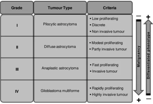
histological grade corresponds to a less differentiated phenotype and to an increasing malignancy (Park and Rich, 2009). Regarding astrocytomas, the main focus of the present work, they can be classified in low grade/WHO Grades I and II or high-grade or malignant/WHO Grades III and IV (Louis
et al., 2007; Siebzehnrubl et al., 2011) (Table I.1.).
Table I. 1 – Astrocytoma grades. Astrocytomas can be divided in four groups based on their malignant grade
and general tumour characteristics, in accordance to World Health Organization.
The latter two tumour subtypes are considered the most malignant gliomas and are associated with a very poor prognosis. Particularly, glioblastoma multiforme (GBM) accounts for 50% of primary brain tumours and only 5% of patients have a 5 years survival rate, meaning that the average survival rate is approximately only 14 to 15 months (Ohgaki and Kleihues, 2005; Stupp et al., 2005; Sutter et al., 2007). The peak of the onset of GBM is between 50 and 55 years, which makes it an age-related
disease, and male are slightly more prone to this pathology than female individuals. Moreover, the black people undergo an incidence 2-3 times higher than white people (DeAngelis, 2001; Stupp et al.,
2005).
Although gliomas are a relatively rare form of cancer, they account for a disproportionately high morbidity and mortality because their location in the brain is generally problematic to surgery and other therapies. Their treatment is a daunting challenge to clinicians due mainly to the lack of effective
Criteria Tumour Type Grade Pilocytic astrocytoma Anaplastic astrocytoma Glioblastoma multiforme
Low proliferating
Discrete
Non invasive tumour
Modest proliferating
Partly invasive tumour
Fast proliferating
Invasive tumour
Rapidly proliferating
I. INTRODUCTION
16
therapeutic options. Gliomas rarely metastasize outside of the brain, but instead, infiltrate extensively into surrounding normal brain tissue. Thus, although the surgery is not curative it can establish the diagnosis. It is taken as the first choice because the resection of the tumour is important to decrease the pressure it exerts and is usually followed by focal external beam radiation (Germano et al., 2010;
Park and Rich, 2009). Subsequently, radiation therapy and chemotherapy increase survival, but disease recurrence is virtually inevitable (Park and Rich, 2009). Besides these difficulties it is observed that the average survival after surgical resection alone is six months with only 7.5% of patients surviving two years post-operatively. Adding radiation therapy, this average survival prolongs to nine months, while systemic chemotherapy provides minimal survival benefits (Siebzehnrubl et al.,
2011).
The latest researches have attempted the use of systemic chemotherapy with alkylating agents such as temozolomide (TMZ). Thus, since 2005, the care standard for newly diagnosed patients with GBM includes resection, fractionated radiation concurrent with TMZ chemotherapy, followed by TMZ alone (DeAngelis, 2001). This association between surgery, radiation and chemotherapy is not able to avoid that the recurrence in high-grade gliomas will occur in more than 90% of cases, frequently within 2 cm of the original site, but 10 to 20% of the cases may develop new lesions at distant sites (Germano et al., 2010). However, it showed to increase the overall survival by 2.5 months, the
progression-free survival by two months and the two-year survival by 16% (DeAngelis, 2001). Despite the important effort that has been made to find therapeutic agents and the development of GMB models representing the features of human malignancy, the poor identification of the malignant tumour initiating cells has limited the development of more effective therapies (Siebzehnrubl et al., 2011).
Overall, the lack of knowledge about the relationship between the normal cell and the tumourigenic cellular transformation, as well as the poor information about the characteristic tumour heterogeneity, have delayed the identification of new molecular targets and the development of novel target therapeutic processes to apply in GBM treatment. Hence, it is necessary a better understanding of the mechanisms involved in the tumour origin and progression, to allow the discovery of new and more effective therapies.
2.2. Cancer stem cell hypothesis
Tumours are currently viewed as a disruption of the cellular organization mechanisms resulting from accumulation of genetic and epigenetic events at the germ line and somatic levels. The cell of origin of cancer has been a strongly debated topic throughout the history of cancer research, and over the last few years the idea that cancer is a disease driven by CSC has emerged (Bapat, 2007; Houghton et al., 2007), as referred on the beginning of this chapter.
Although the concept of CSC has been originally proposed in 1990s, the first evidence of the presence of cells with stem-like characteristics in human brain tumours was only reported later by Ignatova et al. (2002) who isolated clonogenic neurosphere-forming precursors from post-surgery
17
have shown the presence of these cells within brain tumours; for instance, two different groups demonstrated that both GBM and medulloblastoma contain NS-forming cells that can give rise to neuronal and astroglial-like cells (Hemmati et al., 2003; Singh et al., 2003).
The identification of CD133 (prominin 1) immunoreactive cells in brain tumours and their characterization as CSC was based on the fact that the glycosylated epitope of the CD antigen AC133 appeared to be restricted to stem cells. These CD133+ cells in brain tumours were shown to be highly tumourigenic (Germano et al., 2010) and express molecular markers associated with neural
precursors, such as nestin, the transcription factor Sox2, the RNA binding protein Musashi, B-cell-specific Moloney murine leukemia virus integration site 1 (Bmi-1), Notch, the transcription factor Emx2, paired box 6 (Pax6) and the ligand for the receptor notch 1 (Jagged1). However, when exposed to differentiation conditions, these cells downregulate the expression of these immature markers and acquire immunoreactivity for βIII-tubulin (for neurons), GFAP (for astrocytes) and platelet-derived growth factor receptor-α (PDGFRα) (for oligodendrocytes) (Dirks, 2008; Germano et al., 2010; Sutter et al., 2007). Additionally, the CD133+ population is resistant to several chemotherapeutic agents
(such as TMZ, carboplatin, placlitaxel and etoposide) and to radiation therapy (Germano et al., 2010;
Yao et al., 2009). These cells by showing karyotypic and other genetic alterations are indeed
neoplastic and not residual NSC entrapped in the tumour (Fig. I.7) (Germano et al., 2010).
Altogether, these findings demonstrate that: a) different brain tumours are composed by transformed, undifferentiated neural precursors that respond to the same mitogens and activate adult NSC; b) tumour stem-like cells possess some of the molecular features of NSC; c) CD133, a cell-surface protein that is a marker of normal human neural precursors, can be used for the enrichment of tumour stem-like cells from brain tumours; and d) tumour stem-like cells have the ability to maintain stem cell function and to promote tumour growth. These findings and assumptions led to the cancer stem cell hypothesis in GBM and in other solid tumours, such as medulloblastomas and ependymomas (Taylor et al., 2005).
In the core of the relation between NSC and CSC there is a difference related to self-renewal. While NSC can maintain a controlled balance between self-renewal and differentiation, CSC are more long-lived. Indeed, these tumour-derived NS could be cultured for at least four months without relevant changes in their proliferative properties, whereas normal NS grown under identical conditions persisted no longer than one month in culture (Sutter et al., 2007). Hence, this uncontrolled
proliferation can be in the origin of the brain tumour. So, if a normal NSC, during its life, accumulates enough mutations and undergoes neoplastic transformation, it would then become a CSC, forming and maintaining a brain tumour. Moreover, a recent work developed by Jackson and colleagues (2006), has shown that NSC into the SVZ express PDGFRα and the administration of exogenous PDGF (platelet-derived growth factor) initiates a signalling cascade that induces aberrant proliferation of these cells leading to formation of hyperplastic lesions, with properties like gliomas, which, however, regress upon PDGF withdrawal. So, this deregulation of the PDGF signalling suggests that genetic or epigenetic mechanisms affecting the proliferation of NSC can be an early factor in brain tumour formation (Sutter et al., 2007). Thus, it is hypothesized that CSC can be generated from the oncogenic