From the Department of Dermatology, Hospital das Clínicas, Faculty of Medicine, University of Sao Paulo.
NAIL MANIFESTATIONS IN PEMPHIGUS VULGARIS
Juliana Burihan Cahali, Everton Yuji Soyama Kakuda, Cláudia Giuli Santi and Celina Wakisaka Maruta
RHCFAP/3098
CAHALI JB et al. - Nail manifestations in pemphigus vulgaris. Rev. Hosp. Clín. Fac. Med. S. Paulo 57(5):229-234, 2002.
Nail involvement in pemphigus vulgaris is rare. We describe 5 patients with pemphigus vulgaris presenting nail involvement. In this disease, nail manifestations present, by order of frequency, as chronic paronychia, onychomadesis, onycholysis, Beau’s lines and trachyonychia. All our 5 cases presented with paronychia, and 1 of them also had Beau’s lines. Treatment with prednisone and/or cyclophosphamide controlled mucocutaneous and nail manifestations in all cases.
DESCRIPTORS: Pemphigus vulgaris. Paronychia. Beau’s lines. Prednisone. Cyclophosphamide.
INTRODUCTION
Pemphigus vulgaris is an autoimmune, mucocutaneous vesicu-lobullous disease1. It is characterized
by intraepidermal bullae with suprabasal acantholysis and IgG autoantibody against a desmosomal-associated glycoprotein, desmoglein 12,3. Although pemphigus vulgaris may
occur at any age, its most common on-set is in the fourth, fifth, and sixth dec-ades4,5. Involvement of the nail in
pemphigus vulgaris is rarely de-scribed. We describe 5 patients with chronic paronychia as a manifestation of pemphigus vulgaris.
CASE REPORTS
Case 1
A 65-year-old white woman pre-sented in 1991 with oral erosions and cutaneous lesions on the whole body. Histological examination of the af-fected skin revealed suprabasal
clefting containing acantholytic cells. Epidermal intercellular deposition of IgG was noted on direct immunofluo-rescence. After the diagnosis of pem-phigus vulgaris, she was treated with 1 mg/kg daily of oral prednisone, which resulted in clinical resolution of both skin and oral mucosa lesions. In 1996, free of mucocutaneous lesions, on a daily maintenance dose of 10 mg/ day of prednisone, she developed ery-thema, edema, pustules, inflammation, and suppuration of the proximal and lateral nail folds of several fingernails and toenails associated with Beau’s lines of the 2nd, 3rd, and 4th digits on
the left hand (Fig. 1). Gram stain and potassium hydroxide preparations of nail drainage were negative. Bacterial and fungal cultures disclosed
Staphy-lococcus aureus, but no fungi.
Topi-cal and oral antibiotic therapy pro-duced no beneficial effect. A biopsy
specimen of the nail fold of the right first toe revealed suprabasal clefting and acantholysis. Direct immunofluo-rescence showed positive intercellular deposition of IgG and C3. Indirect im-munofluorescence was positive at a dilution of 1/10240. Thus, we consid-ered the paronychia to be a clinical manifestation of pemphigus vulgaris. Mucocutaneous lesions of pemphigus vulgaris occurred 2 weeks after the nail involvement. She was treated with a new cycle of prednisone 1 mg/kg/day, resulting in clinical resolution of mu-cocutaneous and nail lesions with no residual damage on the nails.
Case 2
A 15-year-old white man with pemphigus vulgaris diagnosed in 1993 presented initially with ulcera-tions in the oral cavity and simultane-ous involvement of the nails. The lat-ter was characlat-terized by erythema, edema, inflammation, and bullae, with purulent drainage on 1st and 4th right
2). Skin lesions of pemphigus vulgaris appeared 1 month later. Histological examination of the affected skin re-vealed suprabasal clefting and acan-tholysis. Direct and indirect immun-ofluorescence of perilesional skin showed epidermal intercellular depo-sition of IgG and C3. Gram stain and potassium hydroxide preparations of the drainage were negative, as were bacterial and fungal cultures. Nail bi-opsy was not performed. He was treated with a 1mg/kg daily dose of oral
pred-nisone. Mucocutaneous and nail le-sions improved simultaneously with this therapy, with no permanent dam-age on the nail.
Case 3
A 27-year-old white woman with pemphigus vulgaris diagnosed in Au-gust 2000 presented initially with skin lesions, oral erosions, and nail in-volvement characterized by erythema, edema, and purulent drainage from the
proximal nail fold of the right first fin-ger (Fig. 3). Gram stain and potassium hydroxide preparations of the drain-age were negative as were bacterial and fungal cultures. She was diag-nosed as having pemphigus vulgaris on the basis of a skin biopsy and di-rect immunofluorescence. Indidi-rect im-munofluorescence was positive at a dilution of 1/1280. Nail biopsy was not performed. There was a rapid re-sponse of the mucocutaneous and nail lesions to prednisone 1 mg/kg daily, with resolution of the nail lesions.
Case 4
A 44-year-old white woman with pemphigus vulgaris diagnosed in Au-gust 2000 presented initially with skin and oral lesions. Nail lesions occurred 3 months later, with erythema and edema on the lateral and posterior nail folds of her 1st, 2nd, and 3rd left and
right fingers (Fig. 4). She noticed ex-acerbation of the mucocutaneous le-sions 2 weeks after the development of the paronychia. She was diagnosed as having pemphigus vulgaris on the ba-sis of a skin biopsy and direct immun-ofluorescence. Indirect immunofluo-rescence was positive at a dilution of 1/2560. Nail biopsy was not per-formed. Gram stain and potassium hy-droxide preparations of the nail were negative, as were bacterial and fungal cultures. Mucocutaneous and nail le-sions improved with oral prednisone 1 mg/kg daily associated with endov-enous cyclophosphamide (900 mg/ dose, monthly) and methylprednisone (300 mg/day during 3 consecutive days in the 2nd month of the treatment).
Case 5
A 39-year-old white woman with pemphigus vulgaris diagnosed in 1990 presented initially with oral ero-sive lesions. Skin lesions appeared 2 months later. One month after that, the Figure 2 - CASE 2: Erythema, edema, and bullous lesions, with purulent drainage on 1st and 4th
right fingernails and 1st and 2nd left fingernails.
nail involvement began. It was char-acterized by erythema and edema on all 20 nails, with purulent drainage of her left and right first fingers. Nail in-volvement preceded an exacerbation of mucocutaneous disease. She was di-agnosed as having pemphigus vulgaris on the basis of a skin biopsy and di-rect immunofluorescence. Nail biopsy was not performed. Gram stain and po-tassium hydroxide preparations were negative. Bacterial and fungal cultures disclosed Staphylococcus aureus, but no fungi. Oral antibiotic therapy pro-duced no beneficial effect on the paro-nychia. Mucocutaneous lesions and
paronychia improved markedly with oral prednisone (1 mg/kg daily) and endovenous cyclophosphamide (0.5 g to 1 g/dose, monthly).
DISCUSSION
Pemphigus vulgaris is an autoimmune, mucocutaneous vesicu-lobullous disease1,5. Nail disease has
been reported in a variety of manifes-tations: chronic paronychia, Beau’s lines, trachyonychia, onychomadesis, onycholysis, subungueal hemorrhage, nail plate discoloration, pitting,
ony-choschizia, and nail dystrophy1,6-15.
Engineer et al.1 summarized all reports
in the English literature on nail in-volvement in patients with pemphigus vulgaris (15 patients). The most fre-quent alterations were paronychia (60%) and onychomadesis (33%). Our 5 patients had paronychia, and 1 of them had Beau’s lines. According to the literature, fingernails were more of-ten involved than toenails (9 had fin-gernails involved, 5 had finger and toenails involved, and 1 had only toe-nails involved). In our series, no one had toenail involvement alone (Table 1). In the previously reported cases in Figure 3 - CASE 3: Erythema, edema, and
purulent drainage on the first right finger.
Figure 4 - CASE 4: Erythema and edema on the lateral and posterior nail folds of her 1st, 2nd, and 3rd left and right fingers, with purulent drainage.
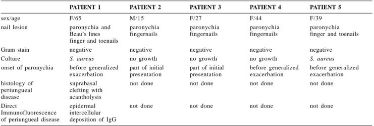
Table 1 - Clinical and laboratory features of the patients.
PATIENT 1 PATIENT 2 PATIENT 3 PATIENT 4 PATIENT 5
sex/age F/65 M/15 F/27 F/44 F/39
nail lesion paronychia and paronychia paronychia paronychia paronychia
Beau’s lines fingernails fingernails fingernails finger and toenails finger and toenails
Gram stain negative negative negative negative negative
Culture S. aureus no growth no growth no growth S. aureus
onset of paronychia before generalized part of initial part of initial before generalized before generalized exacerbation presentation presentation exacerbation exacerbation
histology of suprabasal not done not done not done not done
periungueal clefting with
disease acantholysis
Direct epidermal not done not done not done not done
the literature, the nail changes oc-curred independently of the extent of disease (67% had mucocutaneous le-sions, 27% had cutaneous lele-sions, and just 6% had only mucosal disease). All of our patients had mucocutaneous disease. Nail involvement is due to bullous lesions in the nail bed, nail matrix, or nailfold1 as part of the
dis-ease process8. Nail disease can be part
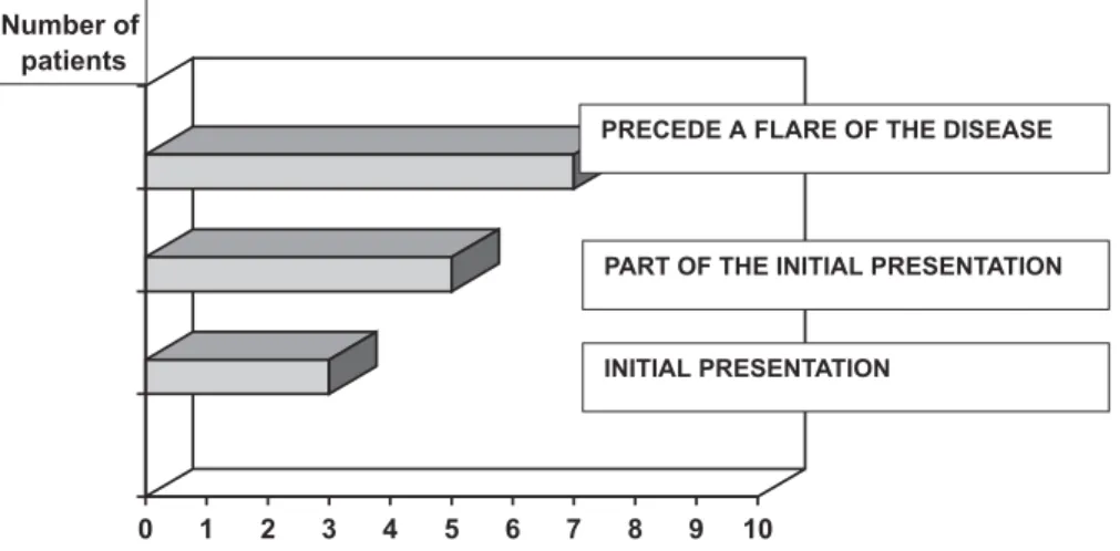
of the initial presentation along with mucosal and cutaneous lesions (47% of all cases reported), can precede a flare of the pre-existing disease (33%), or can be the only sign of the disease (20%). In our study, nail involvement preceded a flare of the disease in 3 cases and was part of the initial pres-entation in 2 cases (Figs. 5 and 6).
We thought bacteria and fungi were secondary invaders6 and the
chronic paronychia would not be re-solved with antibiotic or antifungal therapy. Another possibility that could be included among the differential di-agnosis is herpetic whitlow, since these patients are chronically under the use of immunosuppressive drugs. This di-agnosis could be ruled out once the paronychia was resolved with the proper pemphigus control. In our cases, nail biopsy was performed only in case 1. In the others, we did not take biopsy specimens because the diagno-sis of pemphigus vulgaris had already been made histologically and by direct and indirect immunofluorescence of the affected skin, and there is a known
association of pemphigus vulgaris with nail disease.
CONCLUSION
We present 5 patients with pemphi-gus vulgaris and nail involvement. This involvement can be part of the initial presentation, precede a flare of the pre-existing disease, or can be the only sign of the disease.
It is important in patients with pemphigus vulgaris and paronychia to be aware that the nail changes can be a manifestation of the autoimmune disease, and in these cases, treatment directed against fungi, bacteria, and viruses may not be effective.
Figure 6 - Onset of nail disease in this series.
REFERENCES
1. ENGINEER L, NORTON LA & AHMED R - Nail involvement in pemphigus vulgaris. J Am Acad Dermatol 2000; 43: 529-35. 2. CHAMPION RH, BURTON JL, BURNS DA et al. - Textbook of Dermatology. 6th ed. Oxford, Blackwell Science, 1998. p. 1849-1855.
3. EYRE RW & STANLEY JR - Identification of pemphigus vulgaris antigen extracted from normal human epidermis and comparison with pemphigus foliaceus antigen. J Clin Invest 1988; 81: 807-12.
4. SAMPAIO SAP & RIVITTI EA - Dermatologia. Brasil. 2th ed. São Paulo, Artes Médicas, 2000. p. 229-248.
RESUMO RHCFAP/3098
CAHALI JB e col. - Manifestações ungueais no pênfigo vulgar. Rev. Hosp. Clín. Fac. Med. S. Paulo 57(5):229-234, 2002.
O acometimento ungueal no pênfigo vulgar é um achado raro. Des-crevemos cinco doentes com pênfigo vulgar e alterações ungueais. As
alte-rações ungueais descritas no pênfigo vulgar são, em ordem decrescente de ocorrência: paroníquia crônica, onicomadese, onicólise, linhas de Beau e traqueoníquia. Paroníquia crô-nica foi a forma de acometimento ungueal encontrada em todos os doen-tes deste estudo e um deles apresen-tou, também, linhas de Beau. As
ma-nifestações cutâneo-mucosas e ungueais foram controladas, em todos os casos, com o uso de prednisona e/ ou ciclofosfamida.
DESCRITORES: Pênfigo vulgar. Paroníquia. Linhas de Beau. Prednisona. Ciclofosfamida.
5. MARTINS CR, SQUIQUERA HL & DIAZ LA - Pemphigus vulgaris and pemphigus foliaceus. Curr Probl Dermatol 1989; 1: 33-61.
6. KIM BS, SONG KY, YOUN JI et al. - Paronychia – A manifestation of pemphigus vulgaris. Clin Exp Dermatol 1996; 21: 315-7. 7. De BERKER D, DALZIEL K, DAWBER RPR et al. - Pemphigus associated with nail dystrophy. Br J Dermatol 1993; 129: 461-4.
9. BAUMAL A & ROBINSON M - Nail bed involvement in pemphigus vulgaris. Arch Dermatol 1973; 107 :751.
10.DHAWAN SS, ZAIAS N & PENA J - The nail fold in pemphigus vulgaris. Arch Dermatol 1990; 126: 1374-5.
11.AKIYAMA C, SOU K, FURUYA T, SAITOH A el al. - Paronychia: a sign heralding an exacerbation of pemphigus vulgaris. J Am Acad Dermatol 1993; 29: 494-6.
12.RIVERA DIAZ R, LLAMAZARES JA, PERALTO JLR et al. - Nail involvement in pemphigus vulgaris. Int J Dermatol 1996; 35: 581-2.
13.FULTON RA, CAMPBELL I, CARLYLE D et al. - Nail bed immunofluorescence in pemphigus vulgaris. Acta Derm Venereol (Stockh) 1983; 63: 170-2.
14.LEROY D, LEBRUN J, MAILLARD V et al. - Pemphigus végétant a type clinique de dermatite pustuleuse chronique de hallopeau. Ann Dermatol Venereol 1982; 109: 549-55.
15.STONE OJ & MULLINS JF - Vegetative lesions in pemphigus. Dermatol Int 1966; 5: 137-40.