Article
Printed in Brazil - ©2012 Sociedade Brasileira de Química0103 - 5053 $6.00+0.00A
*e-mail: [email protected]
#Current address: Department of Biochemistry and Microbiology, Institute of Biosciences, Universidade Estadual Paulista, Campus de Rio Claro, Av. 24A, 1515, Bela Vista, 13506-900 Rio Claro-SP, Brazil
§Current address: Embrapa Instrumentação Agropecuária, Rua XV de Novembro, 1452, 13560-970 São Carlos-SP, Brazil
β
-Lactam Antibiotics Epitope Mapping with STD NMR Spectroscopy:
a Study of Drug-Human Serum Albumin Interaction
Cíntia D. F. Milagre,# Luís F. Cabeça,§ Wanda P. Almeida and Anita J. Marsaioli*
Institute of Chemistry, University of Campinas, CP 6154, 13083-970 Campinas-SP, Brazil
Eventos de reconhecimento molecular são questões chave em muitos processos biológicos. O experimento de STD NMR (ressonância magnética nuclear por diferença de transferência de saturação) é uma das técnicas usadas para se entender tais interações biológicas. Neste trabalho, foram investigadas as interações entre quatro antibióticos β-lactâmicos pertencentes às classes das cefalosporinas e penicilinas com a albumina de soro humano (HSA) através de STD NMR de 1H. Nossos resultados indicam que a interação entre o anel aromático destes antibióticos e HSA é responsável pela eficiência da ligação. Assim, as diferenças estruturais entre os tio-anéis de cinco dos seis membros das penicilinas e cefalosporinas parecem não influenciar as interações do antibiótico com a albumina.
Molecular recognition events are key issues in many biological processes. STD NMR (saturation transfer difference nuclear magnetic resonance spectroscopy) is one of the techniques used to understandsuch biological interactions. Herein, we have investigated the interactions of four
β-lactam antibiotics belonging to two classes (cephalosporins and penicillins) with human serum albumin (HSA) by 1H STD NMR revealing that the interaction between the aromatic moietyand HSA is responsible for the binding efficiency. Thus, the structural differences from the five to six-membered thio ring in penicillins and cephalosporins do not seem to influence antibiotic-albumin interactions.
Keywords: STD NMR, ligand-macromolecules interaction, albumin, β-lactam antibiotics
Introduction
Understanding the molecular processes that govern the behavior and pharmacokinetics of drug-drug and drug-plasma protein in vivo is of upmost interest with an increasing appreciation of the role of human serum albumin (HSA) on the effective activity of drugs at their site of action.1 Albumin possesses several different sites for
interaction with small acids and neutral molecules. One of their most outstanding properties is its ability to reversibly bind to an incredible variety of ligands in blood, which is an important factor in the transport and release of various drugs and hormones.2 Therefore, drug-albumin complexes
have been extensively studied with respect to drug storage, drug delivery control systems and drug preservation
from rapid metabolization.3 The most used methods for
measuring plasma protein binding are equilibrium dyalysis, ultrafiltration and ultracentrifugation.4 These methods are
highly reliable, however they require a very laborious experimental protocol with chemical modification of the amino acids in HSA to determine the binding sites, but fail in the identification of the drug epitope. This issue can be addressed using nuclear magnetic resonance (NMR) spectroscopic technique which is a powerful tool to probe and understand at molecular level weak binding processes that are in fast exchange on the NMR time scale.5 In doing so, NMR relaxation time measurements
(T1 and/or T2) and STD (saturation transfer difference) NMR experiments are among the most suitable.6
We report herein the architecture and topology of a binary complex of drug-albumin, based on 1H NMR
The drugs used in these studies belong to two classes of β-lactam antibiotics, cephalosporins and penicillins (Figure 1), which display broad spectrum action, are currently employed in clinical medicine and show relatively low affinities to albumins, which are difficult to characterize with most existing methods due to the short lifetime of the complex of antibiotic-albumin.
Experimental
Materials
Both cephalosporins (Cephalexine and Cefaclor) were obtained from EMS (São Bernardo do Campo, Brazil). Amoxicillin (Amoxil®) and Pencillin V (Pen-Ve-Oral®)
were purchased from GlaxoSmithKline (Rio de Janeiro, Brasil) and Eurofarma (São Paulo, Brasil). Deuterated water (D2O, 99.9%) was obtained from Acros Organics and HSA
(fraction V) from Sigma Aldrich.
Sample preparation
Each antibiotic was previously extracted with distilled water, filtered, lyophilized and used without further purification. Antibiotic solutions (10 mmol L-1) were
prepared in deuterated water (D2O, 99.9%), buffered at
pH 7.4 (phosphate buffer saline). Albumin (90 µmol L-1)
was added to the antibiotic solution and gently mixed to avoid foam formation. For the protein, the ligand ratios were 1:100 and ligand:ligand ratios 1:1.
Acquisition of NMR spectra
All NMR experiments were recorded at a temperature of 298 K with a spectral width of 10 ppm on a Varian INOVA-500 spectrometer operating at 11.74 T, observing
1H at 499.89 MHz. The spectrometer was equipped with
a 1H{13C/15N/31P} 5 mm PFG Penta Probe with inverse
detection and linear pulsed gradient on z-axis. The 1H NMR
chemical shifts are given in ppm related to the residual HDO signal at 4.68 ppm. For the pulse sequence PRESAT (water suppression using presaturation),7 Water package from
Varian was used to selectively suppress the water signal. In STD experiments, selective saturation of the protein was achieved by a train of Gaussian shaped pulses of 50 ms each, truncated at 1% and separate by a 1 ms delay. The duration of the presaturation of 2.55 s was adjusted using n = 150 cycles. With a T1ρ-filter, 30-ms spin-lock pulse
was utilized to remove residual protein resonances. All STD experiments were selectively saturated using the Gaussian train pulses at −0.5 ppm for the on-resonance (Ion) and at
30 ppm for (Ioff) in which no protein signal was detected and
for the STD control. The subtraction was performed after every scan by phase cycling. Spectrum processing was performed on a Sun workstation using VnmrJ software (Varian package). The STD NMR spectrum provides the epitope mapping with values obtained from the individual signal intensities in the STD NMR spectrum (Ion) and in the
reference STD NMR spectrum (Ioff). The relative amount
of saturation transferred is measured by A = ∆I/Ioff and
normalized using the largest STD effect as reference (100),
i.e., to that of the H-7 proton of 1 as determined from STD NMR spectra at a 100-fold excess.
Results and Discussions
1H NMR chemical shift variations of cephalosporins
(1 and 2) and penicillins (3 and 4) in the presence and absence of HSA were the first evidence of formation of these antibiotic-albumin complexes (Tables 1 and 2). The interaction of 1-4 with HSA was delineated by applying STD NMR, an experiment based on NOE transference from
the macromolecule to the ligand. It consists in applying a selective radio frequency pulse on the macromolecule. The magnetization is transferred to the entire macromolecule in less than 0.1 s by spin diffusion, this saturation is transferred to bound ligands and is detected in the free-ligand solution. The ligand hydrogen most tightly bound to the macromolecule will receive the most intense magnetization-transfer and the amplitude of these signals will accordingly change due to NOE effects.8 Therefore, NOE effects are
observed for the protons in close contact with the protein, allowing direct observation of the ligand moiety involved in the protein-ligand interaction, i.e., the epitope. The saturation degrees were calculated by determining individual signal intensities in the STD spectrum (ISTD) and in the reference
STD spectrum (Ioff).5 The relative amount of transferred
saturation is measured by A = ISTD/Ioff and normalized using
the largest STD effect as reference.
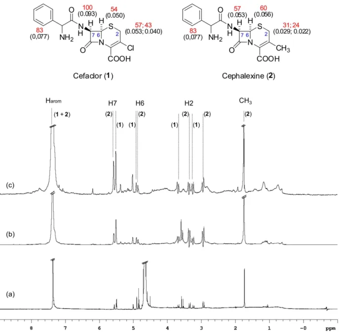
The orientation of antibiotics 1 to 4 inside the HSA receptor was revealed by signal differences in the STD NMR spectra (Tables 1 and 2). As a rule, large STD effects were observed for the aromatic moieties of 1 to 4 indicating that these protons have the most intimate contact with albumin. The other protons displayed secondary STD effects, placing them at a slightly longer distance from the protein interacting site. Among the cephalosporins,
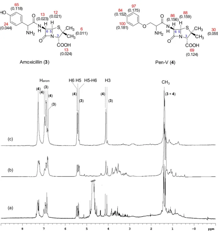
the substitution of methyl group in 2 by a chlorine atom in 1 caused a 50% enhancement in the H-2 proton signal. Likewise, among the penicillins, the change in R group caused a substantial decrease (ca. 50%) in H-3, H-5 and H-6 proton signal intensities from 3 to 4. However, according to these STD NMR experiments, the structural difference between the six-membered thio ring containing sulfur fused to β-lactam ring in cephalosporins and 5-membered thiazole ring of penicillin has no influence on the antibiotic-albumin interactions.
Aiming at revealing interaction differences between HSA in a mixture of cephalosporins 1 + 2, a STD experiment was set up and it could be observed that 1 is more tightly bound to HSA than 2. The epitope map profiles show slight changes (Figure 2). However, in a similar experiment performed with a mixture of penicillins 3 + 4, the interaction differences between 3 and 4 with HSA were significant (as shown in Figure 3), with 4 much more tightly bound to HSA than 3. Signals between 3 to 4 ppm in Figure 3 are from antibiotic impurities and did not interfere in these analyses since they do not interact with albumin as shown on Figure 3c.
In addition, a STD NMR experiment with all four antibiotics 1 to 4 and HSA was carried out and as expected, most signals overlapped due to the structural similarity.
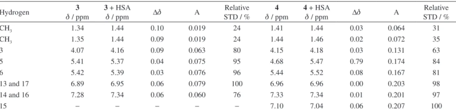
Table 2. 1H NMR spectral parameters of 3 and 4 in the presence and absence of HSA with their relative degrees of saturation
Hydrogen 3
d / ppm
3 + HSA
d / ppm ∆d A
Relative STD / %
4
d / ppm
4 + HSA
d / ppm ∆d A
Relative STD / %
CH3 1.34 1.44 0.10 0.019 24 1.41 1.44 0.03 0.064 31
CH3 1.35 1.44 0.09 0.019 24 1.44 1.46 0.02 0.072 35
3 4.07 4.16 0.09 0.063 80 4.15 4.18 0.03 0.131 63
5 5.41 5.37 0.04 0.075 95 4.68 5.47 0.79 0.174 84
6 5.42 5.39 0.03 0.076 96 5.44 5.52 0.08 0.167 81
13 and 17 6.89 6.95 0.06 0.079 100 6.96 6.96 0.00 0.203 98
14 and 16 7.28 7.34 0.06 0.060 76 7.33 7.34 0.01 0.201 97
15 − − − − − 7.10 7.04 0.06 0.207 100
The experiments were done at 298 K and pH 7.4 using 499.89 MHz in D2O, and residual HDO signal was used as reference at 4.68 ppm. The STD spectrum provided epitope mapping with values obtained from the individual signal intensities in the STD spectrum on resonance (Ion) and in the reference NMR spectrum off resonance (Ioff). The relative degrees of saturation were measured by A = (ISTD/Io) and were normalized using the largest STD effect as reference.
Table 1. 1H NMR spectral parameters of 1 and 2 in the absence and presence of HSA with their relative degrees of saturation
Hydrogen 1
d / ppm
1 + HSA
d / ppm ∆d A
Relative STD / %
2
d / ppm
2 + HSA
d / ppm ∆d A
Relative STD / %
2α 3.28 3.21 0.07 0.088 65 2.97 2.95 0.02 0.040 44
2β 3.73 3.65 0.08 0.089 66 3.40 3.36 0.04 0.031 34
6 5.08 4.98 0.10 0.116 86 4.96 4.91 0.05 0.065 71
7 5.65 5.56 0.67 0.115 85 5.56 5.51 0.05 0.074 81
Aromatics 7.44-7.48 7.33 0.11 0.135 100 7.44-7.46 7.38 0.06 0.091 100
CH3 − − − − − 1.78 1.74 0.04 0.043 47
However, the H-5 and H-6 protons from the penicillins (3, 4) and the H-7 protons from the cephaloporins (1, 2) were well resolved, allowing individual binding information of 1, 2, 3 and 4 to HSA (Figure 4), revealing the order 3 < 2 < 1< 4, corresponding to 20, 52, 78 and 100%, respectively, of relatively bound ligand (normalized values).
The STD NMR technique used in those experiments proved its efficiency in detecting low binding affinity ligands even in mixtures of ligands with high structural similarities, revealing its power as a screening tool for detecting and characterizing ligand binding. Albumins have more than one active binding site, consequently
compounds 1-4 could interact with different regions of the protein. However, the results presented herein suggest that all four antibiotics compete for the same binding site of the albumin, displaying a similar epitope mapping.
Conclusions
The epitope maps of 1, 2, 3 and 4 to HSA determined in this study indicate that in all four antibiotics, the aromatic moiety plays an important role in drug-protein anchorage and showed that the change from five to six-membered thio ring does not seem to influence those
antibiotic-HSA interactions. We have further evaluated the relative binding stability of a mixture of these antibiotic (1, 2, 3, 4)-HSA complexes revealing that 4 (Pen-V) is the best binding drug followed by 2 (Cefaclor), 3 (Amoxicillin) and 1 (Cephalexine). Moreover, apparently compounds 1-4 compete for the same active binding site. Apart from the fact that non-specific binding is one of the limitations of the STD technique, when used to compare
results obtained with different ligands, the competition STD NMR methods are valuable and are important tools in drug discovery. This can be used for compound library screening to identify ligands with relative affinities and to derive structure-activity relationships, which are used to optimize the NMR hits into viable drug leads. Finally, STD competition experiments allow the determination of binding constants when employing titration experiments or one of
Figure 4. (a) STD NMR spectrum of HSA and a mixture of 1 + 2 + 3 + 4, (b) STD NMR control spectrum (off resonance) of the mixture cited before. Spectra (c) and (d) correspond to amplification of circled regions in spectra (a) and (b) respectively and refers to H-7 (1), H7 (2), H-6 and H-5 (3), and H-6 and H-5 (4).
the employed compound has a known KD (dissociation
constant) and all compounds have the same binding site.8,9
Supplementary Information
Supplementary data are available free of charge at http://jbcs.sbq.org.br as pdf file
Acknowledgements
The authors thank the Brazilian science foundations Fundação de Amparo à Pesquisa do Estado de São Paulo (FAPESP, CInAPCe Program) and the Conselho Nacional de Desenvolvimento Científico e Tecnológico (CNPq) for their financial support. We acknowledge Prof. Carol Collins from IQ-Unicamp for the text revision.
References
1. Lin, J. H.; Curr. Drug Metab. 2006, 7, 39; Trainor, G. L.; Expert Opin. Drug Discovery 2007, 2, 51; Paxton, J. W.; Methods Find. Exp. Clin. Pharmacol. 1983, 5, 635.
2. Goodman, D. S.; J. Am. Chem. Soc. 1958, 80, 3892; Daughaday, W. H.; Physiol. Rev. 1959, 39, 885; Jacobsen, J.; FEBS Lett.
1969, 5, 112; Jacobsen, J.; Int. J. Pept. Protein Res. 1977, 9, 235; Burke, C. W.; Lewis, B.; Panveliwalla, D.; Tabaqchali, S.; Clin. Chim. Acta 1971, 32, 207; Beaven, G. H.; Chen, S. H.; D‘Albis, A.; Gratzer, W. D.; Eur. J. Biochem. 1974, 41, 539; Adams, P. A.; Berman, M. C.; Biochem. J. 1980, 191, 95; Brodersen, R.; J. Biol. Chem. 1979, 254, 2364; Klopfenstein, W. E.; Biochim. Biophys. Acta 1969, 181, 323; Richardson, J. S.; Nature 1977, 268, 495; Roda, A.; Cappelleri, G.; Aldini, R.;
Roda, E.; Barbara, L.; J. Lipid Res. 1982, 23, 490; Savu, L.; Benassasyag, C.; Vallette, G.; Christeff, N.; Nuney, E.; J. Biol. Chem. 1981, 256, 9414; Yates, F. E.; Urguhart, J.; Physiol. Rev.
1962, 42, 359; Unger, W. G.; J. Pharm. Pharmacol. 1972, 24, 470; Kragh-Hansen, U.; Pharmacol. Rev. 1981, 33, 17. 3. Ji, Z-S.; Li, C-G.; Mão, X-A.; Liu, M-L.; Hu, J.-M.; Chem.
Pharm. Bull. 2002, 50, 1017.
4. Pacifici, G. M.; Viani, A.; Clin. Pharmacokinet. 1992, 23, 449. 5. Mayer, M.; Meyer, B.; J. Am. Chem. Soc.2001, 123, 6108;
Meyer, B.; Peters, T.; Angew. Chem., Int. Ed. 2003, 42, 864; Carlomango, T.; Annu. Rev. Biophys. Biomol. Struct.2005, 34, 245; Krishna, N. R.; Jayalakshmi, V.; Prog. Nucl. Magn. Reson. Spectrosc. 2006, 49, 1; Milagre, C. D. F.; Cabeça, L. F.; Martins, L. G.; Marsaioli, A. J.: J. Braz. Chem. Soc. 2011, 22, 286. 6. Figueroa-Villar, J. D.; Tinoco, L. W.: Curr. Top. Med. Chem.
2009, 9, 811 and references cited herein.
7. Varian NMR Spectrometer Systems; User guide: Liquids NMR, Pub. No. 01-999161-00 Rev. B0801; Varian Inc.: Palo Alto, USA, 2001, available online at http://www.zhoulab.biochem. duke.edu/VNMR_manual/VNMR_Liquids.pdf accessed in October 2011.
8. Pons, J.; Todeschi, N. E.; Bertho, G.; Benarous, J. G.; Tanchou, V.; Benarous, R.; Girault, J. P.; Biochemistry2008, 47, 14. 9. Fielding. L.; Prog. Nucl. Magn. Reson. Spectrosc.2007, 51,
219; Viegas, A.; Manso, J.; Nobrega, F. L.; Cabrita, E. J.; J. Chem. Educ.2011, 88, 990; Wang, Y. S.; Liu, D. J.; Wyss, D. F.; Magn. Reson. Chem. 2004, 42, 485.
Submitted: September 7, 2011
Published online: December 20, 2011