S
CIENTIFICA
RTICLESThe effect of time interval between electrical
stimulation on the denervated rat muscle
O efeito do intervalo da estimulação elétrica no músculo desnervado de rato
Caierão QM1, Betini J1, Teodori RM1,2, Minamoto VB1,2
Abstract
Objective: To compare the effect of electrical stimulation (ES) applied daily and on alternate days, on the area density of the connective tissue (CT) and on the cross-sectional area (CSA) of the denervated muscle fi bers. Methods: Thirty-fi ve rats were divided into the following groups: control (C), denervated (D), denervated + daily electrical stimulation (D+DES) and denervated + alternate-day electrical stimulation (D+ES). The application of ES on the gastrocnemius was started 24 hours after nerve damage of axonotmesis type and was applied for 20 and 30 days. Cross-sections were stained with hematoxylin-eosin to measure the CSA and area density of CT. The statistical analysis consisted of the Shapiro Wilk test followed by analysis of variance (ANOVA) F (one-way) and the Tukey test (p≤ 0.05). Results: Analysis of the area density of CT showed that only the D+DES Group presented values similar to those of the C Group, for the two periods analyzed. There was no difference in CSA in the 20-day Group between the ES Groups and the D Group (p> 0.05). After 30 days, all the experimental groups reached CSA values similar to the C Group. Conclusions: The ES was ineffi cient for minimizing the muscle fi ber atrophy. However, the CT was responsive to ES, and daily applications were more benefi cial for the muscle than were alternate-day applications, thus suggesting that the interval for applying ES to denervated muscle is an important variable for CT adaptation.
Key words: denervation; gastrocnemius; connective tissue; hypertrophy; electrical stimulation.
Resumo
Objetivo: Comparar o efeito da estimulação elétrica (EE) aplicada diariamente e em dias alternados na densidade de área do tecido conjuntivo (TC) e na área de secção transversa (AST) das fi bras do músculo desnervado. Materiais e métodos: Trinta e cinco ratos foram divididos em grupos controle (C), desnervado (D), desnervado + eletroestimulado diariamente (EED) e desnervado + eletroestimulado em dias alternados (EEA). A aplicação da EE no músculo gastrocnêmio teve início 24 horas após lesão nervosa do tipo axioniotmese, sendo a mesma aplicada durante 20 e 30 dias. Cortes transversais foram corados com HE para mensurações da AST e densidade de área de TC. Análise estatística: teste Shapiro Wilk, seguido pela análise de variância (ANOVA) F (one-way) e teste de Tukey (5%).
Resultados: Na análise da densidade de área do TC, observou-se que somente o Grupo EED apresentou valores similares ao Grupo C nos dois períodos analisados. No Grupo 20 dias, não houve diferença na AST quando comparados os grupos submetidos à EE com o Grupo D (p> 0,05), e após 30 dias todos os grupos experimentais alcançaram valores similares ao Grupo C. Conclusões: A EE não foi efi ciente para minimizar a atrofi a das fi bras musculares. Entretanto, o TC foi responsivo à EE, sendo a aplicação diária mais benéfi ca ao músculo do que a aplicação em dias alternados, sugerindo que o intervalo de aplicação da EE em músculo desnervado é variável importante para as adaptações do TC.
Palavras-chave: desnervação; gastrocnêmio; tecido conjuntivo; hipertrofi a; estimulação elétrica.
Received: 18/10/2007 – Revised: 19/11/2007 – Accepted: 13/02/2008
1 Graduate Program in Physical Therapy, Universidade Metodista de Piracicaba – Piracicaba (SP), Brazil 2 Department of Physical Therapy, Universidade Metodista de Piracicaba – Piracicaba (SP), Brazil
Correspondence to: Prof. Viviane B. Minamoto, Universidade Metodista de Piracicaba, Master’s Program in Physical Therapy, Rodovia do Açúcar Km 156 - Bloco 7, CEP 13400-911 - Piracicaba-SP,
e-mail: vbminamo@unimep.br
Introduction
Muscle fibers have a close relationship with their con-nective tissue, and this characteristic is important for the maintenance of the integrity and properties of the skel-etal muscle, including the production of movements and forces1-3. Both muscle fibers and the extracellular matrix
(ECM), respond directly to electrical stimuli, by means of contraction of the actin and myosin filaments and regulation of protein synthesis in the ECM, respectively4.
Therefore, impairments of muscle innervation affect both muscle fiber, as evidenced by the decrease in the fiber’s cross sectional area (CSA)5,6, and the ECM, observed by the
increase of intramuscular connective tissue (CT)6,7. As a
result, the denervated muscle shows a deficient function, as demonstrated by the decreases in force production and increases in passive resistance8-10.
These modifications occur immediately after denerva-tion and persist while the muscle lacks nervous supply. If the denervation remains for a long period, the CT will substitute the muscle’s contractile elements, inhibiting muscle regeneration completely11. Electrical stimulation
(ES) is a therapeutic modality used, after a nervous injury, to minimize muscle degeneration and weakness, while the nerve is regenerating12,13.
Studies related to the therapeutic use of ES are contro-versial as a consequence of the use of different parameters and protocols in the application of ES. However, these stud-ies are important for the understanding of the effects of this therapy on denervated muscles. It is known that some vari-ables, such as different frequencies of ES applications over 24 hours14 and the quantity of stimuli per day15 are crucial
for the results of this type of intervention. These studies have shown that, whereas 100 muscle contractions applied daily with constant duration were efficient for maintaining muscle mass and strength, the same number of contractions during a four-hour ES session, followed by 20 hours of rest, were not sufficient to maintain the same muscular proper-ties14. Furthermore, one study observed that the number of
200 contractions was ideal to obtain significant effects on muscle mass and strength15. The results of this study also
made it possible to conclude that, although a minimal num-ber of contraction is necessary, excessive contractions can increase tissue damage due to the great amount of energy applied to the muscle. A recent study16 demonstrated that
20 electrically-induced contractions per day were not suf-ficient to prevent atrophy of muscle fibers. It is important to note that Dow et al.15 and Russo et al.16 used different
physi-cal parameters of the electriphysi-cal current, which confounds these results.
h e ef ects of ES on denervated muscle may also depend on the regeneration phase in which the nerve exists. h is hy-pothesis is based on previous studies, which showed that the innervated muscle does not respond to ES17,18. h us, distinct
muscle responses are expected when the ES is applied during dif erent phases of nerve regeneration.
The aim of the present study was to compare mor-phological adaptations of the denervated gastrocnemius muscle after the administration of two ES application pro-tocols. The hypothesis was that such adaptations would be influenced by the frequency of the ES applications, either on alternate days or daily. Moreover, the effects of this modality during different periods of the nerve injury were studied, either 20 or 30 days after the lesion. It was believed that the results of the present study could help in the selection of the most appropriate therapy for the treatment of denervated muscles.
Methods
Thirty-five Wistar rats (200 ± 50g) were randomly di-vided into seven experimental groups (n= 5/group): con-trol (C); denervated and analyzed after 20 and 30 days after denervation (D-20, D-30); denervated + electrostimulated daily during 20 and 30 days (EED-20, EED-30), and den-ervated + electrostimulated in alternate days (EEA-20, EEA-30).
The animals were maintained in plastic cages, at a tem-perature of 23 ± 2 0C, with free access to food and water,
and submitted to a 12 hour light-dark cycle. The experiment was approved by the Ethics in Animal Experimentation Committee of Universidade Federal de São Carlos, with the protocol number of 008-06. For the procedures on nerve injury, ES applications and muscle collection, the animals were anesthetized with intramuscular injection of Ketalar®
(Cloridrato de Ketamina; 50 mg/mL) and Rompun®
(Clo-ridrato de Tiazina; 2 g/100mL), applied to the right gluteus, in a proportion of 1:1, with a dose of 0.3 mL/100g of body weight. After muscle removal the animals were sacrificed through cervical dislocation.
Protocol of nerve injury and electrical stimulation
h e procedure for nerve injury was conducted according to a previously described modii ed protocol19. h e animals
were submitted to an incision of 15 mm, approximately, in the left gluteus region, to expose and injure the sciatic nerve. h e nerve was crushed using hemostatic tweezers, applying four pinches with a duration of 20 secs at intervals of one sec. h e
pinches pressure was standardized for all animals, using the second tooth of the rack7, and all pinches were carried out by
the same person.
Electrical stimulation began 24h after nerve injury, and the animals were electrostimulated daily from Monday to Friday or three times a week, during 20 or 30 days, depend-ing on the experimental group. Electrical current was gen-erated by the DUALPEX 961 system (QUARK, Brazil), using two percutaneous self-adhesive electrodes, with an area of 1 cm2, attached to the inguinal region and on the left
gastrocnemius. The stimulus variables were: a symmetri-cal biphasic quadratic impulse with phase duration=3 ms, frequency=10 Hz, intensity=5 mA, standardized by the observation of a vigorous muscle contraction. To avoid muscle accommodation to the stimulus, 1 mA was added every five mins of the current application, for a total of 10mA at the end of 30 mins of electrostimulation9. In every
ES session, gel was applied to the electrode, to facilitate the coupling between the electrode and the skin.
Muscle Collection
After muscle collection, the belly of the medial portion of the left gastrocnemius muscle was frozen in isopentane, previously cooled in liquid nitrogen, and the samples were stored in liquid nitrogen for subsequent analyses. To obtain the histological sections, the muscles were glued with traga-canth gum on a wooden plate. Sections of a 12μm thickness were obtained from all muscles, using a cryostat microtome (Mod. 300, Ancap, Brazil), and stained with hematoxylin and eosin (H&E). The best histological section, without artifacts or blood vessels, was chosen and photographed in all its extensions with a 20x objective lens. The images were obtained using an optical microscope BX-41 (Olympus, Japan) coupled to a digital camera C5050 (Olympus, Japan). The density of the connective tissue area was measured by point-counting planimetry20 using the software Image Pro
Plus 4.0 (Media Cybernetics, USA). A cross-sectional area (CSA) of approximately 200 fibers/muscle was measured us-ing the software, Motic Image Advanced 3.2 (Motic Instru-ments - Canada).
Data analysis
At i rst, the Shapiro-Wilk test was used to test for data normality. Subsequently, the Leven test was used to verify the homogeneity of the data. As the all variables showed normal distributions and homogeneity, an ANOVA-F (One-Way) was conducted, followed by Tukey HSD tests. For all analyses the level of signii cance was set at 5%. h e software used to carry
out the analyses was the Statistical Package for Social Sci-ences (Version.11.0).
Results
Density of connective tissue areas
h e distribution of CT’s density can be observed in Figure 1. Proliferation of CT was more evident in the perimysium. Consid-ering the morphological patterns of the muscle, the group submit-ted to 30 days of daily-applied ES (Figure 1D), was the one which demonstrated morphological characteristics most similar to the control muscle (Figure 1A). Groups D and EEA, evaluated after 20 days, showed higher CT area densities when compared to the group C (p≤ 0.0008) and the group EED had values similar to the values of group C (Table 1). Related to the animals evaluated after 30 days, group D showed higher CT area densities in comparison to groups C and EED (p≤ 0.0008; Table 1). Furthermore, there were no dif erences between the groups submitted to ES and group C.
145
Table 1. Percentage of connective tissue densities of all experimental groups
Groups
Control 12 ± 1.37 (%)
20 days (%) 30 days (%)
D 18 ± 3.50† 15 ± 2.83*
AES 17 ± 2.70† 13 ± 2.46
DES 15 ± 2.47 11 ± 1.59
* compared to C and EED; p≤ 0.0008; † compared to C; p< 0.005; Denervated (D),
alter-nate electrical stimulated (AES) and daily electrical stimulated (DES).
Figure 1. Cross sections of the gastrocnemius muscle from control (A), denervated 20 days (B) denervated 30 days (C) and daily electrical stimulated 30 days (D) groups. Observed perimysial connective tissue densities (asterisk) on the denervated groups. H&E staining, Bar 50 µm, 20x.
*
B
*
C A
*
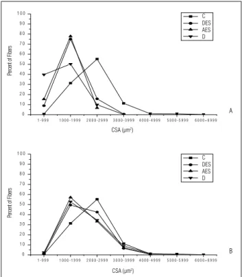
Cross-sectional areas of the muscle fi bers
h e animals of groups D, EED and EEA, evaluated after 20 days, showed a decrease in CSA i bers when compared to the group C (p≤ 0.0000; Table 2). Both EED and EEA were not ef ective in preventing muscle atrophy, since there were no dif erences between electrostimulated groups and group D (p> 0.05; Table 2). Considering the total 30-day period, all groups demonstrated similar CSA values (p> 0.09; Table 2).
Group C showed the highest concentrations of i bers with CSA values of 1000-2999 μm2 (86%). In contrast, the groups
D, EEA, and EED, analyzed after 20 days, showed the highest
concentrations of i bers with CSAs of 1-1999 μm2(84 to 93%),
demonstrating the atrophy of the denervated muscles, in-dependently of the use of ES (Figure 2A). h e groups D, EEA, and EED, analyzed after 30 days, showed a distribution in the number of i bers similar to those observed in group C, with the highest concentration being between 1000-2999 μm2
(87 to 92%), which indicated the recovering of the i bers CSA at this period (Figure 2B).
Discussion
The effectiveness of ES in the treatment of denervated muscles is influenced by several factors and, although there are a great number of studies that have investigated the best treatment protocol in the literature14,15, the effects of daily
and alternate applications on muscle adaptations were not found. The comparison of these modalities of ES is impor-tant, since it is unknown if the frequency of ES applications is a relevant variable in the treatment of denervated mus-cles. Another aim of the present study was to analyze the effects of ES during different phases of nerve regeneration. Therefore, based on previous studies21,22,muscle analyses
were conducted after 20 and 30 days, when the muscle was in the polyinnervation, or in the neuromuscular remodeling phase, and during the period when the muscle recovery was almost complete.
Although CT and muscle fibers are responsive to elec-trical activity, the results suggest that the responses of these tissues to ES depend on the amount of differenti-ated contraction stimuli, since only the CT was responsive to the treatment. One possible hypothesis is that simple muscle contractions regulate ECM expression, whereas hypertrophy stimuli are dependent on muscle overload, which was not reached by the applied ES. Another hypoth-esis is that there was a differentiated temporal response of these tissues to ES, and, thus, the muscle fiber hypertrophy could be observed over the long term, or after 30 days of treatment.
At the 20 day period, it was observed that the frequency of ES application was crucial for CT adaptation, once the daily applied treatment was ef ective for the maintenance of the tissue area densities within normal values. In contrast, in the groups treated over 30 days, which reached more advanced nerve maturation, the frequency of the treatment sessions did not appear to inl uence this maintenance, since both daily and alternate ES applications maintained the tissue area densities similarly to the control values. However, only the group which had submitted to daily ES demonstrated dif erent results in comparison to the denervated group.
146
Table 2. Muscle fi bers cross sectional areas of all experimental groups
Groups
Control 2337 ± 323 (µm2)
20 days (µm2) 30 days (µm2)
D 1246 ± 368* 2052 ± 430
AES 1394 ± 179* 1999 ± 347
DES 1579 ± 207* 2078 ± 203
* compared to C; p< 0.0000; Denervated (D), alternate electrical stimulated (AES) and daily electrical stimulated (DES).
0 1 0 2 0 3 0 4 0 5 0 6 0 7 0 8 0 9 0 1 0 0
Pe
rc
ent
of
Fi
ber
s
A
0 1 0 2 0 3 0 4 0 5 0 6 0 7 0 8 0 9 0 1 0 0
Pe
rc
ent
of
Fi
ber
s
B
C DES AES D
C DES AES D
CSA (μm2) CSA (μm2)
1 - 9 9 9 1 0 0 0 - 1 9 9 9 2 0 0 0 - 2 9 9 9 3 0 0 0 - 3 9 9 9 4 0 0 0 - 4 9 9 9 5 0 0 0 - 5 9 9 9 6 0 0 0 = 6 9 9 9
1 - 9 9 9 1 0 0 0 - 1 9 9 9 2 0 0 0 - 2 9 9 9 3 0 0 0 - 3 9 9 9 4 0 0 0 - 4 9 9 9 5 0 0 0 - 5 9 9 9 6 0 0 0 = 6 9 9 9
The importance of the contractile activity for avoiding proliferation of the intramuscular CT, as was observed in the present study, was also found in a previous publication23.
This mechanism is probably related to the fact that the gene expression in the ECM is regulated by physical stimuli4. The
maintenance of the CT area density is important, since an increase in the quantity of this tissue works as a mechanical barrier which blocks blood supply to the muscle fibers, what may contribute to muscular atrophy24 and impair the process
of muscle reinnervation25. Moreover, the increased density
of the CT area makes the collagen fibers to be configured in a closer contact between themselves, possibly stimulating the formation of abnormal cross-links and resulting in a loss of extensibility and increased tissue stiffness26.
Aside from its inl uences on the ECM density, physical stimuli are responsible for the organization of the ECM4,27. h is
organi-zation is important, since muscle i bers do not always extend from one muscle extremity to the other and this coni guration is crucial for adequate force transmission of the muscle3.
h e rapid loss of muscle proteins after denervation occurs mainly in the myoi brillar components, which represent 60% of the proteins in the muscle28, and the contractile activity is
important to maintain the CSA of the muscle i bers15.However,
in the present study, the daily or alternate electrostimulated groups did not show, at the analyzed periods, dif erent results from the denervated group (D-20 and D-30). Some factors are likely related to these results, such as an insuffi cient amount of electrical activity or inappropriate stimuli, which were proba-bly incapable of producing muscle hypertrophy, and the period of analysis, which was too short to allow for alterations in the CSA i bers. A previous study15 demonstrated that the ES was
capable of maintaining the CSA of denervated muscles, how-ever, the adopted ES protocol, dif erent from the one used in the present study, is not viable for physical therapy treatments, since the authors used implanted electrodes.
In spite of the atrophy observed after 20 days of nerve injury, a progressive increase in the CSA could be noted and, hence, it was spontaneously restored in all groups after 30 days of nerve injury. h ese results suggest that, after an axonotmesis
nerve injury, in which the denervation period is short (reinner-vation begins at the 14º day after lesion)21, the restoration of
voluntary electric stimuli was suffi cient to reverse atrophy of the rat muscle.
It is important to stress that, although the electrical stimu-lation used was not ef ective in minimizing the reduction of the CSA of the muscle i bers, other i bers’ adaptations, not analyzed in the present study, may have occurred due to ES applications. Previous studies demonstrated that electrical stimulation im-proved the metabolism of muscle i bers29, which contributes
to maintain their energetic patterns, thus favoring muscle con-tractions. Furthermore, it was observed that, when a muscle is submitted to contractions, for at least 30 minutes per day, an increase in the mitochondrial volume and enzymatic capacity occurs, thus reducing muscle fatigue30.
Conclusions
h e injury of a peripheral nerve produced signii cant changes in the muscle i bers and intramuscular connective tissue of the gastrocnemius muscle of the rat. h e application of electrical stimulation af ected only the adaptations of the connective tis-sue. h e daily application of the electric stimulus was crucial for an appropriate connective tissue response, thus showing that the frequency of the stimulus application, during the pe-riod of treatment, was an important variable for these tissue adaptations. h is information is relevant for the choice of the most appropriate electrical stimulation protocol for denervated muscles. Although electrical stimulation is important to main-tain muscle mass, the protocols used in the present study were not ef ective in minimizing muscle atrophy and the restoration of the i bers’ cross-sectional areas occurred spontaneously over the 30-day period after the axonotmesis.
Acknowledgements
FAPESP (05/52720-0) and FAP–UNIMEP (384/05).
148
References
1. Huijing PA. Muscle as a collagen fi ber reinforced composite: a review of force transmission in muscle and whole limb. J Biomech. 1999;32(4):329-45.
2. Lieber RL.Skeletal muscle structure, function, & plasticity: the physiological basis of rehabilitation. 2a ed. Philadelphia: Lippincott; 2002.
3. Järvinen TA, Józsa L, Kannus P, Järvinen TL, Järvinen M. Organization and distribution of intramuscular connective tissue in normal and immobilized skeletal muscles. An immunohistochemical, polarization and scanning electron microscopic study. J Muscle Res Cell Motil. 2002;23(3):245-54.
4. Aaron RK, Ciombor DM, Wang S, Simon B. Clinical biophysics: the promotion of skeletal repair by physical forces. Ann N Y Acad Sci. 2006;1068:513-31.
5. Soić-Vranić T, Bobinac D, Bajek S, Jerković R, Malnar-Dragojević D,
Nikolić M. Effect of salbutamol on innervated and denervated rat soleus
muscle. Braz J Med Biol Res. 2005;38(12):1799-805.
6. Fernandes KCBG, Polacow MLO, Guirro RRJ, Campos GER, Somazz MC, Pinto VF, et al. Análise morfométrica dos tecidos muscular e conjuntivo após desnervação e estimulação elétrica de baixa freqüência. Rev Bras Fisioter. 2005;9(2):235-41.
7. Polacow MLO, Silva CA, Guirro RRJ, Campos MR, Borges JP. Estudo morfométrico do músculo sóleo denervado de ratos tratados pela associação de metformina e estimulação elétrica. Rev Bras Fisioter. 2003;7(1):77-84.
8. Salonen V, Lehto M, Kalimo M, Penttinen R, Aro H. Changes in intramuscular collagen and fi bronectin in denervation atrophy. Muscle Nerve. 1985;8(2):125-31.
9. Ashley Z, Sutherland H, Lanmüller H, Russold MF, Unger E, Bijak M, et al. Atrophy, but not necrosis, in rabbit skeletal muscle denervated for periods up to one year. Am J Physiol Cell Physiol. 2007;292(1):440-51.
10. Dow DE, Carlson BM, Hassett CA, Dennis RG, Faulkner JA. Electrical stimulation of denervated muscles of rats maintains mass and force, but not recovery following grafting. Restor Neurol Neurosci. 2006;24(1):41-54.
11. Lehto M, Alanen A. Healing of a muscle trauma. Correlation of sonographical and histological fi ndings in a experimental study in rats. J Ultrasound Med. 1987;6:425-9.
12. Eberstein A, Eberstein S. Electrical stimulation of denervated muscle: is it worthwhile? Med Sci in Sports Exerc. 1996;28(12):1463-9.
13. Lomo T, Westgaard RH, Dahl HA. Contractile properties of muscle: control by pattern of muscle activity in the rat. Proc R Soc Lond B Biol Sci. 1974;187(1086):99-103.
14. Dow DE, Faulkner JA, Dennis RG. Distribution of rest periods between electrically generated contractions in denervated muscles of rats. Artifi cial Organs. 2005;29(6):432-5.
15. Dow DE, Cederna PS, Hassett CA, Kostrominova TY, Faulkner JA, Dennis RG. Number of contractions to maintain mass and force of a denervated rat muscle. Muscle Nerve. 2004;30(1):77-86.
16. Russo TL, Peviani SM, Freria CM, Gigo-Benato D, Geuna S, Salvini TF. Electrical stimulation based on chronaxie reduces atrogin-1 and MyoD gene expressions in denervated rat muscle. Muscle Nerve. 2007;35(1):87-97.
17. Noronha MA, Camargo LC, Minamoto VB, Castro CES, Salvini TF. O efeito da estimulação elétrica neuromuscular (NMES) no músculo tibial anterior do rato. Rev Bras Fisioter. 1997;2(2):71-6.
18. Brasileiro JS, Salvini TF. Limites da estimulação elétrica neuromuscular no fortalecimento de músculos esqueléticos saudáveis e com défi cit de força. Fisioterapia Brasil. 2004;5(3):224-30.
19. Bridge PM, Ball DJ, Jackinnon SE, Nakao Y, Brandt K, Hunter DA, et al. Nerve crush injuries--a model for axonotmesis. Exp Neurol. 1994;127(2):284-90.
20. Mathieu O, Cruz-Orive LM, Hoppeler H, Weibel ER. Measuring error and sampling variation in stereology: comparison of the effi ciency of various methods for planar image analysis. J Microsc. 1981;121(1):75-88.
21. Carmignoto G, Finesso M, Siliprandi R, Gorio A. Muscle reinnervation--I. Restoration of transmitter release mechanisms. Neuroscience. 1983;8(3):393-401.
22. Gorio A, Carmignoto G, Finesso M, Polato P, Nunzi MG. Muscle reinnervation-II. Sprouting, synapse formation and repression. Neuroscience. 1983;8(3):403-16.
23. Williams PE, Catanese T, Lucey EG, Goldspink G. The importance of stretch and contractile activity in the prevention of connective tissue accumulation in muscle. J Anat. 1988;158:109-14.
24. Lu DX, Huang SK, Carlson BM. Electron microscopic study of long-term denervated rat skeletal muscle. Anat Rec. 1997;248(3):355-65.
25. Carter AJ, Kristmundsdottir F, Gilmour J, Glasby MA. Changes in muscle cytoarchitecture after peripheral nerve injury and repair. J Hand Surg. 1998;23(3):365-9.
26. Borisov AB, Huang SK, Carlson BM. Remodeling of the vascular bed and progressive loss of capillaries in denervated skeletal muscle. Anat Rec. 2000;258(3):292-304.
27. Chiquet M. Regulation of extracellular matrix gene expression by mechanical stress. Matrix Biology. 1999;18(5):417-26.
28. Furuno K, Goodman MN, Goldberg AL. Role of different proteolytic systems in the degradation of muscle proteins during denervation atrophy. J Biol Chem. 1990;265(15):8550-7.
29. Forti F, Cancelliero KM, Guirro RRJ, Silva CA. Efeitos da glutamina e da estimulação elétrica sobre o perfi l metabólico de músculos desnervados. Rev Bras Educ Fis Esp. 2004;18(3):273-81.